. 2021 Dec 2;5(3):e202101164.
doi: 10.26508/lsa.202101164. Print 2022 Mar.

Bis-choline tetrathiomolybdate prevents copper-induced blood-brain barrier damage

Affiliations

Bis-choline tetrathiomolybdate prevents copper-induced blood-brain barrier damage

Sabine Borchard et al. Life Sci Alliance. .

Abstract

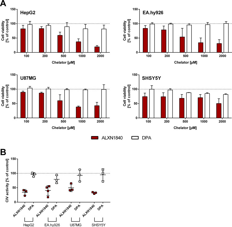
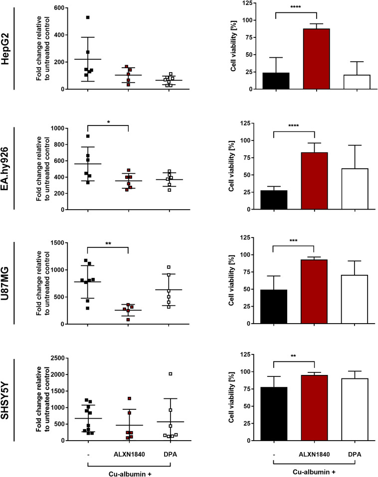
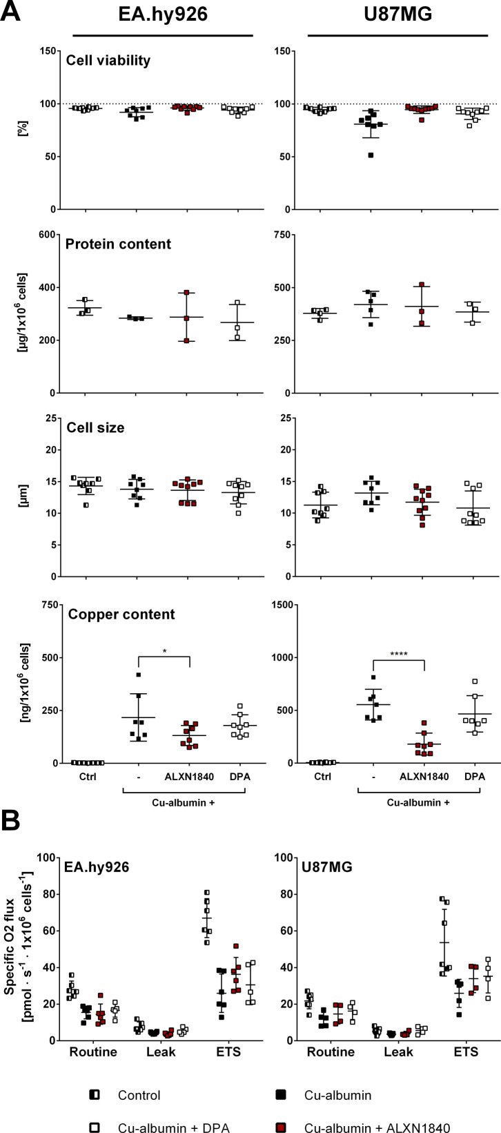
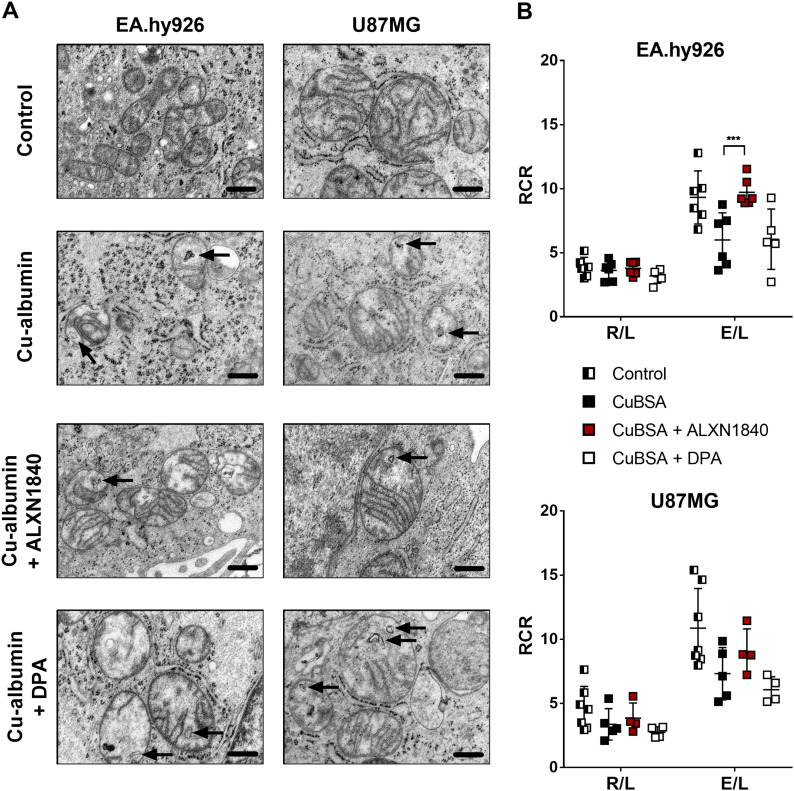
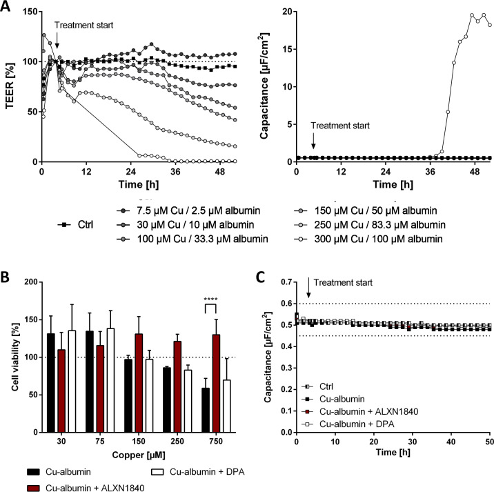
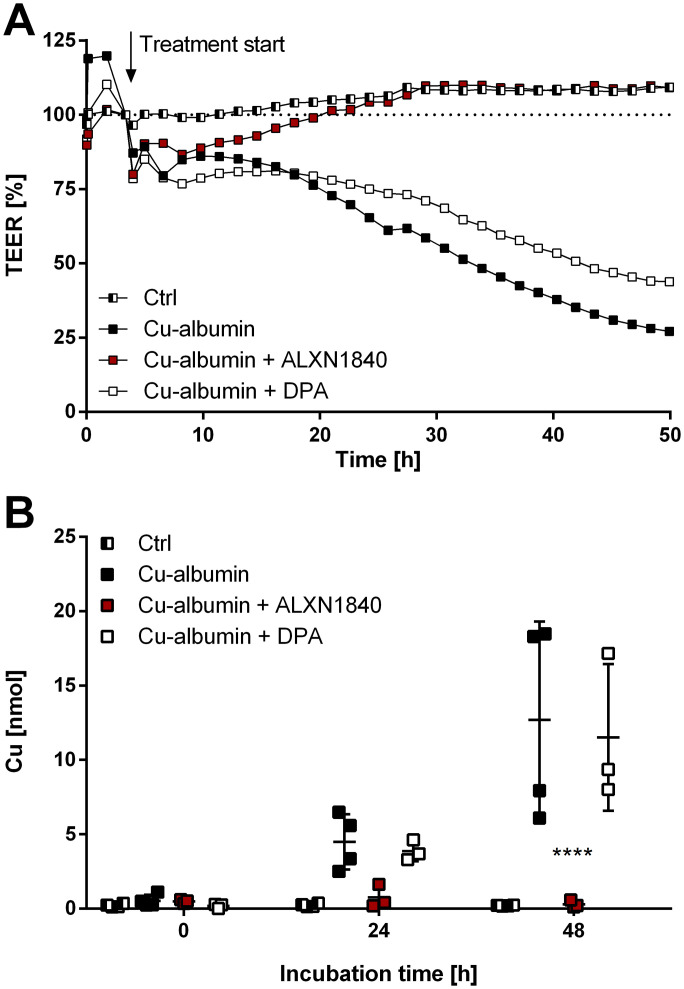
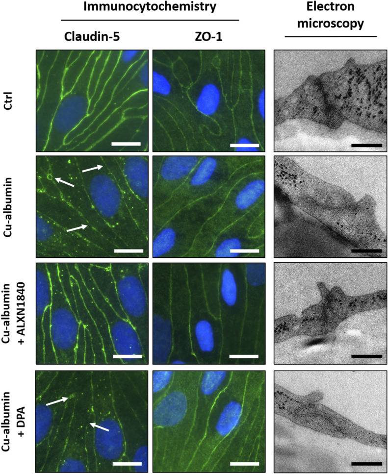
In Wilson disease, excessive copper accumulates in patients' livers and may, upon serum leakage, severely affect the brain according to current viewpoints. Present remedies aim at avoiding copper toxicity by chelation, for example, by D-penicillamine (DPA) or bis-choline tetrathiomolybdate (ALXN1840), the latter with a very high copper affinity. Hence, ALXN1840 may potentially avoid neurological deterioration that frequently occurs upon DPA treatment. As the etiology of such worsening is unclear, we reasoned that copper loosely bound to albumin, that is, mimicking a potential liver copper leakage into blood, may damage cells that constitute the blood-brain barrier, which was found to be the case in an in vitro model using primary porcine brain capillary endothelial cells. Such blood-brain barrier damage was avoided by ALXN1840, plausibly due to firm protein embedding of the chelator bound copper, but not by DPA. Mitochondrial protection was observed, a prerequisite for blood-brain barrier integrity. Thus, high-affinity copper chelators may minimize such deterioration in the treatment of neurologic Wilson disease.


Conflict of interest statement

AH Coles and R Viana are employees and shareholders of Alexion AstraZeneca Rare Disease. T Plitz was an employee of Wilson Therapeutics.

Figures

Figure 1.
Figure 1.. ALXN1840 and DPA increase blood copper levels.
(A) Positron emission tomography scan of wild-type rats with 64Cu injected either i.v. or i.p. I.v. injection results in a fast and high 64Cu signal in brain proximate vessels in contrast to i.p.–injected rats. (B) Significantly increased serum copper levels are detected in Atp7b−/− rats treated with ALXN1840 (for 4 d) upon euthanasia, in contrast to DPA treatment (N = 3). (C) During DPA treatment of Atp7b−/− rats, a significantly increased urinary copper excretion is detected (N = 3). (D) No increased fecal copper excretion is observed during ALXN1840 and DPA treatments (N = 3). One-way ANOVA with Dunnett’s multiple comparisons test was used for statistical analysis. *P < 0.05, **P < 0.01, ***P < 0.001, ****P < 0.0001.
Figure S1.
Figure S1.. Intravenous injection causes fast 64Cu signals in brain proximate vessels.
Positron emission tomography scans of wild-type rats i.v. injected with 64Cu show its distribution in large blood vessels in the brain proximity already 15 min post injection and a subsequent time-dependent decrease of the positron emission tomography signal. In contrast, i.p. injection of copper causes very low signal intensities over 120 min.
Figure 2.
Figure 2.. ALXN1840 forms a stable complex with albumin and copper.
(A) Size-exclusion chromatography demonstrates that a Cu–albumin mixture of a molar ratio of 3:1 causes the formation of a Cu–albumin complex as well as a second peak representing unbound copper. In the additional presence of ALXN1840, a single peak is encountered, suggesting the formation of an albumin–Cu–ALXN1840 complex, in contrast to the addition of DPA (N = 2). (B) Structural analysis of albumin (upper panel) and its Sudlow site I (SsI). The lower panels present close-ups of SsI with calculated difference map (Fobs–Fcalc, colored green) before (left) and after (right) refinement. ALXN1840 and copper atoms are covered by calculated 2Fobs–Fcalc map (colored blue), indicating the presence of these molecules inside SsI. (C) Electron paramagnetic resonance measurements reveal a partial reduction of Cu2+ in the albumin/Cu/ALXN1840 tripartite complex. Complete Cu2+ reduction is achieved by excess sodium dithionite (Na2S2O4).
Figure 3.
Figure 3.. Cu–albumin ratio dependent toxicity.
(A, B, C, D) Increasing molar Cu–albumin ratios cause a ratio dependent decrease in CellTiter-Glo-assessed cell viability in (A) HepG2, (B) EA.hy926, (C) U87MG, and (D) SHSY5Y cells. Such cytotoxicity is largely avoided by ALXN1840 but to a very minor part by DPA (both 750 μM, N = 3–5, n = 6–10). Two-way ANOVA with Dunnett’s multiple comparisons test was used for statistical analysis. *P < 0.05, **P < 0.01, ***P < 0.001, ****P < 0.0001.
Figure S2.
Figure S2.. Partial rescue of Cu–albumin induced cell toxicity.
(A, B, C, D) A low dose of ALXN1840 (250 μM) partially rescues Cu–albumin–induced cell toxicity in (A) HepG2 and (B) EA.hy926 cells, but not (C) U87MG and (D) SHSY5Y cells (CellTiter-Glo assay) in contrast to low-dose DPA (N = 3–5, n = 6–10). Two-way ANOVA with Dunnett’s multiple comparisons test was used for statistical analysis. *P < 0.05, **P < 0.01, ***P < 0.001, ****P < 0.0001.
Figure S3.
Figure S3.. High-affinity chelators may be cell- and mitochondria-toxic in the absence of copper accumulations.
(A) In the absence of Cu–albumin, all investigated cell lines show a dose-dependent reduction in cell viability (CellTiter-Glo) by the high-affinity chelator ALXN1840, but not by DPA (N = 3, n = 6). (B) Complex IV activity is already reduced at a non-toxic ALXN1840 concentration (200 μM) in all cell lines, whereas 200 μM DPA has no effect on complex IV activity (N = 3–4, n = 6–10).
Figure 4.
Figure 4.. Massive cellular copper accumulations are partially resolved by ALXN1840.
(Left panels) Cu–albumin incubation at a molar ratio of 3:1 (i.e., 750 μM Cu2+ and 250 μM albumin) leads to massive copper accumulation in all investigated cell lines. In the co-presence of ALXN1840, U87MG, and EA.hy926 cells, but not HepG2 and SHSY5Y cells, present with significantly lower copper content, not observed in the co-presence of DPA (N = 4–12). One-way ANOVA with Dunnett’s multiple comparisons test was used for statistical analysis. (Right panels) Such Cu–albumin incubations lead to massive cell viability loss of HepG2, EA.hy926, U87MG, and SHSY5Y as assessed by trypan blue staining. Co-presence of ALXN1840, but not of DPA, significantly protects all tested cell lines (N = 4–12). One-way ANOVA with Sidak’s multiple comparisons test was used for statistical analysis. *P < 0.05, **P < 0.01, ***P < 0.001, ****P < 0.0001.
Figure S4.
Figure S4.. Cellular parameters of EA.hy926 and U87MG cells subjected to high-resolution respirometry measurements.
(A) Cell viability assessed by trypan blue (N = 5–13), cell size (N = 5–13), and cellular protein (N = 3, n = 9) content of EA.hy926 and U87MG cells are comparable in the investigated cells. In contrast, cellular copper content is strongly elevated upon Cu–albumin treatment, partially depleted by co-presence of ALXN1840 (N = 5–13). (B) Mitochondrial respiration is decreased in EA.hy926 and U87MG cells upon Cu–albumin incubation, partially rescued by co-presence of (N = 4–7; electron transport system, capacity of the electron transport system). Two-way ANOVA with Dunnett’s multiple comparisons test was used for statistical analysis. *P < 0.05, **P < 0.01, ***P < 0.001, ****P < 0.0001.
Figure 5.
Figure 5.. Cu–albumin–induced structural and functional mitochondrial alterations.
(A) Cu–albumin incubation causes membranous inclusions and unorganized/shortened cristae in mitochondria of EA.hy926 and U87MG cells. In the co-presence of ALXN1840, but not of DPA, these alterations are partially resolved (Scale bars 500 nm). (B) Respiratory control ratios (RCR), defined as routine to leak respiration (R/L) or electron transport system to leak respiration (E/L). Co-presence of ALXN1840, but not of DPA, significantly/markedly augments the Cu–albumin induced E/L ratio drop in EA.hy926 and U87MG cells, respectively (N = 4–7). Two-way ANOVA with Dunnett’s multiple comparisons test was used for statistical analysis. *P < 0.05, **P < 0.01, ***P < 0.001, ****P < 0.0001.
Figure S5.
Figure S5.. Cu–albumin causes a dose-dependent leakiness of PBCEC monolayers already at non–cell-toxic copper concentrations.
(A) Exemplary curves of transendothelial electrical resistance (TEER) and capacitance changes of PBCEC monolayers in the presence of increasing Cu–albumin (Cu/albumin ratio 3:1 in all cases). Already low Cu–albumin concentrations cause progressive TEER decreases, whereas a capacitance increase, indicative of cell death, is only detectable at the highest tested Cu–albumin concentration. (B) Neutral red assay of PBCECs reveals no toxicity below 250 μM copper and 83.3 μM albumin upon 48 h of incubation. However, 750 μM copper (and 250 μM albumin) causes reduction in cell viability, which can be rescued by the presence of 750 μM ALXN1840 (N = 3, n = 12). (C) Capacitance values of PBCEC monolayers are unaffected by Cu–albumin treatment in the absence or presence of ALXN1840 or DPA (N = 2, n = 4).
Figure 6.
Figure 6.. Cu–albumin permeabilizes blood–brain barrier constituting endothelial cell monolayers.
(A) Cu–albumin (250 μM copper, 83.3 μM albumin), either alone or in the co-presence of DPA, leads to a time-dependent reduction in the transepithelial electrical resistance (TEER) of primary porcine brain capillary endothelial cell monolayers that is avoided by the co-presence of 250 μM ALXN1840 (N = 2, n = 4). (B) Such decreased resistance is paralleled by progressive copper appearance in the basolateral compartment (resembling the brain parenchyma) (N = 2, n = 4). Two-way ANOVA with Dunnett’s multiple comparisons test was used for statistical analysis. *P < 0.05, **P < 0.01, ***P < 0.001, ****P < 0.0001.
Figure 7.
Figure 7.. Cu–albumin disrupts tight junctions in blood brain barrier constituting endothelial cell monolayers.
(Left panels) Immunocytochemistry staining against the tight junction protein Claudin-5 shows a continuous staining of the cell margins in control PBCECs, being disrupted upon Cu–albumin treatment (250 μM copper and 83.3 μM albumin). Co-presence of ALXN1840 (250 μM), but not of DPA, alleviates these morphologic alterations. (Middle panels) The tight junction–associated protein Zonula occludens-1 (ZO-1) reveals a plasma membrane associated or more diffuse cytosolic localization in either untreated control or Cu–albumin–treated PBCECs, respectively. Co-presence of ALXN1840, but not of DPA, avoids such diffuse localization. Scale bars equal 10 μm. Electron micrographs of Cu–albumin–treated versus control PBCECs reveal less electron-dense tight junction structures. Tight junctions appear electron dense upon co-presence of ALXN1840 but not of DPA. Scale bars equal 250 nm.

References

    1. Wilson SAK (1912) Progressive lenticular degeneration: A familial nervous disease associated with cirrhosis of the liver. Brain 34: 295–507. 10.1093/brain/34.4.295 - DOI - PubMed
    1. Bull PC, Thomas GR, Rommens JM, Forbes JR, Cox DW (1993) The Wilson disease gene is a putative copper transporting P-type ATPase similar to the Menkes gene. Nat Genet 5: 327–337. 10.1038/ng1293-327 - DOI - PubMed
    1. Petrukhin K, Fischer SG, Pirastu M, Tanzi RE, Chernov I, Devoto M, Brzustowicz LM, Cayanis E, Vitale E, Russo JJ (1993) Mapping, cloning and genetic characterization of the region containing the Wilson disease gene. Nat Genet 5: 338–343. 10.1038/ng1293-338 - DOI - PubMed
    1. Tanzi RE, Petrukhin K, Chernov I, Pellequer JL, Wasco W, Ross B, Romano DM, Parano E, Pavone L, Brzustowicz LM (1993) The Wilson disease gene is a copper transporting ATPase with homology to the Menkes disease gene. Nat Genet 5: 344–350. 10.1038/ng1293-344 - DOI - PubMed
    1. Bearn AG, Kunkel HG (1954) Abnormalities of copper metabolism in Wilson[R8S2Q1M7]s disease and their relationship to the aminoaciduria. J Clin Invest 33: 400–409. 10.1172/JCI102912 - DOI - PMC - PubMed

Publication types

MeSH terms