

Secreted frizzled related-protein 2 (Sfrp2) deficiency decreases adult skeletal stem cell function in mice
- PMID: 34857734
- PMCID: PMC8639730
- DOI: 10.1038/s41413-021-00169-7
Secreted frizzled related-protein 2 (Sfrp2) deficiency decreases adult skeletal stem cell function in mice
Abstract
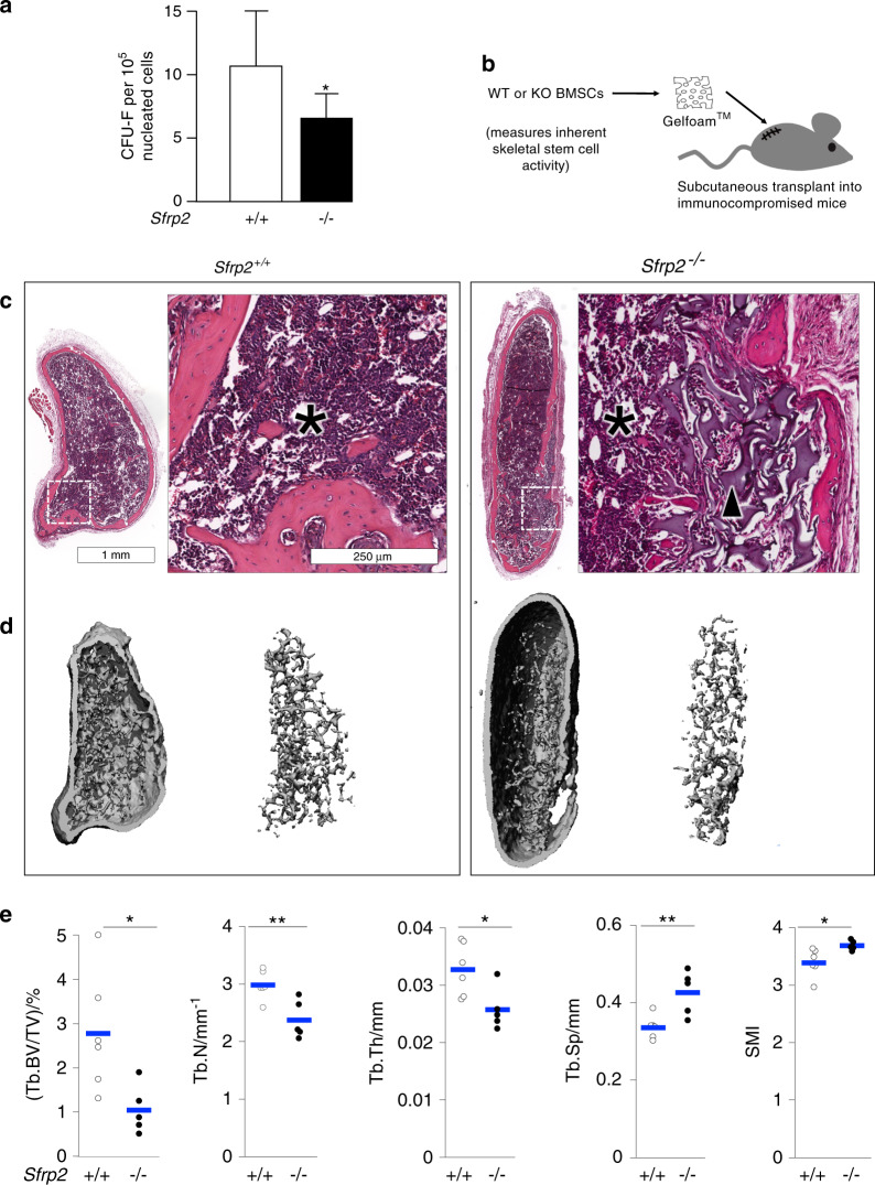
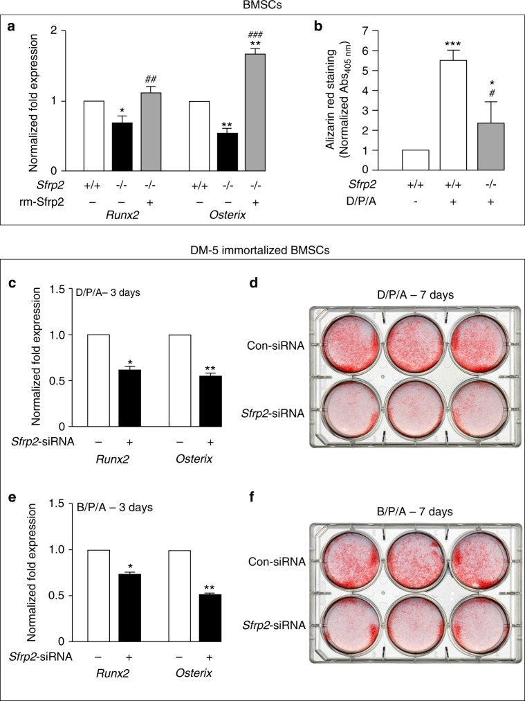
In a previous transcriptomic study of human bone marrow stromal cells (BMSCs, also known as bone marrow-derived "mesenchymal stem cells"), SFRP2 was highly over-represented in a subset of multipotent BMSCs (skeletal stem cells, SSCs), which recreate a bone/marrow organ in an in vivo ectopic bone formation assay. SFRPs modulate WNT signaling, which is essential to maintain skeletal homeostasis, but the specific role of SFRP2 in BMSCs/SSCs is unclear. Here, we evaluated Sfrp2 deficiency on BMSC/SSC function in models of skeletal organogenesis and regeneration. The skeleton of Sfrp2-deficient (KO) mice is overtly normal; but their BMSCs/SSCs exhibit reduced colony-forming efficiency, reflecting low SSC self-renewal/abundancy. Sfrp2 KO BMSCs/SSCs formed less trabecular bone than those from WT littermates in the ectopic bone formation assay. Moreover, regeneration of a cortical drilled hole defect was dramatically impaired in Sfrp2 KO mice. Sfrp2-deficient BMSCs/SSCs exhibited poor in vitro osteogenic differentiation as measured by Runx2 and Osterix expression and calcium accumulation. Interestingly, activation of the Wnt co-receptor, Lrp6, and expression of Wnt target genes, Axin2, C-myc and Cyclin D1, were reduced in Sfrp2-deficient BMSCs/SSCs. Addition of recombinant Sfrp2 restored most of these activities, suggesting that Sfrp2 acts as a Wnt agonist. We demonstrate that Sfrp2 plays a role in self-renewal of SSCs and in the recruitment and differentiation of adult SSCs during bone healing. SFRP2 is also a useful marker of BMSC/SSC multipotency, and a factor to potentially improve the quality of ex vivo expanded BMSC/SSC products.
© 2021. The Author(s).
Conflict of interest statement
The authors declare no competing interests.
Figures


Similar articles
-
IGF-1 enhances BMSC viability, migration, and anti-apoptosis in myocardial infarction via secreted frizzled-related protein 2 pathway.Stem Cell Res Ther. 2020 Jan 9;11(1):22. doi: 10.1186/s13287-019-1544-y. Stem Cell Res Ther. 2020. PMID: 31918758 Free PMC article.
-
Comparative effect of skeletal stem cells versus bone marrow mesenchymal stem cells on rotator cuff tendon-bone healing.J Orthop Translat. 2024 Jun 20;47:87-96. doi: 10.1016/j.jot.2024.05.005. eCollection 2024 Jul. J Orthop Translat. 2024. PMID: 39007033 Free PMC article.
-
DNA methylation and gene expression of sFRP2, sFRP4, Dkk 1, and Wif1 during osteoblastic differentiation of bone marrow derived mesenchymal stem cells.J Oral Biosci. 2020 Dec;62(4):349-356. doi: 10.1016/j.job.2020.08.001. Epub 2020 Aug 22. J Oral Biosci. 2020. PMID: 32835781
-
The use of adult stem cells in rebuilding the human face.J Am Dent Assoc. 2006 Jul;137(7):961-72. doi: 10.14219/jada.archive.2006.0317. J Am Dent Assoc. 2006. PMID: 16803822 Review.
-
Recent updates on the biological basis of heterogeneity in bone marrow stromal cells/skeletal stem cells.Biomater Transl. 2022 Mar 28;3(1):3-16. doi: 10.12336/biomatertransl.2022.01.002. eCollection 2022. Biomater Transl. 2022. PMID: 35837340 Free PMC article. Review.
Cited by
-
Disturbance of calcium homeostasis and myogenesis caused by TET2 deletion in muscle stem cells.Cell Death Discov. 2022 Apr 30;8(1):236. doi: 10.1038/s41420-022-01041-1. Cell Death Discov. 2022. PMID: 35490157 Free PMC article.
-
Decoding SFRP2 progenitors in sustaining tooth growth at single-cell resolution.Stem Cell Res Ther. 2025 Feb 7;16(1):58. doi: 10.1186/s13287-025-04190-z. Stem Cell Res Ther. 2025. PMID: 39920788 Free PMC article.
-
IL-27 deficiency inhibits proliferation and invasion of trophoblasts via the SFRP2/Wnt/β-catenin pathway in fetal growth restriction.Int J Med Sci. 2023 Feb 5;20(3):392-405. doi: 10.7150/ijms.80684. eCollection 2023. Int J Med Sci. 2023. PMID: 36860682 Free PMC article.
-
Distinct fibroblast progenitor subpopulation expedites regenerative mucosal healing by immunomodulation.J Exp Med. 2023 Mar 6;220(3):e20221350. doi: 10.1084/jem.20221350. Epub 2022 Dec 30. J Exp Med. 2023. PMID: 36584405 Free PMC article.
-
The pivotal role of the Hes1/Piezo1 pathway in the pathophysiology of glucocorticoid-induced osteoporosis.JCI Insight. 2024 Dec 6;9(23):e179963. doi: 10.1172/jci.insight.179963. JCI Insight. 2024. PMID: 39641269 Free PMC article.
References
-
- Owen M, Friedenstein AJ. Stromal stem cells: marrow-derived osteogenic precursors. Ciba Found. Symp. 1988;136:42–60. - PubMed
-
- Bianco, P. & Robey, P. G. in Handbook of Adult and Fetal Stem Cells. (ed Lanza, R. P.) 415–424 (Academic Press, 2004).
Grants and funding
LinkOut - more resources
Full Text Sources
Other Literature Sources
Research Materials
