. 2021 Dec 2;12(1):7020.
doi: 10.1038/s41467-021-27345-x.

Repression of germline genes by PRC1.6 and SETDB1 in the early embryo precedes DNA methylation-mediated silencing

Affiliations

Repression of germline genes by PRC1.6 and SETDB1 in the early embryo precedes DNA methylation-mediated silencing

Kentaro Mochizuki et al. Nat Commun. .

Abstract

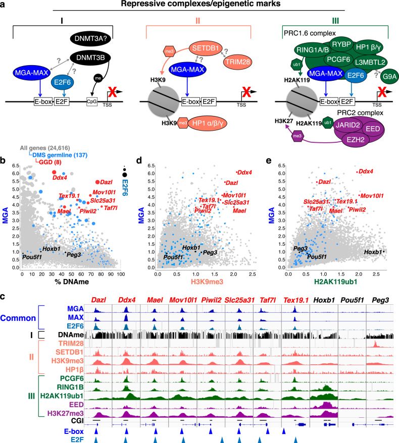
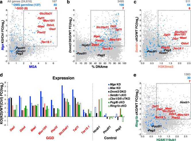
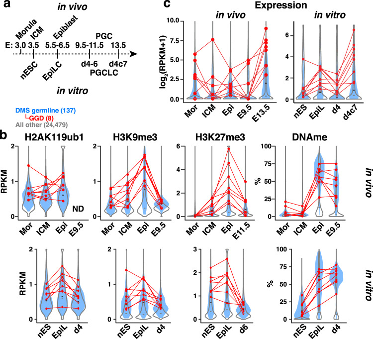
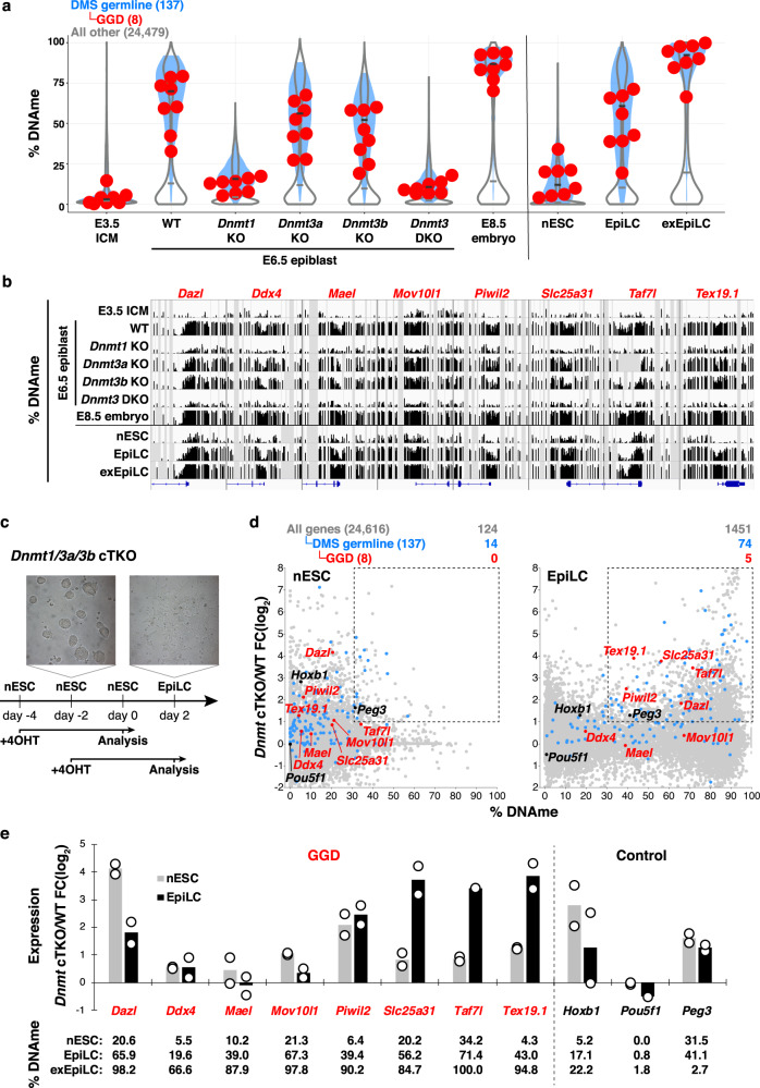
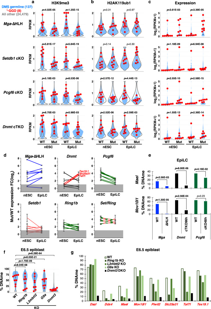
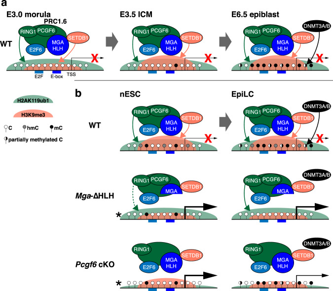
Silencing of a subset of germline genes is dependent upon DNA methylation (DNAme) post-implantation. However, these genes are generally hypomethylated in the blastocyst, implicating alternative repressive pathways before implantation. Indeed, in embryonic stem cells (ESCs), an overlapping set of genes, including germline "genome-defence" (GGD) genes, are upregulated following deletion of the H3K9 methyltransferase SETDB1 or subunits of the non-canonical PRC1 complex PRC1.6. Here, we show that in pre-implantation embryos and naïve ESCs (nESCs), hypomethylated promoters of germline genes bound by the PRC1.6 DNA-binding subunits MGA/MAX/E2F6 are enriched for RING1B-dependent H2AK119ub1 and H3K9me3. Accordingly, repression of these genes in nESCs shows a greater dependence on PRC1.6 than DNAme. In contrast, GGD genes are hypermethylated in epiblast-like cells (EpiLCs) and their silencing is dependent upon SETDB1, PRC1.6/RING1B and DNAme, with H3K9me3 and DNAme establishment dependent upon MGA binding. Thus, GGD genes are initially repressed by PRC1.6, with DNAme subsequently engaged in post-implantation embryos.


Conflict of interest statement

The authors declare no competing interests.

Figures

Fig. 1
Fig. 1. Repressive complexes and associated epigenetic marks acting on DMS germline genes in pESCs.
a Three repressive complexes (I–III) which act on a subset of germline genes have been identified previously in primed (serum-grown) ESCs (pESCs) and/or somatic cells. The promoter regions of these genes are bound by heterodimers of the transcription factors MGA and MAX, which are recruited to E-Box sites as well as E2F6, which binds to E2F sites. Complex I: The de novo DNA methyltransferase DNMT3B is recruited to germline genes via MGA/MAX and/or E2F6. Complex II: The lysine (K)MTase SETDB1, which deposits H3K9me3, is recruited to germline genes dependent upon MAX. SETDB1 is known to interact with the co-repressor TRIM28. HP1 proteins are recruited to H3K9me3 marked regions. Complex III: The non-canonical PRC1 complex PRC1.6, including PCGF6, L3MBTL2, RYBP, HP1β/γ, and RING1A/B subunits, is recruited to germline genes by MGA/MAX and E2F6. RING1A/B deposit H2AK119ub1, which in turn is recognized by the PRC2 complex, promoting H3K27me3 deposition. The KMTase G9A/EHMT2 was previously reported to associate with PRC1.6 complex via L3MBTL2. b Scatterplot showing the relationship between the enrichment (RPKM) of MGA or E2F6 (dot size) and % DNAme (TSS −0.9/+0.4 kb). c Screenshots of ChIP-seq and WGBS tracks showing RPM values of the indicated subunits of each of the complexes described in (a) as well as histone PTMs and % DNAme at the TSSs (±3 kb) of genes in pESCs. E-Box motifs and E2F motifs are shown as arrowheads. Annotated CpG islands (CGIs) are also shown. d, e Scatterplots showing the relationship between (d) MGA and H3K9me3, or (e) MGA and H2AK119ub1 (RPKM), around the TSSs (±2 kb) of all annotated genes in pESCs. In panels (b, d, e), all genes are labeled in (gray) while the 137 DMS germline genes upregulated in Dnmt3a/3b DKO embryonic day 8.5 embryos and subset categorized as germline “genome-defence” (GGD) genes (also upregulated in DNAme-deficient MEFs) are labeled in blue and red, respectively. Control genes Hoxb1 (a canonical PRC1/2 target), Pou5f1 (an actively transcribed gene), and Peg3 (a TRIM28 target) are labeled in black. See Supplementary Data 2 for details of published datasets analyzed.
Fig. 2
Fig. 2. Depletion of key repressive complex subunits impacts expression of DMS germline genes in pESCs.
ac Scatterplots showing the fold-change (FC) of gene expression in knock-out (KO) or knock-down (KD) vs WT/control (Ctrl) pESCs (y-axis) for: a Mga KD versus MGA enrichment (RPKM, TSS ±2 kb), b Dnmt3a/3b Double KO (Dnmt3 DKO) vs % DNAme (TSS −0.9/+0.4 kb), c Setdb1 conditional KO (cKO) vs H3K9me3 enrichment (TSS ±2 kb). For (ac), ChIP-seq data are presented as RPKM values. d Bar graph showing the FC of gene expression in repressor complex subunit KO/KD pESCs relative to WT/Ctrl at GGD and control genes. e Scatterplot showing the FC of gene expression in Ring1b cKO vs H2AK119ub1 enrichment (RPKM, TSS ±2 kb). The total number of genes in each category (in parentheses), as well as the number of genes showing a >2-fold increase in expression and MGA enrichment (RPKM > 2), DNAme level >30%, H3K9me3 enrichment (RPKM > 0.8), and H2AK119ub1 enrichment (RPKM > 0.8), respectively, is shown at the top of each plot. All, DMS germline and GGD genes are color-coded as in panel (a).
Fig. 3
Fig. 3. H2AK119ub1 and H3K9me3 precede DNAme at DMS germline genes during embryonic development and EpiLC induction.
a Application of an in vitro differentiation system to model pre-implantation (ICM), post-implantation (epiblast) and primordial germ cell (PGC) development. Naïve (n) ESCs (cultured in 2i), EpiLCs, day (d) 4–6 and d4 plus 7 days of culture (d4c7) PGCLCs correspond to embryonic day (E) 3.5 inner cell mass (ICM), E5.5-6.5 epiblast (Epi), E9.5-11.5, and E13.5 PGCs, respectively. b Violin plots showing in vivo and in vitro dynamics of H2AK119ub1, H3K9me3, and H3K27me3 enrichment (TSS ±2 kb) as well as DNAme levels (TSS −0.9/+0.4 kb) at 137 DMS germline (blue filled), 8 GGD (red data points) and all other gene loci (open). ND = no data. CUT&RUN and ChIP-seq data are presented as RPKM values. Epi = E6.5 epiblast. c Violin plots showing gene expression (log2(RPKM + 1)) during embryonic development (in vivo) and in vitro, labeled as shown in panel (a). Epi = E5.5 epiblast. See Supplementary Data 2 for details of published datasets analyzed.
Fig. 4
Fig. 4. DNAme plays a more important role in silencing of DMS germline genes in EpiLCs than nESCs.
a Violin plots showing the distribution of DNAme levels in the promoter regions (TSS −0.9/+0.4 kb) of the 137 DMS germline (blue filled), 8 GGD (red data points) and all other gene loci (open) in E3.5 ICM, WT, Dnmt1-, Dnmt3a-, Dnmt3b-, or Dnmt3a/3b double KO (DKO) E6.5 epiblast cells and E8.5 embryos. Data for nESCs, EpiLCs, and exEpiLCs are also shown. b Genome browser tracks showing % DNAme in the promoter regions (TSS ±3 kb) of GGD gene loci at the developmental time points shown in panel (a). For each WGBS track, regions highlighted in gray reflect the absence of DNAme data. c A schematic representation of the induction of Dnmt1/3a/3b conditional triple KO (cTKO) in nESCs and derived EpiLCs. To induce Dnmt cTKO, 4-hydroxytamoxifen (4OHT) was added at the indicated time points and cells were harvested for RNA-seq analysis at the time points indicated. d Scatterplots showing the fold-change (FC) of gene expression in Dnmt cTKO nESCs (n = 2) and EpiLC (n = 2) vs % DNAme in the promoter region (TSS −0.9/+0.4 kb). All, DMS germline and GGD genes are color-coded as shown and the number of genes in each category showing % DNAme >30 and a concomitant >2-fold increase in expression is included at the top right of each plot. e Bar graph showing the mean FC of expression of GGD genes (red) and control genes (black) in nESCs and EpiLCs with Dnmt cTKO relative to WT cells. Data points show biological duplicates. % DNAme in nESCs, EpiLCs and exEpiLCs in the promoter regions (TSS −0.9/+0.4 kb) of each gene are also shown.
Fig. 5
Fig. 5. Roles of MGA, SETDB1, and PRC1.6 in silencing and DNAme of DMS germline genes in nESCs and EpiLCs.
ac Violin plots showing the enrichment (RPKM) of H3K9me3 (TSS ±1 kb) and H2AK119ub1 (TSS ±2 kb) at 137 DMS germline (filled), 8 GGD (red data points) and all other gene loci (open) (a, b), as well as their expression levels (log2(RPKM + 1)) (n = 2) (c) in control (WT) nESCs and EpiLCs vs mutants (Mut) of the indicated repressor complex subunits. Two-tailed paired-samples t-tests were performed for each mutant/cKO and WT pair of all DMS germline gene values. To induce Setdb1 cKO or Pcgf6 cKO, 4OHT was added for 48 h during ESC(2i) culture or EpiLC induction (cKO48h). d Line graphs showing the FC (log2) in expression of GGD genes in nESCs and EpiLCs for each of the mutant/cKO lines indicated. Gray area represents FC < 2. Error bars show SE of biological replicates. e Bar graphs showing the levels of DNAme in the promoter regions of Mael and Mov10l1 in Mga-ΔHLH, Dnmt cTKO, or Pcgf6 cKO vs WT EpiLCs (see Supplementary Fig. 6). Two-tailed Mann-Whitney U-tests were performed between mutant and WT. f Violin plots showing DNAme profiles of the promoter regions (TSS ±0.3 kb) of DMS germline, GGD and all other genes (labeled as in panel a) in WT, Ring1b-, L3mbtl2-, or G9a KO E6.5 epiblast cells. Two-tailed paired-sample t-tests were performed for each KO and WT pair of all DMS germline gene values. g Graph showing the levels of DNAme in the promoter regions (TSS ±0.3 kb) of GGD genes in WT vs Ring1b-, L3mbtl2-, or G9a KO E6.5 epiblast cells.
Fig. 6
Fig. 6. The order and crosstalk among repressive mechanisms acting at GGD genes during embryogenesis.
a The CGI promoter regions of GGD genes with E2F and E-box motifs are bound by MGA and E2F6 (as determined in ESCs). Prior to implantation (E3.5 ICM), these GGD genes are sequentially marked by H2AK119ub1, dependent in part upon PRC1.6, H3K9me3, dependent upon SETDB1 and H3K27me3, dependent upon PRC2 (not shown). DNMT3A/3B-dependent de novo DNAme occurs at these regions only after implantation (~E6.5 epiblast) and accumulates further thereafter. Single-nucleotide resolution data for hmC has not been determined in ICM or epiblast. MGA/PRC1.6 binding may be disrupted once the CpG site embedded within the E-box motif is methylated (not shown), leaving DNAme as the only repressive mark. b In vitro modeling of this transition recapitulates H3K9me3 and H2AK119ub1 enrichment, as well as de novo DNAme of these promoter regions following induction of EpiLCs from nESCs. While nESCs show lower levels of DNAme than EpiLCs, hmC levels at GGD gene promoters are detected at similar levels in both cell types, likely reflecting TET1 activity (not shown). The absence of the bHLHZ DNA-binding domain of MGA (Mga-ΔHLH) is associated with a reduction of H3K9me3 at these loci in nESCs and disruption of de novo DNAme following EpiLCs induction. PCGF6, a subunit of PRC1.6 is required for efficient deposition of H3K9me3 as well as H2AK119ub1 in nESCs and EpiLCs, but its deletion has only a modest impact on de novo DNAme in EpiLCs relative to Mga-ΔHLH. Germline genes are derepressed to a greater extent in Pcgf6 cKO nESCs than EpiLCs, perhaps due to the accumulation of DNAme in the latter. *The status of mC and hmC of GGD gene promoters was not determined in Mga-ΔHLH or Pcgf6 cKO nESCs. Setdb1 and Ring1b Double KO also leads to robust derepression of GGD genes in nESCs (not shown), confirming the importance of both H3K9me3 and H2AK119ub1 in their transcriptional repression prior to the onset of de novo DNAme.

Similar articles

Cited by

References

    1. Smith ZD, Meissner A. DNA methylation: roles in mammalian development. Nat. Rev. Genet. 2013;14:204–220. - PubMed
    1. Greenberg, M. V. C. & Bourc’his, D. The diverse roles of DNA methylation in mammalian development and disease. Nat. Rev. Mol. Cell Biol. 20, 590–607 (2019). - PubMed
    1. Borgel J, et al. Targets and dynamics of promoter DNA methylation during early mouse development. Nat. Genet. 2010;42:1093–1100. - PubMed
    1. Auclair G, Guibert S, Bender A, Weber M. Ontogeny of CpG island methylation and specificity of DNMT3 methyltransferases during embryonic development in the mouse. Genome Biol. 2014;15:545–16. - PMC - PubMed
    1. Hon GC, et al. Epigenetic memory at embryonic enhancers identified in DNA methylation maps from adult mouse tissues. Nat. Genet. 2013;45:1198–1206. - PMC - PubMed

Publication types

MeSH terms

Substances