. 2021 Dec 2;12(1):7018.
doi: 10.1038/s41467-021-27305-5.

Conformational changes in Lassa virus L protein associated with promoter binding and RNA synthesis activity

Affiliations

Conformational changes in Lassa virus L protein associated with promoter binding and RNA synthesis activity

Tomas Kouba et al. Nat Commun. .

Abstract

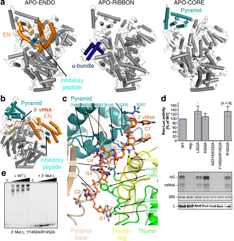
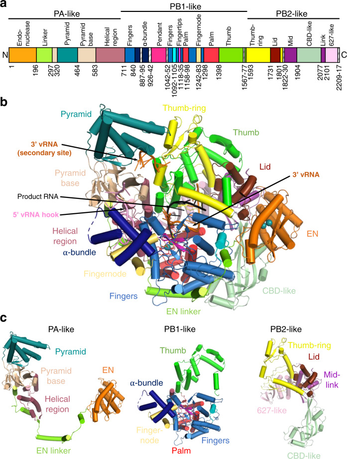
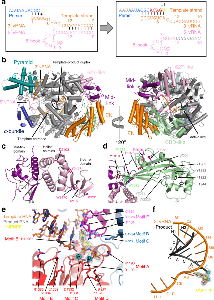
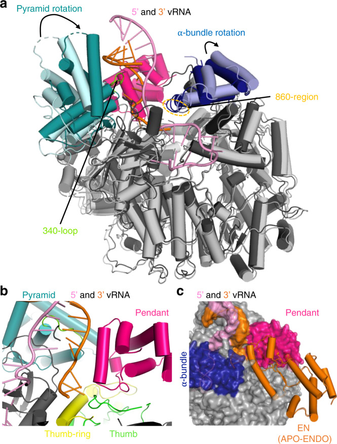
Lassa virus is endemic in West Africa and can cause severe hemorrhagic fever. The viral L protein transcribes and replicates the RNA genome via its RNA-dependent RNA polymerase activity. Here, we present nine cryo-EM structures of the L protein in the apo-, promoter-bound pre-initiation and active RNA synthesis states. We characterize distinct binding pockets for the conserved 3' and 5' promoter RNAs and show how full-promoter binding induces a distinct pre-initiation conformation. In the apo- and early elongation states, the endonuclease is inhibited by two distinct L protein peptides, whereas in the pre-initiation state it is uninhibited. In the early elongation state, a template-product duplex is bound in the active site cavity together with an incoming non-hydrolysable nucleotide and the full C-terminal region of the L protein, including the putative cap-binding domain, is well-ordered. These data advance our mechanistic understanding of how this flexible and multifunctional molecular machine is activated.


Conflict of interest statement

The authors declare no competing interests.

Figures

Fig. 1
Fig. 1. L protein in the apo-state and with 3′ viral RNA bound in the secondary binding site.
a Ribbon diagram presentations of the structures APO-ENDO, APO-RIBBON and APO-CORE. Each of the respective experimental maps resolves distinct regions of the L protein better than the others and those regions are shown in colour and indicated by name in the respective structures. b Overall structure of L protein 3END-CORE as a ribbon diagram with the 3′ vRNA bound below the pyramid domain. Pyramid (teal), pyramid base (wheat), EN domain (orange) and the inhibitory peptide (cyan) are highlighted. c Close-up of the secondary 3′ vRNA binding site with the 3′ vRNA nucleotides 1–7 (orange), pyramid domain (teal), pyramid base (wheat) as well as thumb (green) and thumb-ring (yellow) domains. Important amino acids in the RNA-protein interface are shown as sticks with respective labels. Hydrogen bonds are indicated by dotted lines. For selected regions, secondary structure depiction was disabled to enhance visibility. d LASV mini-replicon data for L proteins with mutations in the secondary 3′ RNA binding site presenting luciferase reporter activity (in standardised relative light units relative to the wild-type L protein (WT)). Data were presented as mean values ±  SD of at least six biological replicates (n = 6), although for most mutants seven biological replicates were included. All biological replicates are shown as blue dots (top panel). Middle panels present Northern blotting results with signals for antigenomic viral RNA (AG), viral mRNA (mRNA) and 28 S ribosomal RNA (28 S) as a loading control, and the bottom panel shows Western blot detection of FLAG-tagged L proteins (L) to demonstrate general expressibility of the mutants. Source data are provided in a Source Data file. e Electrophoretic mobility shift assay of wild-type L protein (WT L) and mutant Y1450A/R1452A (Mut L) with 10 nt 3′ viral RNA. L protein concentrations ranging from 0–1 µM and 0.2 µM of fluorescently labelled 3′ vRNA (Supplementary Table 1) were used (see methods).
Fig. 2
Fig. 2. L protein in the pre-initiation state.
a Ribbon diagram of the PRE-INITIATION structure with pendant domain (pink), α-bundle (dark blue), pyramid (teal), 3′ vRNA nts 7–19 (orange) and 5′ vRNA nts 0–19 (pink) highlighted in colour and indicated by name. b Interactions of the L protein pyramid (teal), pendant (pink) thumb-ring (yellow) and thumb (green) domains with the 3′ vRNA are shown. Important amino acid side chains and the RNA nucleotides of 3′ and 5′ vRNA are shown as sticks with respective labels. c Viral RNA observed in this structure with a 5′ vRNA hook structure composed of 5′ vRNA nts 0–9 and a distal duplex region involving 5′ vRNA nts 12–19 and 3′ vRNA nts 12–19. The 3′ vRNA nts 1–11 are directed towards the RdRp active site (towards AS) but not resolved. d Close-up of the 5′ RNA hook binding site involving the fingers domain (blue), finger node (light yellow), pyramid base (wheat) and helical region (raspberry). Residues important for the RNA-protein interface and nucleotides are shown as sticks and are labelled. e Schematic presentation of the promoter RNA (3′ vRNA in orange, 5′ vRNA in pink) in the PRE-INITIATION structure. Nucleotides 1–6 of the 3′ vRNA, which are not resolved, are coloured in grey. Distal duplex and 5′ hook regions are labelled.
Fig. 3
Fig. 3. Overview of the L protein structure.
a Schematic linear presentation of the L protein domain structure. b Complete ELONGATION structure of the L protein presented as a ribbon diagram in front view. Domains are coloured according to (a) and labelled. 3′ vRNA is coloured in orange, 5′ vRNA in pink and product RNA in black. See also Supplementary Movie 3 for a 3D impression of the L protein and its domains. c Separate presentation of the PA-like, PB1-like and PB2-like regions of the L protein in the ELONGATION structure.
Fig. 4
Fig. 4. Elongation state of the L protein.
a Schematic presentation of the primed reaction was carried out to obtain the ELONGATION structure with the L protein stalled in an early elongation state. Nucleotides that are not visible or not clearly assignable from the experimental map are shown in grey italics. b ELONGATION structure of the L protein presented as a ribbon diagram in two views as indicated. EN, pyramid, α-bundle, mid-link and 627-like domains are coloured. 3′ vRNA is shown in orange, 5′ RNA in pink and product RNA in black. A dashed circle (hot pink) indicates the putative product exit. c Close-up on the mid-link and 627-like domains with the respective structural features labelled and the side chains of amino acids shown to be selectively important for viral transcription by Lehmann et al. 2014 shown as pink sticks. d Close-up on the CBD-like domain with side chains of amino acids that have been tested in the LASV mini-replicon system shown as sticks (pink—selective role in viral transcription; light grey—no significant reduction of L protein activity upon mutation shown by Lehmann et al. 2014; green—no or weak effect on L protein function upon mutation; dark red—potential selective role in viral transcription; dark grey—general defect of L protein activity upon mutation). Corresponding mini-replicon data are presented in Supplementary Fig. 8. e Close-up of the polymerase active site with the template RNA (orange), the product RNA (dark grey), the non-hydrolysable UTP (UMPNPP, yellow) and catalytic manganese ions (teal, A and B) involving the palm (red), fingers (blue), fingertips (magenta) and thumb (green) domains of the L protein. Important side chains are shown as sticks and conserved RdRp active site motifs (A–G) are labelled. The map around the UMPNPP and the manganese ions is shown as a grey mesh. f Template-product duplex of the polymerase active site is shown as a ribbon diagram with the product in black, the 3′ template in orange, the non-hydrolysable UTP (UMPNPP) in yellow and the catalytic manganese ions as teal spheres. The map around the ions and the UMPNPP is shown as a grey mesh.
Fig. 5
Fig. 5. Conformational flexibility of the endonuclease domain.
a Overview of the three different conformations of the EN (orange) observed in the 3END-CORE, MID-LINK and ELONGATION structures. The EN linker (green) and the inhibitory peptide (cyan) are highlighted as well. b In the middle panel a superimposition of the EN domains of 3END-CORE (orange), corresponding also to the overall conformation of the EN domain in the MID-LINK structure, and ELONGATION (brown) with the respective positions of the inhibitory peptides in teal, cyan and light blue, respectively, is shown. Connections to the fingers domain are indicated in blue. The position of the inhibitory peptide of 3END-CORE is the same as in the APO-ENDO structure, similarly is the same position of the EN observed in both MID-LINK and DISTAL-PROMOTER structures. Right and left panels show close-ups of the autoinhibited EN active sites in the ELONGATION and 3END-CORE structures, respectively. Important residues of the protein-protein interactions are labelled and side chains are shown as sticks. For a focus on the inhibitory loop in the different conformations see Supplementary Fig. 11.
Fig. 6
Fig. 6. Global rearrangements upon promoter binding.
a PRE-INITIATION (dark grey, teal, dark blue, hot pink) and APO-RIBBON (light grey, light cyan, light blue) structures are superimposed. The pyramid and α-bundle rotations between apo- and promoter-bound structures are indicated as well as the 340-loop and the 860-region. Promoter RNA is shown in pink and orange. b Close-up of the interaction site between promoter RNA (pink and orange) and the pendant (hot pink), thumb-ring (yellow) and thumb (green) domains in the PRE-INITIATION structure. The pyramid domain is shown in teal. c Superposition of the PRE-INITIATION and APO-ENDO structures. PRE-INITIATION is presented as a transparent surface in grey with the pendant (hot pink) and α-bundle (blue) as well as the 5′ (pink) and 3′ (orange) vRNA highlighted in colour. The EN domain of the APO-ENDO structure is shown as an orange ribbon, which overlaps with the pendant domain volume of the PRE-INITIATION structure.
Fig. 7
Fig. 7. Schematic diagram of conformational changes in Lassa virus L protein associated with promoter binding and RNA synthesis activity.
a On the surface of the inactive/resting L protein core, there is mutually exclusive positioning (black double-arrow) of either the α-bundle and pendant (APO-RIBBON structure) or the EN domain (APO-ENDO, 3END-ENDO structures). When placed on the core, the inhibitory peptide (cyan) autoinhibits the EN domain by binding in its active site. In the absence of the 5′ vRNA, the 3′ vRNA binds preferentially, base-specifically, into a distinct secondary 3′ RNA binding site between the pyramid and thumb domains (3END-ENDO/CORE structures). b Upon full promoter binding, major conformational changes occur (based on PRE-INITIATION, DISTAL-PROMOTER, MID-LINK structures). The 5′ end nucleotides 0–9 are bound in a hook-like conformation in a specific pocket outside the active site. The distal promoter (formed by 3′ and 5′ vRNA nucleotides 12–19) is positioned by tight association with the α-bundle and pendant, with concomitant rotation of the pyramid domain. This forces release of the 3′ vRNA from the secondary binding site, allowing it to be directed towards the RNA synthesis active site (marked by the white A in the teal circle). Positioning of the pendant domain next to the distal promoter displaces the EN domain, which relocates to the other end of the L protein core with the inhibitory peptide contacting its surface leaving the EN active site accessible. In this configuration, the EN is presumed to be in close vicinity to the CBD-like domain and could potentially cleave an incoming capped RNA to generate a transcription primer by ‘cap-snatching’. How the capped primer associates with the CBD-like domain and how it is navigated towards the active site to initiate transcription remains elusive. c Upon transition to the elongation state, the distal promoter duplex melts and the pendant domain is released, which allows the pyramid to rotate back and re-establish the availability of the secondary 3′ end binding site (ELONGATION structure). We presume the 3′ vRNA template, after exiting the active site, wraps around the L protein core and rebinds to the secondary 3′ end binding site, as described for influenza virus polymerase complex. The EN repositions once again and together with the mid-link and CBD-like domains form a highly structured ring around the putative product exit channel. The EN active site is again autoinhibited, this time by the relocation of its C-terminal helix (181–188) to the active site.

References

    1. Akpede GO, et al. Caseload and case fatality of Lassa fever in Nigeria, 2001–2018: a specialist center’s experience and its implications. Front. Public Health. 2019;7:170. - PMC - PubMed
    1. Basinski AJ, et al. Bridging the gap: using reservoir ecology and human serosurveys to estimate Lassa virus spillover in West Africa. PLoS Comput. Biol. 2021;17:e1008811. - PMC - PubMed
    1. Olschewski S, Cusack S, Rosenthal M. The cap-snatching mechanism of Bunyaviruses. Trends Microbiol. 2020;28:293–303. - PubMed
    1. Raju R, et al. Nontemplated bases at the 5’ ends of Tacaribe virus mRNAs. Virology. 1990;174:53–59. - PubMed
    1. Polyak SJ, Zheng S, Harnish DG. 5’ termini of Pichinde arenavirus S RNAs and mRNAs contain nontemplated nucleotides. J. Virol. 1995;69:3211–3215. - PMC - PubMed

Publication types

MeSH terms