. 2021 Dec 2;11(1):23293.
doi: 10.1038/s41598-021-02780-4.

Electrospun tube reduces adhesion in rabbit Achilles tendon 12 weeks post-surgery without PAR-2 overexpression

Affiliations

Electrospun tube reduces adhesion in rabbit Achilles tendon 12 weeks post-surgery without PAR-2 overexpression

Gabriella Meier Bürgisser et al. Sci Rep. .

Abstract

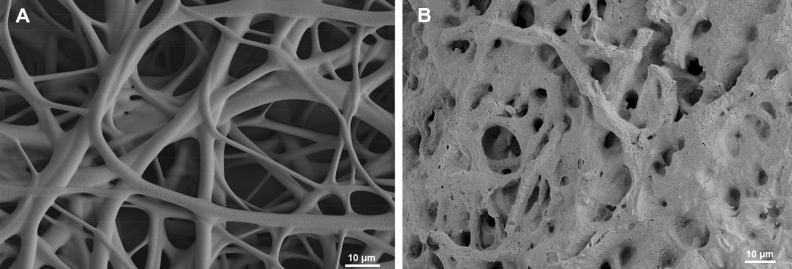
One great challenge in surgical tendon repair is the minimization of peritendinous adhesions. An electrospun tube can serve as a physical barrier around a conventionally sutured tendon. Six New Zealand White rabbits had one Achilles tendon fully transsected and sutured by a 4-strand suture. Another six rabbits had the same treatment, but with the additional electrospun DegraPol tube set around the sutured tendon. The adhesion formation to the surrounding tissue was investigated 12 weeks post-operation. Moreover, inflammation-related protease-activated receptor-2 (PAR-2) protein expression was assessed. Finally, rabbit Achilles tenocyte cultures were exposed to platelet-derived growth factor-BB (PDGF-BB), which mimicks the tendon healing environment, where PAR-2 gene expression was assessed as well as immunofluorescent staining intensity for F-actin and α-tubulin, respectively. At 12 weeks post-operation, the partially degraded DegraPol tube exhibited significantly lower adhesion formation (- 20%). PAR-2 protein expression was similar for time points 3 and 6 weeks, but increased at 12 weeks post-operation. In vitro cell culture experiments showed a significantly higher PAR-2 gene expression on day 3 after exposure to PDGF-BB, but not on day 7. The cytoskeleton of the tenocytes changed upon PDGF-BB stimulation, with signs of reorganization, and significantly decreased F-actin intensity. An electrospun DegraPol tube significantly reduces adhesion up to twelve weeks post-operation. At this time point, the tube is partially degraded, and a slight PAR-2 increase was detected in the DP treated tendons, which might however arise from particles of degrading DegraPol that were stained dark brown. PAR-2 gene expression in rabbit tenocytes reveals sensitivity at around day 10 after injury.


Conflict of interest statement

The authors declare no competing interests.

Figures

Figure 1
Figure 1
SEM images of electrospun DP fiber mesh freshly produced (A) and after 12 weeks in vivo as implant material intended to reduce adhesion formation (B).
Figure 2
Figure 2
Adhesion formation as assessed by histology via contact region to the surrounding tissue. Adhesions were determined at 12 weeks. Quantitative determination: percentage of contact region (A); typical Picosirius Red stained cross-sections, including white dashed areas that are magnified below each image (B). Key: DP = DegraPol. Stars, with p-values, indicate pairwise comparison probability: p < 0.05 (*), p < 0.01 (**) and p < 0.001 (***). Scale bars indicate 500 µm. Arrows depict areas of adhesion (contact to surrounding tissue).
Figure 3
Figure 3
PAR-2 protein expression in DP treated tendon sections (n = 3) compared to contralateral side (no treatment; n = 3); quantified by the red/green ratio in histograms with 5 FOVs per section (A) and representative samples stained for PAR-2 (B), with areas of higher magnification, taken from the middle of the AT (C). The dark brown staining at 12 weeks does not come from an overexpression of PAR-2 by the tenocytes; it is the partially degraded DegraPol polymer that stains like this (Supporting Information SI Fig. 3). Nevertheless, the mid portion of the AT showed a significant slight increase in PAR-2 expression (+ 4.9%). Scale bars indicate 500 µm; in inserts they indicate 100 µm. Stars, with p-values, indicate pairwise comparison probability: p < 0.05 (*), p < 0.01 (**) and p < 0.001 (***).
Figure 4
Figure 4
PAR-2 gene expression with 20 ng/mL PDGF-BB and without PDGF-BB after 3, 7 and 14 days under in vitro culture; manifold expression compared to cell culture without PDGF-BB; for tenocytes of four rabbits. (A). Manifold PAR-2 gene expression in a 3-day experiment, using different inhibitors in a 40 µM concentration and 5% FBS (B). Key: SB203580 = inhibitor for MAPK; LY-294002 = inhibitor for PI3K; PD153035 = inhibitor for EGFR (same family as PDGFR); DMSO = dimethyl sulfoxide (solvent of inhibitors); Medium = basal culture medium (with 10% FCS).
Figure 5
Figure 5
Images of tenocyte cultures without PDGF-BB (A) and with 20 ng/mL PDGF-BB (B) on day 3, showing F-actin (green), α-tubulin (red) and DAPI (blue, cell nuclei). PDGF-BB accompanying PAR-2 induction changes the F-actin cytoskeleton. Scale bars are equivalent to 50 µm. Achilles tenocytes of one rabbit are shown in the upper row, and of another rabbit in the lower row. Size of cells (C), F-actin green intensity (D) and α-tubulin red intensity (E), and aspect ratio of tenocytes on day 3 after addition of an inhibitor (40 µM, 5% FBS) with or without PDGF-BB (F). Key: SB203580 = inhibitor for MAPK; LY-294002 = inhibitor for PI3K; PD153035 = inhibitor for EGFR; DMSO = dimethyl sulfoxide (solvent of inhibitors); Medium = basal culture medium DMEM (with 10% FCS).

References

    1. Goldenberg BT, et al. Biologics to improve healing in large and massive rotator cuff tears: A critical review. Orthop. Res. Rev. 2020;12:151–160. - PMC - PubMed
    1. Serra CI, Navarro P, Guillem R, Soler C. Use of frozen tendon allograft in two clinical cases: Common calcaneal tendon and patellar ligament rupture. J. Am. Anim. Hosp. Assoc. 2020;56:315. - PubMed
    1. Elliot D, Giesen T. Primary flexor tendon surgery: The search for a perfect result. Hand Clin. 2013;29:191–206. - PubMed
    1. Galatz LM, Gerstenfeld L, Heber-Katz E, Rodeo SA. Tendon regeneration and scar formation: The concept of scarless healing. J. Orthop. Res. 2015;33:823–831. - PMC - PubMed
    1. Jackson JE, Kopecki Z, Anderson PJ, Cowin AJ. Increasing the level of cytoskeletal protein Flightless I reduces adhesion formation in a murine digital flexor tendon model. J. Orthop. Surg. Res. 2020;15:362. - PMC - PubMed

Publication types

MeSH terms