. 2021 Dec;53(12):1850-1865.
doi: 10.1038/s12276-021-00713-9. Epub 2021 Dec 2.

The impact of fine particulate matter (PM) on various beneficial functions of human endometrial stem cells through its key regulator SERPINB2

Affiliations

The impact of fine particulate matter (PM) on various beneficial functions of human endometrial stem cells through its key regulator SERPINB2

Se-Ra Park et al. Exp Mol Med. 2021 Dec.

Abstract

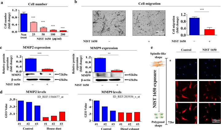
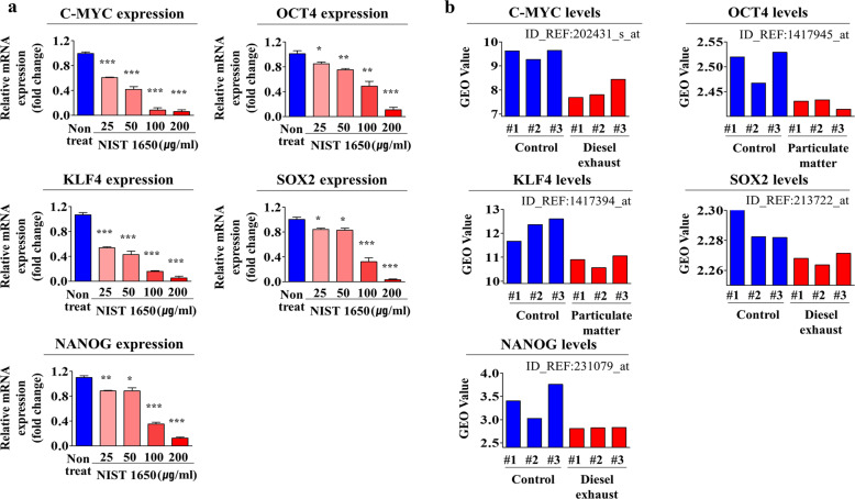
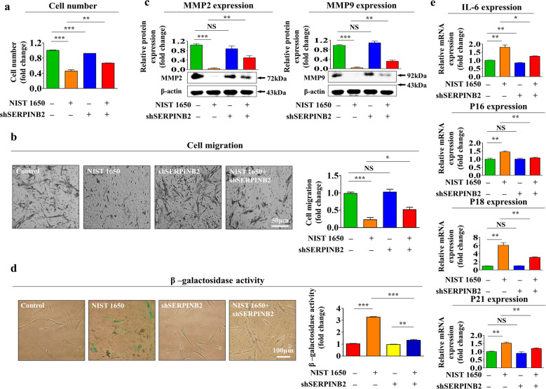
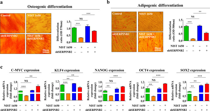
Fine particulate matter (PM) has a small diameter but a large surface area; thus, it may have broad toxic effects that subsequently damage many tissues of the human body. Interestingly, many studies have suggested that the recent decline in female fertility could be associated with increased PM exposure. However, the precise mechanisms underlying the negative effects of PM exposure on female fertility are still a matter of debate. A previous study demonstrated that resident stem cell deficiency limits the cyclic regenerative capacity of the endometrium and subsequently increases the pregnancy failure rate. Therefore, we hypothesized that PM exposure induces endometrial tissue damage and subsequently reduces the pregnancy rate by inhibiting various beneficial functions of local endometrial stem cells. Consistent with our hypothesis, we showed for the first time that PM exposure significantly inhibits various beneficial functions of endometrial stem cells, such as their self-renewal, transdifferentiation, and migratory capacities, in vitro and in vivo through the PM target gene SERPINB2, which has recently been shown to be involved in multiple stem cell functions. In addition, the PM-induced inhibitory effects on the beneficial functions of endometrial stem cells were significantly diminished by SERPINB2 depletion. Our findings may facilitate the development of promising therapeutic strategies for improving reproductive outcomes in infertile women.


Conflict of interest statement

The authors declare no competing interests.

Figures

Fig. 1
Fig. 1. PM exposure significantly inhibits the self-renewal and reduces the migratory ability of endometrial stem cells in vitro.
We hypothesized that PM exposure inhibited various beneficial functions of endometrial stem cells, such as self-renewal, transdifferentiation, and migration. The inhibition of endometrial stem cell self-renewal by treatment with various concentrations of PM (diesel particulate matter, NIST 1650; 25, 50, 100, and 200 μg/ml) was evaluated at 72 h by an MTT assay. Stem cell viability (%) was calculated as a percent of the viability of cells treated with vehicle control (a). Endometrial stem cells were treated with PM (25 μg/ml) for 72 h, and the inhibitory effect of PM exposure on the stem cell migratory capacity was then analyzed using a Transwell migration assay. PM exposure markedly reduced the ability of cells to migrate across the membrane compared with that of control cells (b). The relative expression of important regulators of cell migration (MMP-2/9) with and without PM exposure was evaluated by western blot analysis (c). The GEO database was further analyzed to verify the correlations between increased expression levels of MMP-2/9 and exposure to various air pollutants (d). PM-induced morphological changes and dynamic actin filament disorganization in endometrial stem cells were analyzed by staining actin filaments with phalloidin (e). DAPI was used to label nuclei. β-Actin was used as the internal control. The bar graphs show the average of three independent experiments. Significant differences are presented. *p < 0.05, **p < 0.005, and ***p < 0.001 (two-sample t-test).
Fig. 2
Fig. 2. PM exposure markedly induces senescence in endometrial stem cells in vitro.
The effects of PM exposure on endometrial stem cell aging in vitro were analyzed by evaluating senescence-associated β-galactosidase (SA-β-Gal) activity. Endometrial stem cells were pretreated with PM (25 μg/ml) for 72 h, and changes in stem cell aging were determined by evaluating SA-β-Gal activity (a). The effects of PM exposure on the expression of multiple cellular senescence markers (p16, p18, p21, and IL-6) were evaluated by real-time PCR (b). The Gene Expression Omnibus (GEO) database (https://www.ncbi. nlm.nih.gov/geo/) was used to further analyze the correlations between increased expression levels of these senescence markers and exposure to various air pollutants (c). The bar graphs show the average of three independent experiments. Significant differences are presented. *p < 0.05, **p < 0.005, and ***p < 0.001 (two-sample t-test).
Fig. 3
Fig. 3. PM exposure markedly suppresses the multilineage differentiation potential and induces the apoptosis of endometrial stem cells in vitro.
Endometrial stem cells were cultured in adipocyte or osteoblast differentiation medium with or without PM (25 μg/ml). The inhibitory effects of PM exposure on adipocyte (a) and osteoblast (b) differentiation were evaluated by oil red O and alizarin red staining, respectively. The relative quantification of calcium mineral content and lipid droplet formation in differentiated cells was performed by measuring the absorbance at 500 nm and 570 nm, respectively. Endometrial stem cells were cultured with or without PM (25 μg/ml) for 72 h, and the increase in the level of cleaved caspase-3 following PM exposure was then analyzed by western blotting (c). PM-induced apoptotic DNA condensation and fragmentation after incubation in the absence of PM (25 μg/ml) for 72 h were analyzed using DAPI staining (d). The decrease in cell viability following PM exposure (5 and 10 μg/ml) for 72 h was detected via an MTT assay in both endometrial stem cells and terminally differentiated cells (fibroblasts and vascular endothelial cells). Cell viability (%) was calculated as the percent of the viability of cells treated with the vehicle control (e). DAPI was used to label nuclei. β-Actin was used as the internal control. The bar graphs show the average of three independent experiments. Significant differences are presented. *p < 0.05, **p < 0.005, and ***p < 0.001 (two-sample t-test).
Fig. 4
Fig. 4. PM exposure reduces the expression of various stem cell markers in endometrial stem cells in vitro.
The inhibitory effects of PM exposure on the expression of the stem cell markers C-MYC, KLF4, NANOG, OCT4, and SOX2 were evaluated by real-time PCR (a). The GEO database was also analyzed to further verify the correlations between increased expression levels of these pluripotency-associated factors and exposure to air pollutants (b). The bar graphs show the average of three independent experiments. Significant differences are presented. *p < 0.05, **p < 0.005, and ***p < 0.001 (two-sample t-test).
Fig. 5
Fig. 5. PM exposure significantly elevates the mitochondrial respiratory and glycolytic rates in endometrial stem cells.
To evaluate the effect of PM exposure on the bioenergetic status of endometrial stem cells, the mitochondrial respiration and glycolytic rates were analyzed with or without PM exposure (a). Respiratory flux profiles of endometrial stem cells with or without exposure to PM (25 μg/ml) for 72 h were determined by collecting twelve consecutive oxygen consumption rate (OCR) measurements using a Seahorse XF analyzer. Cells were detached using trypsin-EDTA and reseeded in licensed cell culture miniplates at a density of 2 × 104 cells per well in complete growth medium supplemented with 10% FBS. Cells were incubated overnight and washed 3 times with Seahorse XF medium. The ATP synthase inhibitor oligomycin (a complex V blocker, 1.5 μM) was added to inhibit ATP-coupled respiration. FCCP (a mitochondrial uncoupler, 2 μM) was added to decrease the mitochondrial membrane potential (Δψm). Rotenone (an inhibitor of complex I in the electron transport chain, 0.5 µM) and antimycin A (an inhibitor of complex III in the electron transport chain, 0.5 µM) were added to block mitochondrial respiration completely. The inhibitors were applied automatically in the analyzer, and the OCR was measured at 15-min intervals (b). PM exposure significantly increased the basal respiration rate (c), spare respiratory capacity (d), and maximal respiration rate (e) in endometrial stem cells. Overall ATP production in endometrial stem cells was significantly enhanced by PM exposure (f). The schematic of the procedure for real-time analysis of glycolysis in endometrial stem cells using the Seahorse XF analyzer is shown (g). For real-time analysis of glycolytic rates, the Seahorse XF glycolytic rate assay utilizes both ECAR (extracellular acidification rate) and OCR measurements to evaluate the glycolytic proton efflux rate (glycoPER) of the cells; in this assay, cells are incubated in glucose-free medium to which rotenone, antimycin A (1.67 μM), and finally 2-deoxyglucose (2-DG, glycolysis inhibitor, 50 mM) are sequentially added. The inhibitors were applied automatically in the analyzer, and the ECAR was measured at 15-minute intervals. The percentage of PER from glycolysis represents the contribution of the glycolytic pathway to the total ECAR (h). Compensatory glycolysis is the rate of glycolysis in cells following the inhibition of oxidative metabolism and refers to the ability of cells to drive compensatory energy production by using glycolysis to meet their energy demands (i). The ECAR values were normalized to the number of cells in each well. The bar graphs show the average of three independent experiments. Significant differences are presented. *p < 0.05, **p < 0.005, and ***p < 0.001 (two-sample t-test).
Fig. 6
Fig. 6. PM exposure stimulates SERPINB2 expression in endometrial stem cells in vitro.
We hypothesized that SERPINB2 could act as a key mediator of PM-induced toxic effects in endometrial stem cells. Endometrial stem cells (a), fibroblasts (b), and vascular endothelial cells (c) were treated with or without PM (25 μg/ml) for 72 h. Real-time PCR and western blotting were performed to confirm the increase in SERPINB2 expression by PM exposure in vitro. The GEO database was also analyzed to further verify the correlations between the increased SERPINB2 level and exposure to air pollutants (d). β-Actin was used as the internal control. The bar graphs show the average of three independent experiments. Significant differences are presented. *p < 0.05, **p < 0.005, and ***p < 0.001 (two-sample t-test).
Fig. 7
Fig. 7. SERPINB2 mediates the inhibitory effect of PM exposure on the self-renewal, migration, and replicative senescence of endometrial stem cells in vitro.
Schematic representation showing the functions of SERPINB2 as a gene that regulates PM-induced effects in endometrial stem cells. Endometrial stem cells were transfected with shRNA targeting SERPINB2 and were treated with or without PM (25 μg/ml) for 72 h; subsequent changes in cell viability were measured by an MTT assay (a). The ability of SERPINB2 depletion to abolish the PM-induced inhibitory effects on the migratory capacity of endometrial stem cells was analyzed via a Transwell assay (b) and western blotting with anti-MMP-2 and anti-MMP-9 antibodies (c). The attenuating effects of SERPINB2 depletion on PM-induced replicative senescence were evaluated by measuring SA-β-Gal activity (d) and the expression levels of the senescence markers IL-6, p16, p18, and p21 (e). β-Actin was used as the internal control. The bar graphs show the average of three independent experiments. Significant differences are presented. *p < 0.05, **p < 0.005, and ***p < 0.001 (two-sample t-test).
Fig. 8
Fig. 8. SERPINB2 mediates the inhibitory effect of PM exposure on the multilineage differentiation potential and pluripotency of endometrial stem cells.
Endometrial stem cells were transfected with a shRNA targeting SERPINB2 and treated with or without PM (25 μg/ml); subsequent changes in adipocyte (a) and osteoblast (b) differentiation were evaluated by oil red O and alizarin red staining, respectively. The relative quantification of calcium mineral content and lipid droplet formation in differentiated cells was evaluated by measuring the absorbance at 500 nm and 570 nm, respectively. The attenuating effects of SERPINB2 depletion on PM-induced suppression of the pluripotency-related genes C-MYC, KLF4, NANOG, OCT4, and SOX2 were analyzed by real-time PCR (c). The bar graphs show the average of three independent experiments. Significant differences are presented. *p < 0.05, **p < 0.005, and ***p < 0.001 (two-sample t-test).
Fig. 9
Fig. 9. PM exposure suppresses the secretion of various growth factors or cytokines that are related to toxicity networks.
Human growth factor antibody array analysis was performed using PM-treated or untreated protein samples. The membrane was printed with antibodies against 40 growth factors, cytokines, and receptors. The levels of eleven growth factors (AR, HGF, NT-4, TGF-β3, EGF, IGFBP4/6, PDGFR β, VEGFR3, and FGF4/7) were significantly decreased in the PM-treated protein samples (a, b). The bar graphs show the average of three independent experiments. Significant differences are presented. *p < 0.05, **p < 0.005, and ***p < 0.001 (two-sample t-test).
Fig. 10
Fig. 10. The correlations between the eleven downregulated growth factors and exposure to various toxicants.
The GEO database was analyzed to further verify the correlations between the eleven downregulated growth factors and exposure to various toxicants.
Fig. 11
Fig. 11. PM exposure significantly inhibits various beneficial functions of endometrial stem cells in vivo.
Schematic representation of the experimental protocol as described in the materials and methods section (a). Mice were treated two times with a low (20 mg/kg) or high (40 mg/kg) dose of PM or with vehicle (PBS) by intraperitoneal injection. Endometrial stem cells were isolated from the mouse uterine endometrium, and changes in stem cell viability were evaluated by an MTT assay. Stem cell viability (%) was calculated as a percent of the viability of cells treated with vehicle control (b). Changes in cell migration were evaluated via a Transwell assay (c) and western blotting with anti-MMP-2 and anti-MMP-9 antibodies (d). The effects of PM exposure on adipocyte (e) and osteoblast (f) differentiation in vivo were evaluated by oil red O and alizarin red staining, respectively. The relative quantification of calcium mineral content and lipid droplet formation in differentiated cells was analyzed by measuring the absorbance at 500 nm and 570 nm, respectively. β-Actin was used as the internal control. The bar graphs show the average of three independent experiments. Significant differences are presented. *p < 0.05, **p < 0.005, and ***p < 0.001 (two-sample t-test).
Fig. 12
Fig. 12. Schematic summary of this study.
PM exposure significantly inhibited various beneficial functions of endometrial stem cells, such as their self-renewal, transdifferentiation, and migratory capacity, in vitro and in vivo through its target gene SERPINB2, which has recently been shown to be involved in multiple stem cell functions.

Similar articles

Cited by

References

    1. Orru H, Ebi KL, Forsberg B. The interplay of climate change and air pollution on health. Curr. Environ. Health Rep. 2017;4:504–513. - PMC - PubMed
    1. Wolkoff P. Indoor air humidity, air quality, and health - an overview. Int. J. Hyg. Environ. Health. 2018;221:376–390. - PubMed
    1. Brook RD, et al. Particulate matter air pollution and cardiovascular disease: an update to the scientific statement from the American Heart Association. Circulation. 2010;121:2331–2378. - PubMed
    1. Brunekreef B, Forsberg B. Epidemiological evidence of effects of coarse airborne particles on health. Eur. Respir. J. 2005;26:309–318. - PubMed
    1. WHO’s global air-quality guidelines. Lancet. 2006;368:1302. - PubMed

Publication types

Substances