. 2022 Jun;29(6):1187-1198.
doi: 10.1038/s41418-021-00910-z. Epub 2021 Dec 2.

Ferroptosis mediates selective motor neuron death in amyotrophic lateral sclerosis

Affiliations

Ferroptosis mediates selective motor neuron death in amyotrophic lateral sclerosis

Taide Wang et al. Cell Death Differ. 2022 Jun.

Abstract

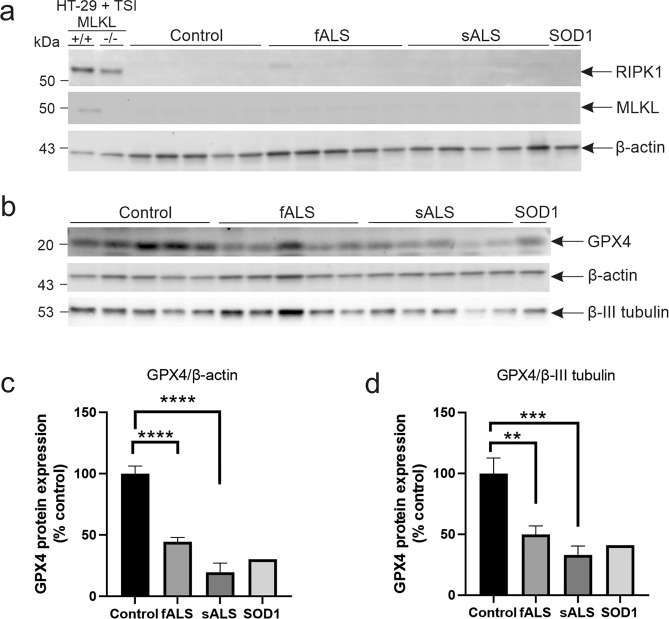
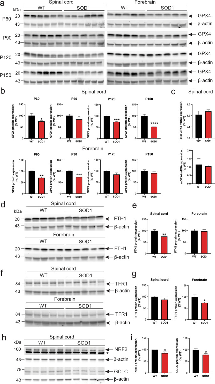
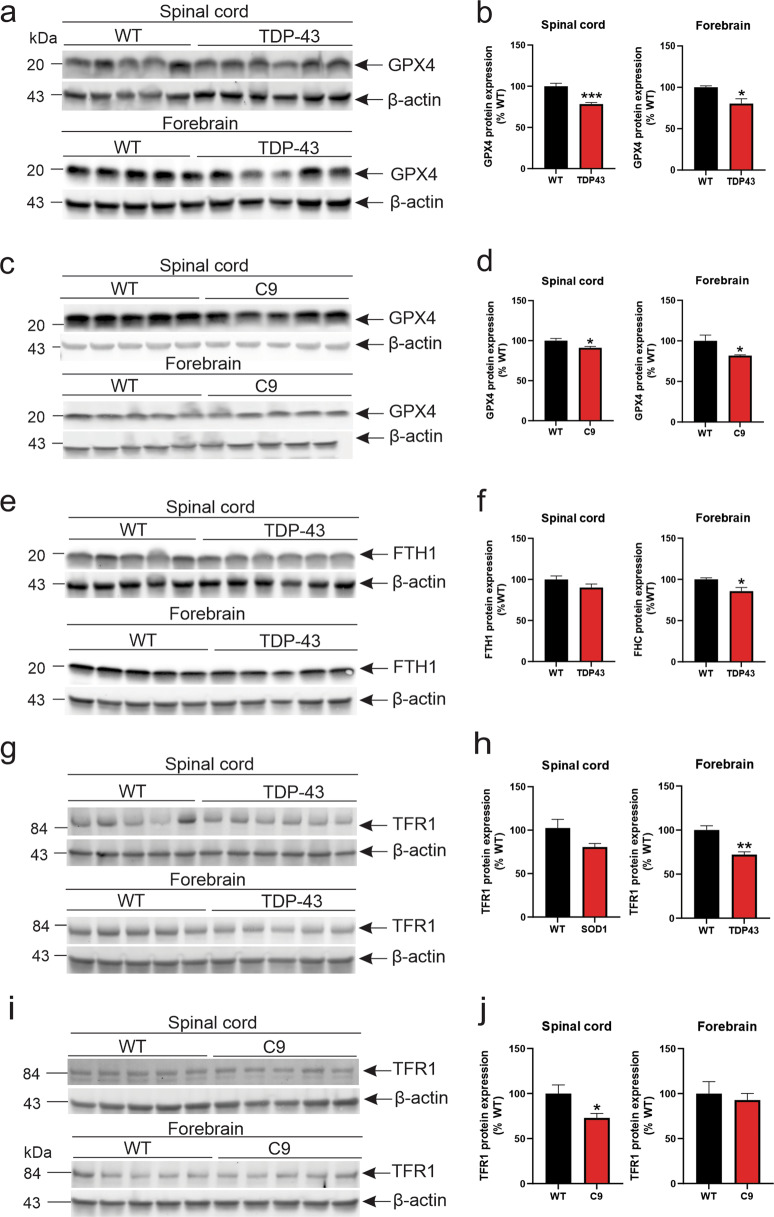
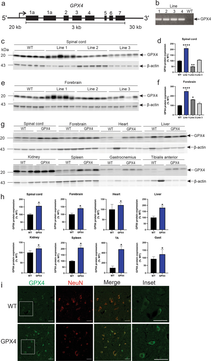
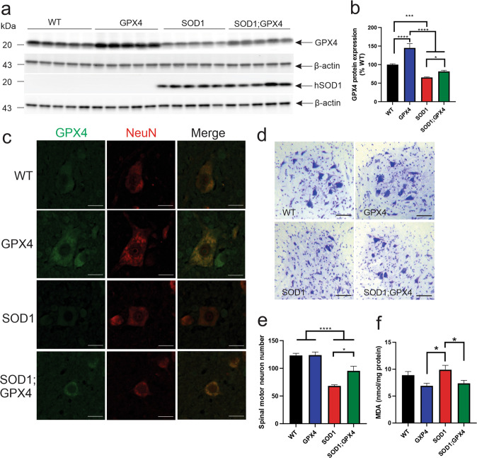
Amyotrophic lateral sclerosis (ALS) is caused by selective degeneration of motor neurons in the brain and spinal cord; however, the primary cell death pathway(s) mediating motor neuron demise remain elusive. We recently established that necroptosis, an inflammatory form of regulated cell death, was dispensable for motor neuron death in a mouse model of ALS, implicating other forms of cell death. Here, we confirm these findings in ALS patients, showing a lack of expression of key necroptotic effector proteins in spinal cords. Rather, we uncover evidence for ferroptosis, a recently discovered iron-dependent form of regulated cell death, in ALS. Depletion of glutathione peroxidase 4 (GPX4), an anti-oxidant enzyme and central repressor of ferroptosis, occurred in post-mortem spinal cords of both sporadic and familial ALS patients. GPX4 depletion was also an early and universal feature of spinal cords and brains of transgenic mutant superoxide dismutase 1 (SOD1G93A), TDP-43 and C9orf72 mouse models of ALS. GPX4 depletion and ferroptosis were linked to impaired NRF2 signalling and dysregulation of glutathione synthesis and iron-binding proteins. Novel BAC transgenic mice overexpressing human GPX4 exhibited high GPX4 expression localised to spinal motor neurons. Human GPX4 overexpression in SOD1G93A mice significantly delayed disease onset, improved locomotor function and prolonged lifespan, which was attributed to attenuated lipid peroxidation and motor neuron preservation. Our study discovers a new role for ferroptosis in mediating motor neuron death in ALS, supporting the use of anti-ferroptotic therapeutic strategies, such as GPX4 pathway induction and upregulation, for ALS treatment.


Conflict of interest statement

ALS and JMM contribute to a programme developing inhibitors of necroptosis with Anaxis Pharma Pty Ltd.

Figures

Fig. 1
Fig. 1. Necroptotic and ferroptotic mediator expression in the spinal cords of familial and sporadic ALS patients.
a Immunoblot of necroptotic markers, RIPK1 and MLKL, in spinal cords of non-neurological, familial (fALS) and sporadic ALS (sALS) patients. Specificity of the MLKL antibody was confirmed using lysates from wild-type of MLKL–/– HT-29 cells treated with necroptotic stimuli (TSI). b Immunoblot and quantification of GPX4 in spinal cords of fALS and sALS patients, compared to non-neurological controls, relative to c β-actin and d β-III tubulin levels. **p < 0.01, ***p < 0.001, ****p < 0.0001; one-way ANOVA with Tukey’s post hoc test. Data represent mean ± SEM, n = 5 cases per group, except for fALS-SOD1 (n = 1).
Fig. 2
Fig. 2. GPX4 loss is common in spinal cords and brains of mouse models of ALS.
a Immunoblot analysis and b quantification of GPX4 protein levels in the spinal cord and forebrain of WT and SOD1G93A mice from postnatal day (P) 60–150. *p < 0.05, **p < 0.01, ***p < 0.001, ***p < 0.0001; Student’s unpaired t-test. Data represent mean ± SEM, n = 5 mice per group. c mRNA expression of total GPX4 and mitochondrial (mGPX4) in WT and SOD1G93A mice at P150. Data represent mean ± SEM, n = 5 mice per group. dg Immunoblot analysis and quantification of FTH1 and TFR1 protein levels in the spinal cord of SOD1G93A mice at P90. **p < 0.01, Student’s unpaired t-test. Data represent mean ± SEM, n = 5 mice per group. h, i Immunoblot analysis and quantification of NRF2 and GCLC in the spinal cords of P120 WT and SOD1G93A mice. *p < 0.05, Student’s unpaired t-test. Data represent mean ± SEM, n = 5 mice per group. *non-specific band.
Fig. 3
Fig. 3. Anti-ferroptosis defence is impaired in affected CNS tissues of TDP-43Q331K and C9orf72500 mice.
Immunoblot analysis and quantification of GPX4 protein levels in the spinal cord and forebrain of WT and a, b TDP-43Q331K at P240, and c, d C9orf72500 mice at P300. Immunoblot analysis and quantification of FTH1 and TFR1 protein levels in the spinal cord of eh TDP-43Q331K and i, j C9orf72500 mice. *p < 0.05, **p < 0.01, ***p < 0.001; Student’s unpaired t-test. Data represent mean ± SEM, n = 5–6 mice per group.
Fig. 4
Fig. 4. Characterisation of transgenic human GPX4 mice.
a Human GPX4 BAC transgene map including 20 kb upstream and 30 kb downstream genomic sequence. b Gel PCR of GPX4 transgene detection in mice. Immunoblot and quantification of GPX4 protein levels normalised to β-actin, relative to WT mice in cd spinal cord and e, f forebrain of GPX4 mice at P30. *p < 0.05, **p < 0.01, ****p < 0.0001; Student’s unpaired t-test. Data represent mean ± SEM, n = 5 mice per group. g, h Immunoblot and quantification of GPX4 protein expression relative to WT mice in various tissues of GPX4 mice at P180–200. *p < 0.05; Student’s unpaired t-test. Data represent mean ± SEM, n = 3 mice per group. i Representative GPX4 immunohistochemical analysis of ventral horn sections of spinal cords of WT and GPX4 mice. Scale bars, 50 µm.
Fig. 5
Fig. 5. GPX4 overexpression delays weight loss, improves locomotor function and survival in SOD1G93A mice.
Body weights of a female and b male WT, GPX4, SOD1G93A and SOD1G93A; GPX4 mice. Age of peak body weight of c female and d male SOD1G93A and SOD1G93A; GPX4 mice. Data represent mean ± SEM, n = 12 mice per group, *p < 0.05, Student’s t-test. Locomotor function of e female and f male WT, GPX4, SOD1G93A and SOD1G93A; GPX4 mice determined by latency to fall using weekly rotarod analysis. *p < 0.05, **p < 0.01, ***p < 0.001, two-way ANOVA, Sidak’s post hoc analysis. Age of peak rotarod performance of g female and h male SOD1G93A and SOD1G93A; GPX4 mice. Data represent mean ± SEM, n = 12 mice per group, *p < 0.05, **p < 0.01, Student’s t-test. Kaplan–Meier survival analysis of i female and j male mice determined by onset of hindlimb paralysis in SOD1G93A and SOD1G93A; GPX4 mice, n = 12 mice per group, **p < 0.01.
Fig. 6
Fig. 6. Protection from ferroptosis rescues motor neuron loss in SOD1G93A mice.
a Immunoblot analysis and b quantification of GPX4 protein levels in the spinal cord of WT, GPX4, SOD1G93A and SOD1G93A;GPX4 mice at clinical endstage. c Representative GPX4 immunohistochemical analysis of spinal motor neurons in mice. Scale bars, 20 µm. d Representative photomicrographs and e quantification of lumbar spinal cords of mice stained with cresyl violet at clinical endstage. *p < 0.05, ***p < 0.001, ****p < 0.0001; one-way ANOVA with Dunnett’s post hoc test. Data represent mean ± SEM, n = 5 per genotype. Scale bars, 50 µm. f Quantification of malondialdehyde (MDA) accumulation in forebrains of mice at clinical endstage. *p < 0.05, one-way ANOVA with Bonferroni post hoc test. Data represent mean ± SEM, n = 5 per genotype.

References

    1. Brown RH, Al-Chalabi A. Amyotrophic lateral sclerosis. N Engl J Med. 2017;337:162–72. doi: 10.1056/NEJMra1603471. - DOI - PubMed
    1. Taylor JP, Brown RH, Cleveland DW. Decoding ALS: from genes to mechanism. Nature. 2016;539:197–206. doi: 10.1038/nature20413. - DOI - PMC - PubMed
    1. Galluzzi L, Vitale I, Aaronson SA, Abrams JM, Adam D, Agostinis P, et al. Molecular mechanisms of cell death: recommendations of the Nomenclature Committee on Cell Death 2018. Cell Death Differ. 2018;25:486–541. doi: 10.1038/s41418-017-0012-4. - DOI - PMC - PubMed
    1. Aebischer J, Bernard-Marissal N, Pettmann B, Raoul C. Death receptors in the selective degeneration of motoneurons in amyotrophic lateral sclerosis. J Neurodegener Dis. 2013;2013:746845. 10.1155/2013/746845. - PMC - PubMed
    1. Re DB, Le Verche V, Yu C, Amoroso MW, Politi KA, Phani S, et al. Necroptosis drives motor neuron death in models of both sporadic and familial ALS. Neuron. 2014;81:1001–8. doi: 10.1016/j.neuron.2014.01.011. - DOI - PMC - PubMed

Publication types

Substances