Cutaneous Mycobacterium chelonae in a Patient with Sjogren's Syndrome
- PMID: 34858038
- PMCID: PMC8631975
- DOI: 10.2147/IDR.S342336
Cutaneous Mycobacterium chelonae in a Patient with Sjogren's Syndrome
Abstract
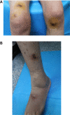
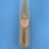
A case of cutaneous Mycobacterium chelonae infection in a patient with Sjogren's syndrome (SS) was misdiagnosed as sporotrichosis. A 56-year-old female patient was admitted to another hospital. Based on results of the histopathological examination and secretion culture obtained at the other hospital, the patient was diagnosed with sporotrichosis and received antifungal therapy. After treatment failure, the patient was admitted to our hospital, and a histopathological examination and secretion culture were performed again. The secretion culture revealed the presence of Mycobacterium chelonae. The antinuclear antibody test suggested SS, and the patient was treated with antibiotics and corticosteroids.
Keywords: Mycobacterium chelonae; cutaneous infection.
© 2021 Guo et al.
Conflict of interest statement
The authors declare no conflicts of interest in this work.
Figures


Similar articles
-
Mycobacterium chelonae keratitis in a patient with Sjögren's syndrome.Eur J Ophthalmol. 2008 Mar-Apr;18(2):294-6. doi: 10.1177/112067210801800221. Eur J Ophthalmol. 2008. PMID: 18320526
-
[Psychiatric manifestations of lupus erythematosus systemic and Sjogren's syndrome].Encephale. 2001 Nov-Dec;27(6):588-99. Encephale. 2001. PMID: 11865567 French.
-
The immunogenetic relationship between anti-Ro(SS-A)/La(SS-B) antibody positive Sjögren's/lupus erythematosus overlap syndrome and the neonatal lupus syndrome.J Invest Dermatol. 1989 Dec;93(6):751-6. doi: 10.1111/1523-1747.ep12284404. J Invest Dermatol. 1989. PMID: 2584740
-
Primary Sjögren's syndrome with diffuse cystic lung changes developed systemic lupus erythematosus: a case report and literature review.Oncotarget. 2017 May 23;8(21):35473-35479. doi: 10.18632/oncotarget.16010. Oncotarget. 2017. PMID: 28415674 Free PMC article. Review.
-
Annular erythema in primary Sjogren's syndrome: description of 43 non-Asian cases.Lupus. 2014 Feb;23(2):166-75. doi: 10.1177/0961203313515764. Epub 2013 Dec 10. Lupus. 2014. PMID: 24326481 Review.
Cited by
-
Difference analysis of cutaneous sporotrichosis between different regions in China: a secondary analysis based on published studies on sporotrichosis in China.Ann Transl Med. 2023 Feb 28;11(4):180. doi: 10.21037/atm-23-448. Ann Transl Med. 2023. PMID: 36923077 Free PMC article.
-
Effects of non-tuberculous mycobacteria on BCG vaccine efficacy: A narrative review.J Clin Tuberc Other Mycobact Dis. 2024 May 4;36:100451. doi: 10.1016/j.jctube.2024.100451. eCollection 2024 Aug. J Clin Tuberc Other Mycobact Dis. 2024. PMID: 38764556 Free PMC article. Review.
References
-
- Wallace RJ Jr, Brown BA, Onyi GO, et al. Skin, soft tissue, and bone infections due to Mycobacterium chelonae chelonae: importance of prior corticosteroid therapy, frequency of disseminated infections, and resistance to oral antimicrobials other than clarithromycin. J Infect Dis. 1992;166:405–412. doi:10.1093/infdis/166.2.405 - DOI - PubMed
Publication types
LinkOut - more resources
Full Text Sources

