

Chronic Venous Leg Ulcer in Klinefelter Syndrome Treated with Platelet-Rich Fibrin: A Case Report
- PMID: 34858066
- PMCID: PMC8630374
- DOI: 10.2147/IMCRJ.S337738
Chronic Venous Leg Ulcer in Klinefelter Syndrome Treated with Platelet-Rich Fibrin: A Case Report
Abstract
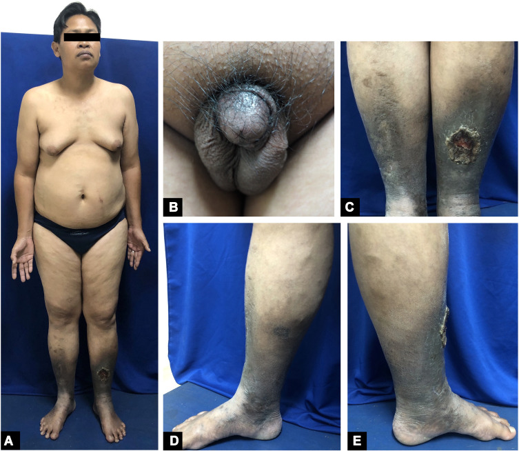
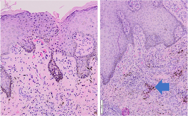
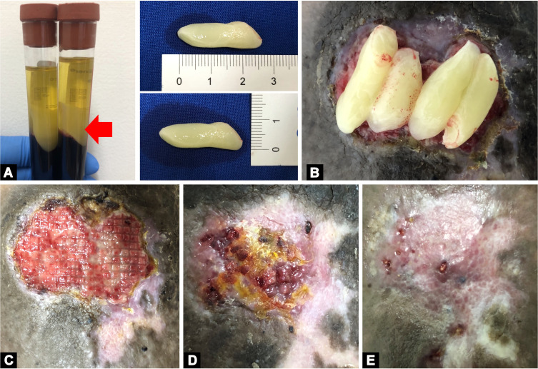
Venous leg ulcers (VLUs) are the most common causes of leg ulcers due to venous insufficiency. Most cases persist for more than 6 weeks, referred to as chronic VLUs. These chronic ulcers have been described as a manifestation of Klinefelter syndrome (KS). Platelet-rich fibrin (PRF) is a second-generation platelet concentrate, which contains growth factors required for chronic wound healing. The use of PRF in the management of VLUs in KS has not been reported, to the best of our knowledge. We report a case of chronic VLU associated with KS in a 41-year-old man treated with PRF. Dermatological examination showed a tender, shallow, irregular ulcer partly covered with hard, yellow necrotic tissue on the anterior side of the lower-left leg and hyperpigmented indurated skin on both lower legs. The diagnosis of venous ulcer was established based on clinical manifestation and supported by the result of Doppler ultrasound showed chronic venous insufficiency. Histopathological examination, which showed epidermal acanthosis, dermal fibrosis, and thickening with hemosiderin deposits consistent with the diagnosis of venous ulcer. The patient presented with eunuchoid features characterized by long extremities, gynecomastia, increased fat distribution around the hips, scanty pubic hairs, and small testes. Laboratory tests found decreased levels of testosterone, increased levels of follicle-stimulating and luteinizing hormone, and bilateral testicular atrophy was found from testicular ultrasound. These physical examinations and laboratory findings supported the diagnosis of KS. The patient was treated with PRF dressing once a week. After 7 weeks of treatment with PRF, the ulcer almost reached complete closure. PRF gives a good result in a chronic VLU with KS.
Keywords: Klinefelter syndrome; platelet-rich fibrin venous leg ulcers.
© 2021 Sutedja et al.
Conflict of interest statement
The authors report no conflicts of interest in this work.
Figures
Similar articles
-
Leucocyte- and platelet-rich fibrin (L-PRF) as a regenerative medicine strategy for the treatment of refractory leg ulcers: a prospective cohort study.Platelets. 2018 Jul;29(5):468-475. doi: 10.1080/09537104.2017.1327654. Epub 2017 Jul 20. Platelets. 2018. PMID: 28727481
-
Comparison of Efficacy of Autologous Platelet-rich Fibrin versus Saline Dressing in Chronic Venous Leg Ulcers: A Randomised Controlled Trial.J Cutan Aesthet Surg. 2017 Jan-Mar;10(1):8-12. doi: 10.4103/JCAS.JCAS_137_16. J Cutan Aesthet Surg. 2017. PMID: 28529414 Free PMC article.
-
Comparison of Efficacy of Autologous Platelet-Rich Fibrin versus Unna's Paste Dressing in Chronic Venous Leg Ulcers: A Comparative Study.Indian Dermatol Online J. 2020 Jan 13;11(1):58-61. doi: 10.4103/idoj.IDOJ_119_19. eCollection 2020 Jan-Feb. Indian Dermatol Online J. 2020. PMID: 32055510 Free PMC article.
-
Klinefelter's syndrome presenting with leg ulcers.Skinmed. 2004 Sep-Oct;3(5):274-8. doi: 10.1111/j.1540-9740.2004.03005.x. Skinmed. 2004. PMID: 15365265 Review.
-
Treatment of vascular leg ulcers with leukocyte- and platelet-rich fibrin (L-PRF): A systematic review.Phlebology. 2024 Sep;39(8):512-520. doi: 10.1177/02683555241256543. Epub 2024 May 23. Phlebology. 2024. PMID: 38782448
Cited by
-
L-PRF in extra-oral wound care.Periodontol 2000. 2025 Feb;97(1):342-362. doi: 10.1111/prd.12605. Epub 2024 Sep 20. Periodontol 2000. 2025. PMID: 39305000 Free PMC article. Review.
-
Dermatologic care of patients with differences of sex development.Int J Womens Dermatol. 2023 Sep 5;9(3):e106. doi: 10.1097/JW9.0000000000000106. eCollection 2023 Oct. Int J Womens Dermatol. 2023. PMID: 37671254 Free PMC article. Review.
References
-
- Hafner J. Ulceration resulting from disorders of the veins and arteries. In: Griffiths CE, Barker J, Bleiker T, Chaimers R, Creamer D, editors. Rook’s Textbook of Dermatology. 9th ed. West Sussex: Willey Blackwell; 2016:104.1‒104.14.
-
- De-morentin HM, Dodiuk-Gad RP, Brenner S. Klinefelter’s syndrome presenting with leg ulcers. Skinmed. 2004;3(5):274‒278. - PubMed
Publication types
LinkOut - more resources
Full Text Sources
