. 2021 Nov 8:2021:1673490.
doi: 10.1155/2021/1673490. eCollection 2021.

Multimodal MRI Analysis of Cervical Cancer on the Basis of Artificial Intelligence Algorithm

Affiliations

Multimodal MRI Analysis of Cervical Cancer on the Basis of Artificial Intelligence Algorithm

Bin Wang et al. Contrast Media Mol Imaging. .

Abstract

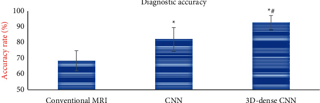
The purpose of this study is to explore the application value of artificial intelligence algorithm in multimodal MRI image diagnosis of cervical cancer. Based on the traditional convolutional neural network (CNN), an artificial intelligence 3D-CNN algorithm is designed according to the characteristics of cervical cancer. 70 patients with cervical cancer were selected as the experimental group, and 10 healthy people were selected as the reference group. The 3D-CNN algorithm was applied to the diagnosis of clinical cervical cancer multimodal MRI images. The value of the algorithm was comprehensively evaluated by the image quality and diagnostic accuracy. The results showed that compared with the traditional CNN algorithm, the convergence rate of the loss curve of the artificial intelligence 3D-CNN algorithm was accelerated, and the segmentation accuracy of whole-area tumors (WT), core tumor areas (CT), and enhanced tumor areas (ET) was significantly improved. In addition, the clarity of the multimodal MRI image and the recognition performance of the lesion were significantly improved. Under the artificial intelligence 3D-CNN algorithm, the Dice values of WT, ET, and CT regions were 0.78, 0.71, and 0.64, respectively. The sensitivity values were 0.92, 0.91, and 0.88, respectively. The specificity values were 0.93, 0.92, and 0.9 l, respectively. The Hausdorff (Haus) distances were 0.93, 0.92, and 0.90, respectively. The data of various indicators were significantly better than those of the traditional CNN algorithm (P < 0.05). In addition, the diagnostic accuracy of the artificial intelligence 3D-CNN algorithm was 93.11 ± 4.65%, which was also significantly higher than that of the traditional CNN algorithm (82.45 ± 7.54%) (P < 0.05). In summary, the recognition and segmentation ability of multimodal MRI images based on artificial intelligence 3D-CNN algorithm for cervical cancer lesions were significantly improved, which can significantly enhance the clinical diagnosis rate of cervical cancer.


Conflict of interest statement

The authors declare no conflicts of interest.

Figures

Figure 1
Figure 1
Residual unit structure.
Figure 2
Figure 2
Network structure pattern of cervical cancer characteristic lesions extraction.
Figure 3
Figure 3
Flowchart based on artificial intelligence 3D-CNN algorithm.
Figure 4
Figure 4
Comparison of DCE-MRI scanning parameters between the two groups of patients. (Note.   represents a significant difference relative to the CNN algorithm (P < 0.05).
Figure 5
Figure 5
Comparison of IVIM scanning parameters between the two groups of patients. (Note.   represents a significant difference relative to the CNN algorithm (P < 0.05).
Figure 6
Figure 6
Loss training results analysis of two algorithms.
Figure 7
Figure 7
Dice analysis of the two algorithms under the training set. (Note.   indicates a significant difference compared with the CNN algorithm (P < 0.05).
Figure 8
Figure 8
MRI images processed by different algorithms. (a) T2W1 image of normal abdominal MRI sagittal position. (b) T1W1 image of normal abdominal MRI transverse position. (c) T2W1 image of normal abdominal MRI transverse position. (d) T2W1 image of abdominal MRI sagittal position of traditional CNN. (e) T1W1 image of abdominal MRI transverse position of traditional CNN. (f) T2W1 image of abdominal MRI transverse position of traditional CNN. (g) T2W1 image of abdominal MRI sagittal position of artificial intelligence 3D-CNN algorithm. (h) T1W1 image of abdominal MRI transverse position of artificial intelligence 3D-CNN algorithm. (i) T2W1 image of abdominal MRI transverse position of artificial intelligence 3D-CNN algorithm.
Figure 9
Figure 9
Visual comparison results of multimodal MRI images in the experiment. (Note. A and E are the transverse and sagittal images of multimodal MRI in patients under normal conditions. B and F are the prediction results of cervical cancer lesions in transverse and sagittal planes delineated by doctors. C and G are the prediction results of cervical cancer lesions in transverse and sagittal positions of the traditional CNN algorithm. D is the prediction of cervical cancer lesions in transverse and sagittal positions by the artificial intelligence 3D-CNN algorithm).
Figure 10
Figure 10
Image processing quality evaluation index comparison chart of different algorithms. (a) Dice value comparison diagram. (b) Sensitivity value comparison chart. (c) Specificity value comparison diagram. (d) Haus distance comparison chart.   represents a significant difference relative to the CNN algorithm (P < 0.05).
Figure 11
Figure 11
Comparison of image diagnostic accuracy of different algorithms. (Note.   represents a significant difference in diagnostic accuracy compared with conventional multimodal MRI images (P< 0.05). # shows a significant difference compared with the CNN algorithm (P< 0.05).

References

    1. Pedersen K., Fogelberg S., Thamsborg L. H., et al. An overview of cervical cancer epidemiology and prevention in Scandinavia. Acta Obstetricia et Gynecologica Scandinavica . 2018;97(7):795–807. doi: 10.1111/aogs.13313. Epub 2018 Feb 23. PMID: 29388202. - DOI - PubMed
    1. Moga M., Dimienescu O., Arvatescu C., Mironescu A., Dracea L., Ples L. The role of natural polyphenols in the prevention and treatment of cervical cancer-an overview. Molecules . 2016;21(8):p. 1055. doi: 10.3390/molecules21081055.PMID:27548122. - DOI - PMC - PubMed
    1. De Strooper L. M. A., Berkhof J., Steenbergen R. D. M., et al. Cervical cancer risk in HPV‐positive women after a negative FAM19A4/mir124‐2 methylation test: a post hoc analysis in the POBASCAM trial with 14 year follow‐up. International Journal of Cancer . 2018;143(6):1541–1548. doi: 10.1002/ijc.31539. - DOI - PMC - PubMed
    1. Tamura K., Hasegawa K., Katsumata N., et al. Efficacy and safety of nivolumab in Japanese patients with uterine cervical cancer, uterine corpus cancer, or soft tissue sarcoma: multicenter, open‐label phase 2 trial. Cancer Science . 2019;110(9):2894–2904. doi: 10.1111/cas.14148. - DOI - PMC - PubMed
    1. Hong D. S., Concin N., Vergote I., et al. Tisotumab vedotin in previously treated recurrent or metastatic cervical cancer. Clinical Cancer Research . 2020;26(6):1220–1228. doi: 10.1158/1078-0432.CCR-19-2962. - DOI - PubMed