

Neurovascular Reactivity in the Aging Mouse Brain Assessed by Laser Speckle Contrast Imaging and 2-Photon Microscopy: Quantification by an Investigator-Independent Analysis Tool
- PMID: 34858312
- PMCID: PMC8631776
- DOI: 10.3389/fneur.2021.745770
Neurovascular Reactivity in the Aging Mouse Brain Assessed by Laser Speckle Contrast Imaging and 2-Photon Microscopy: Quantification by an Investigator-Independent Analysis Tool
Abstract
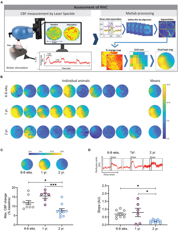
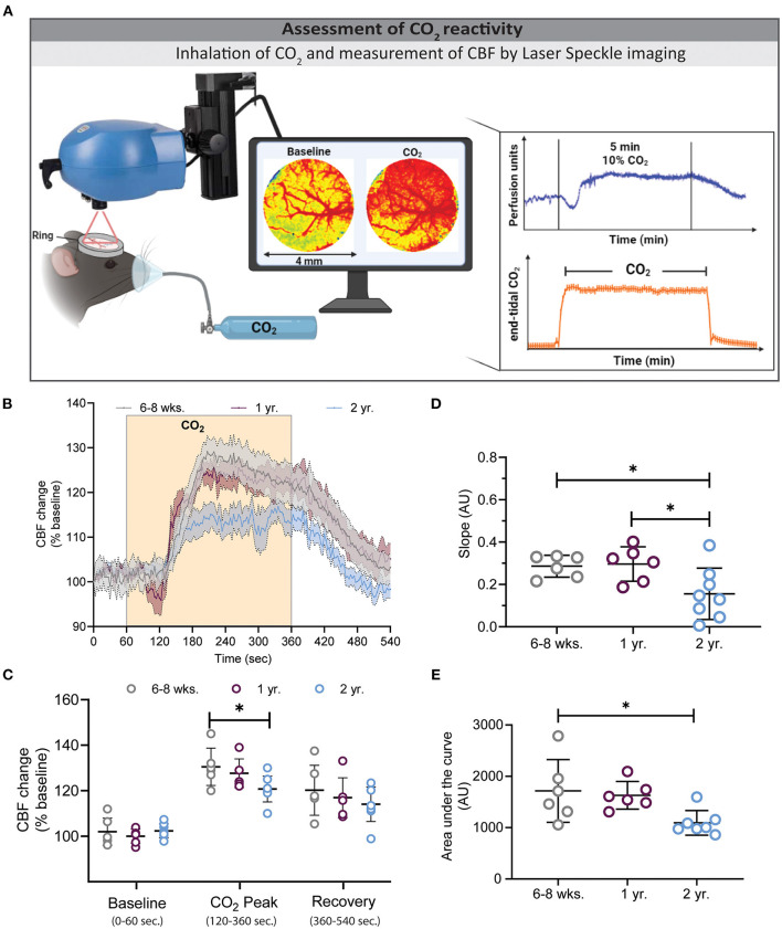
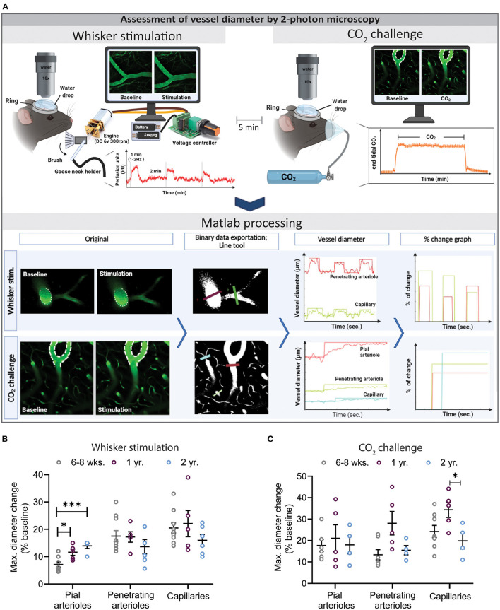
The brain has a high energy demand but little to no energy stores. Therefore, proper brain function relies on the delivery of glucose and oxygen by the cerebral vasculature. The regulation of cerebral blood flow (CBF) occurs at the level of the cerebral capillaries and is driven by a fast and efficient crosstalk between neurons and vessels, a process termed neurovascular coupling (NVC). Experimentally NVC is mainly triggered by sensory stimulation and assessed by measuring either CBF by laser Doppler fluxmetry, laser speckle contrast imaging (LSCI), intrinsic optical imaging, BOLD fMRI, near infrared spectroscopy (NIRS) or functional ultrasound imaging (fUS). Since these techniques have relatively low spatial resolution, diameters of cerebral vessels are mainly assessed by 2-photon microscopy (2-PM). Results of studies on NVC rely on stable animal physiology, high-quality data acquisition, and unbiased data analysis, criteria, which are not easy to achieve. In the current study, we assessed NVC using two different imaging modalities, i.e., LSCI and 2-PM, and analyzed our data using an investigator-independent Matlab-based analysis tool, after manually defining the area of analysis in LSCI and vessels to measure in 2-PM. By investigating NVC in 6-8 weeks, 1-, and 2-year-old mice, we found that NVC was maximal in 1-year old mice and was significantly reduced in aged mice. These findings suggest that NVC is differently affected during the aging process. Most interestingly, specifically pial arterioles, seem to be distinctly affected by the aging. The main finding of our study is that the automated analysis tool works very efficiently in terms of time and accuracy. In fact, the tool reduces the analysis time of one animal from approximately 23 h to about 2 s while basically making no mistakes. In summary, we developed an experimental workflow, which allows us to reliably measure NVC with high spatial and temporal resolution in young and aged mice and to analyze these data in an investigator-independent manner.
Keywords: aging; hypercapnia; investigator-independent analysis; laser speckle contrast imaging; neurovascular coupling; two-photon microscopy.
Copyright © 2021 Seker, Fan, Gesierich, Gaubert, Sienel and Plesnila.
Conflict of interest statement
The authors declare that the research was conducted in the absence of any commercial or financial relationships that could be construed as a potential conflict of interest.
Figures
Similar articles
-
Inversion of neurovascular coupling after subarachnoid hemorrhage in vivo.J Cereb Blood Flow Metab. 2017 Nov;37(11):3625-3634. doi: 10.1177/0271678X16686595. Epub 2017 Jan 23. J Cereb Blood Flow Metab. 2017. PMID: 28112024 Free PMC article.
-
Spatially and temporally mismatched blood flow and neuronal activity by high-intensity intracortical microstimulation.Brain Stimul. 2025 May-Jun;18(3):885-896. doi: 10.1016/j.brs.2025.04.015. Epub 2025 Apr 15. Brain Stimul. 2025. PMID: 40246195
-
Fusogenic liposomes effectively deliver resveratrol to the cerebral microcirculation and improve endothelium-dependent neurovascular coupling responses in aged mice.Geroscience. 2019 Dec;41(6):711-725. doi: 10.1007/s11357-019-00102-1. Epub 2019 Oct 25. Geroscience. 2019. PMID: 31654270 Free PMC article.
-
Neuromodulation of Cerebral Blood Flow: A Physiological Mechanism and Methodological Review of Neurovascular Coupling.Bioengineering (Basel). 2025 Apr 23;12(5):442. doi: 10.3390/bioengineering12050442. Bioengineering (Basel). 2025. PMID: 40428061 Free PMC article. Review.
-
Nitric Oxide Pathways in Neurovascular Coupling Under Normal and Stress Conditions in the Brain: Strategies to Rescue Aberrant Coupling and Improve Cerebral Blood Flow.Front Physiol. 2021 Oct 22;12:729201. doi: 10.3389/fphys.2021.729201. eCollection 2021. Front Physiol. 2021. PMID: 34744769 Free PMC article. Review.
Cited by
-
Programmable scanning diffuse speckle contrast imaging of cerebral blood flow.ArXiv [Preprint]. 2024 Aug 22:arXiv:2408.12715v1. ArXiv. 2024. Update in: Neurophotonics. 2025 Jan;12(1):015006. doi: 10.1117/1.NPh.12.1.015006. PMID: 39253639 Free PMC article. Updated. Preprint.
-
Aerobic exercise reverses aging-induced depth-dependent decline in cerebral microcirculation.bioRxiv [Preprint]. 2023 Feb 13:2023.02.12.528244. doi: 10.1101/2023.02.12.528244. bioRxiv. 2023. Update in: Elife. 2023 Jul 04;12:e86329. doi: 10.7554/eLife.86329. PMID: 36824939 Free PMC article. Updated. Preprint.
-
The effects of time restricted feeding on age-related changes in the mouse retina.Exp Gerontol. 2024 Sep;194:112510. doi: 10.1016/j.exger.2024.112510. Epub 2024 Jul 5. Exp Gerontol. 2024. PMID: 38964431 Free PMC article.
-
Capillary responses to functional and pathological activations rely on the capillary states at rest.J Cereb Blood Flow Metab. 2023 Jun;43(6):1010-1024. doi: 10.1177/0271678X231156372. Epub 2023 Feb 8. J Cereb Blood Flow Metab. 2023. PMID: 36752020 Free PMC article.
-
Programmable scanning diffuse speckle contrast imaging of cerebral blood flow.Neurophotonics. 2025 Jan;12(1):015006. doi: 10.1117/1.NPh.12.1.015006. Epub 2025 Jan 27. Neurophotonics. 2025. PMID: 39872020 Free PMC article.
References
LinkOut - more resources
Full Text Sources