. 2021 Oct 25:12:669447.
doi: 10.3389/fmicb.2021.669447. eCollection 2021.

Fis Connects Two Sensory Pathways, Quorum Sensing and Surface Sensing, to Control Motility in Vibrio parahaemolyticus

Affiliations

Fis Connects Two Sensory Pathways, Quorum Sensing and Surface Sensing, to Control Motility in Vibrio parahaemolyticus

Jessica G Tague et al. Front Microbiol. .

Abstract

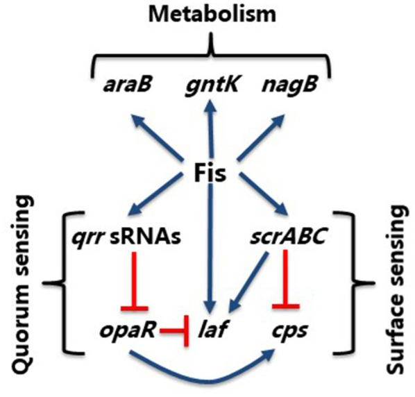
Factor for inversion stimulation (Fis) is a global regulator that is highly expressed during exponential phase growth and undetectable in stationary phase growth. Quorum sensing (QS) is a global regulatory mechanism that controls gene expression in response to changes in cell density and growth phase. In Vibrio parahaemolyticus, a marine species and a significant human pathogen, the QS regulatory sRNAs, Qrr1 to Qrr5, are expressed during exponential growth and negatively regulate the high cell density QS master regulator OpaR. OpaR is a positive regulator of capsule polysaccharide (CPS) formation, which is required for biofilm formation, and is a repressor of lateral flagella required for swarming motility. In V. parahaemolyticus, we show that Fis is a positive regulator of the qrr sRNAs expression. In an in-frame fis deletion mutant, qrr expression was repressed and opaR expression was induced. The Δfis mutant produced CPS and biofilm, but swarming motility was abolished. Also, the fis deletion mutant was more sensitive to polymyxin B. Swarming motility requires expression of both the surface sensing scrABC operon and lateral flagella laf operon. Our data showed that in the Δfis mutant both laf and scrABC genes were repressed. Fis controlled swarming motility indirectly through the QS pathway and directly through the surface sensing pathway. To determine the effects of Fis on cellular metabolism, we performed in vitro growth competition assays, and found that Δfis was outcompeted by wild type in minimal media supplemented with intestinal mucus as a sole nutrient source. The data showed that Fis positively modulated mucus components L-arabinose, D-gluconate and N-acetyl-D-glucosamine catabolism gene expression. In an in vivo colonization competition assay, Δfis was outcompeted by wild type, indicating Fis is required for fitness. Overall, these data demonstrate a global regulatory role for Fis in V. parahaemolyticus that includes QS, motility, and metabolism.

Keywords: Fis; metabolism; motility; quorum sensing; swarming.


Conflict of interest statement

The authors declare that the research was conducted in the absence of any commercial or financial relationships that could be construed as a potential conflict of interest.

Figures

Figure 1
Figure 1
Factor for inversion stimulation binds to the regulatory regions of V. parahaemolyticus regulatory sRNAs (A) qrr1, (B) qrr2, (C) qrr3, (D) qrr4, and (E) qrr5. A putative Fis binding site (BS) is shown as a box in the regulatory region of qrr1 to qrr5. Electrophoretic mobility shift assays (EMSA) using purified Fis and the regulatory regions of qrr1 to qrr5. DNA:protein ratios are as follows: 1:0, 1:1, 1:20, 1:50. (F) Positive Fis BS control in gyrA regulatory region showing complete shift at all concentrations. (G) Fis non-binding negative control using DNA with no putative Fis BS showing weak non-specific binding and no shift at lowest concentration. (H) qrr3 probe containing a single Fis BS (wild type) and a mutated BS demonstrating specificity of Fis binding.
Figure 2
Figure 2
Fis is a positive regulator of the qrr genes. (A) Pqrr1, (B) Pqrr2, (C) Pqrr3, (D) Pqrr4, (E) Pqrr5, and (F) PopaR green fluorescent protein (GFP) transcriptional reporter assays in wild type and the Δfis mutant in cultures grown to OD 0.4–0.45 and measured for specific fluorescence (RFU/OD). Means and standard deviations of three biological replicates are plotted. Statistics calculated using a Student’s t-test (*p<0.05, **p<0.01, ***p<0.001).
Figure 3
Figure 3
Phenotypic analysis of the Δfis mutant. (A) Capsule polysaccharide (CPS) production in wild type (WT), Δfis and ΔopaR mutants was observed after incubation for 48h at 30°C. Images are an example of three biological replicates performed in triplicate. (B) Swimming motility assays and quantification. Two biological replicates were measured, performed in triplicate. Statistics calculated using a Student’s t-test (***p<0.001). (C) Swarming motility assay of V. parahaemolyticus wild type and the Δfis mutant and Δfis complementation strain. Complementation assays were grown in the presence of Cm and IPTG. All images are examples from three biological replicates.
Figure 4
Figure 4
Polymyxin B sensitivity assay. (A) Disk containing 100μg of total polymyxin B was used to identify the zone of inhibition for WT, Δfis and ΔrpoE and was quantified by measuring the diameter of the zone of inhibition. (B) Survival assays were conducted using WT, Δfis, and ΔrpoE after treating the bacterial cultures with polymyxin B (final concentration of 40μg/ml) and calculating percent survival at 30min and 60min. The disk assay was performed in duplicate using two biological replicates. The survival assay was performed in triplicate using two biological replicates. Unpaired t-test was conducted to determine the value of p. ***p<0.001.
Figure 5
Figure 5
Regulation of lateral flagella biosynthesis by Fis. (A) Regulatory region of polar flagellum flh genes with putative Fis binding sites (BS) depicted as blue circles. (B) EMSA of Fis bound to PflhA in a concentration dependent manner. DNA: protein ratios are as follows: 10, 1:1, 1:10, 1:20. (C) Probe 2 containing a single Fis BS (WT) was mutated. DNA: protein ratios are as follows: 0, 1:0.5, 1:1, 1:5. (D) Transcriptional GFP reporter assay of PflhA-gfp in the Δfis mutant relative to wild type. (E) Regulatory region of lateral flagellum laf genes with putative Fis binding sites. (F) EMSA of Fis bound to Plaf DNA probe. (G) Probe 2 containing a single Fis BS and a mutated probe 2 Fis BS. DNA: protein ratios are as follows: 0, 1:0.5, 1:1, 1:5. (H) Transcriptional GFP reporter assay of PlafB-gfp between wild type and Δfis (***p<0.001).
Figure 6
Figure 6
Fis is a positive regulator of the surface sensing operon, scrABC. (A) Putative Fis binding sites identified in the regulatory region of the scrABC surface sensing operon. (B) EMSA using purified Fis protein and probes 1, 2 and 3 encompassing the regulatory region of scrABC. DNA: protein ratios are as follows: 10, 1:1, 1:10, 1:20, 1:50. (C) Probe 3 containing a single Fis BS and a mutated probe 3 Fis BS. DNA: protein ratios are as follows: 0, 1:0.5, 1:1, 1:5. (D) GFP reporter assay of PscrABC-gfp in wild type and the Δfis mutant. Specific fluorescence was calculated (RFU/OD) for three biological replicates and plotted as mean and standard deviation. Statistics were calculated using a Student’s t-test (***p<0.001).
Figure 7
Figure 7
In vitro growth competition assay between wild type and the Δfis mutant. WBWlacZ and Δfis were grown in co-culture (1:1 ratio) for 24h in LBS, M9 supplemented with 100μg/ml intestinal mucus, 10mm of D-glucose, L-arabinose, D-glucosamine, D-gluconate, N-acetyl-D-glucosamine (NAG), or D-ribose. WBWlacZ outcompetes Δfis, <1.00, and Δfis outcompetes WBWlacZ when >1.00. The assay was conducted in two biological replicates in triplicates. Error bar indicates SEM. Unpaired Student’s t-test was conducted. The significant difference is denoted by asterisks (*p<0.05, **p<0.01, ***p<0.001).
Figure 8
Figure 8
Fis is a positive regulator of the L-arabinose operon araBCDA. (A) Fis binding sites identified in the regulatory region of the araBDAC operon depicted as blue circles. (B) ParaB was divided into two probes, probe 1 and probe 2 for EMSA analysis using purified Fis protein. (C) EMSA analysis probe 3 wild type with a single Fis BS and a mutated probe 3. DNA: protein ratios were as follows: 10, 1:1, 1:10, 1:20, 1:50. (D) GFP transcriptional reporter assay of the araBDAC regulatory region in wild type and the Δfis mutant. *p<0.05.
Figure 9
Figure 9
Fis is a positive regulator of gntK. (A) Putative Fis binding sites in the regulatory region of gntK. (B) EMSA analysis with purified Fis protein and PgntK probe 1. DNA: protein ratios were as follows: 10, 1:1, 1:10, 1:20, 1:50. (C) EMSA analysis of probe 2 containing a single Fis BS and a mutated Fis BS probe 2. (D) GFP transcriptional reporter assay of gntK regulatory region in wild type and Δfis. **p<0.01.
Figure 10
Figure 10
Fis is a positive regulator of NAG gene nagB. (A) Fis binding sites identified in the regulatory region of nagB. (B) EMSA analysis of Fis binding to probe 1. (C) EMSA analysis of probe 2 and a mutated Fis BS probe 2. DNA: protein molar ratios were as follows: 10, 1:1, 1:10, 1:20, 1:50. (D) GFP transcriptional reporter assay with PnagB-gfp. Means and standard deviations of two biological replicates are shown. Statistics calculated using a Student’s t-test (**p<0.01).
Figure 11
Figure 11
Analysis of Fis consensus sequence. (A) A Fis DNA binding motif was created using MEME analysis. Sequences containing a putative Fis binding site were used. (B) An alignment of the motifs found in each sequence, along with the corresponding values of p.
Figure 12
Figure 12
Model of Fis integration of QS and surface sensing pathways. Model shows control of swarming (laf) and CPS (cps) production in V. parahaemolyticus. Blue arrows show positive regulation and orange hammers show negative regulation.

References

    1. Auner H., Buckle M., Deufel A., Kutateladze T., Lazarus L., Mavathur R., et al. . (2003). Mechanism of transcriptional activation by FIS: role of core promoter structure and DNA topology. J. Mol. Biol. 331, 331–344. doi: 10.1016/S0022-2836(03)00727-7, PMID: - DOI - PubMed
    1. Azam T. A., Iwata A., Nishimura A., Ueda S., Ishihama A. (1999). Growth phase-dependent variation in protein composition of the Escherichia coli nucleoid. J. Bacteriol. 181, 6361–6370. doi: 10.1128/JB.181.20.6361-6370.1999, PMID: - DOI - PMC - PubMed
    1. Bassler B. (1999). How bacteria talk to each other: regulation of gene expression by quorum sensing. Curr. Opin. Microbiol. 2, 582–587. doi: 10.1016/S1369-5274(99)00025-9, PMID: - DOI - PubMed
    1. Bassler B. L., Greenberg E. P., Stevens A. M. (1997). Cross-species induction of luminescence in the quorum-sensing bacterium Vibrio harveyi. J. Bacteriol. 179, 4043–4045. doi: 10.1128/jb.179.12.4043-4045.1997, PMID: - DOI - PMC - PubMed
    1. Belas R., Simon M., Silverman M. (1986). Regulation of lateral flagella gene transcription in Vibrio parahaemolyticus. J. Bacteriol. 167, 210–218. doi: 10.1128/jb.167.1.210-218.1986, PMID: - DOI - PMC - PubMed

LinkOut - more resources