

Pattern Recognition Receptors (PRRs) in Macrophages Possess Prognosis and Immunotherapy Potential for Melanoma
- PMID: 34858419
- PMCID: PMC8630683
- DOI: 10.3389/fimmu.2021.765615
Pattern Recognition Receptors (PRRs) in Macrophages Possess Prognosis and Immunotherapy Potential for Melanoma
Abstract
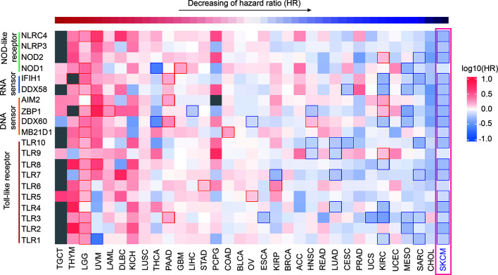
Background: Pattern recognition receptors (PRRs) family plays a vital role in the initial stage of innate immune response and the subsequent activation of adaptive immunity. Increasing evidences have indicated that several PRRs play critical roles in the progress of inflammation and tumorigenesis. However, the comprehensive significance of PRRs family in clinical prognosis of different cancers is still elusive.
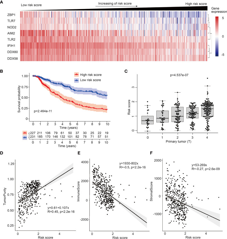
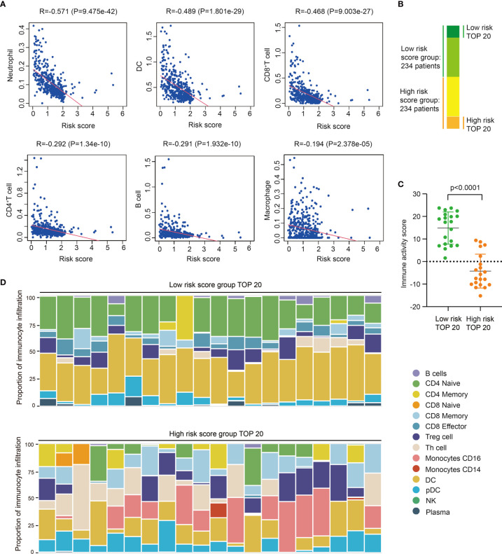
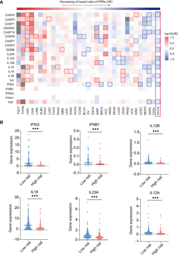
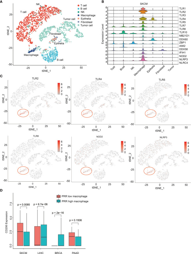
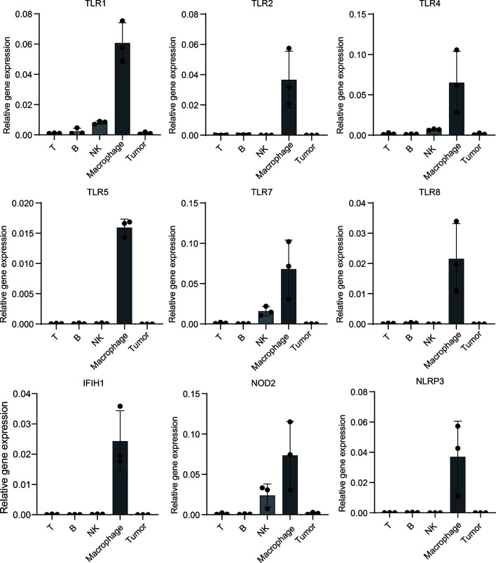
Methods: We analyzed expression of 20 canonical PRRs in tumor samples from 9502 patients of 33 tumor types. Next, we used expression profiles of PRRs in skin cutaneous melanoma (SKCM) to build a Cox prognosis model. Then, we analyzed immune infiltration features and immune activity of high risk score and low risk score patients. Finally, we analyzed the single-cell sequencing data of different cancers and detected the expression of PRRs in mouse melanoma model to identify PRRs-expressing cell types.
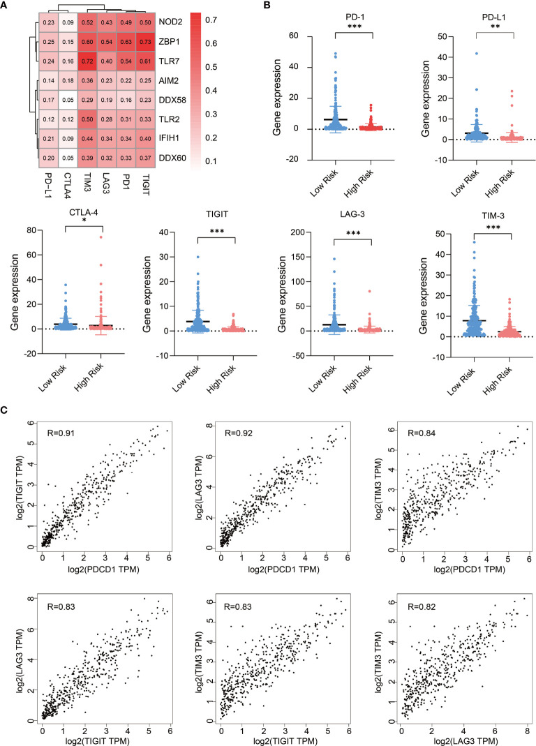
Results: We found PRRs had a significantly positive correlation with prognosis in SKCM rather than other tumors, and PRR-based Cox model had a much better prognosis potential than any single PRR. Further analysis shows risk score could indicate immunocyte infiltration and immune activity in SKCM. We also found the expressions of some PRR genes were highly correlated with the expression of immune checkpoints molecules in SKCM, indicating they could be indicators for clinical immune therapy. Finally, we found only in SKCM samples, the expression of PRRs is especially high in a subpopulation of macrophages with a trait of CD206 low expression, probably explaining why PRRs have prognosis potential in melanoma.
Conclusions: Our study reveals PRR family in macrophages has a positive prognosis potential in melanoma and could be valuable for clinical prognosis and immune therapy.
Keywords: immune-infiltration; macrophage; pattern recognition receptors; prognosis; skin cutaneous melanoma.
Copyright © 2021 Zhao, Wang, Wang, Xu, Li, Liu, Yao and Wang.
Conflict of interest statement
The authors declare that the research was conducted in the absence of any commercial or financial relationships that could be construed as a potential conflict of interest.
Figures
Similar articles
-
Development of a biomarker signature associated with anoikis to predict prognosis and immunotherapy response in melanoma.Arch Dermatol Res. 2024 May 24;316(6):219. doi: 10.1007/s00403-024-03085-y. Arch Dermatol Res. 2024. PMID: 38787413
-
Development and validation of an immune gene set-based prognostic signature in cutaneous melanoma.Future Oncol. 2021 Nov;17(31):4115-4129. doi: 10.2217/fon-2021-0104. Epub 2021 Jul 22. Future Oncol. 2021. PMID: 34291650
-
Circadian rhythm related genes identified through tumorigenesis and immune infiltration-guided strategies as predictors of prognosis, immunotherapy response, and candidate drugs in skin cutaneous malignant melanoma.Front Immunol. 2025 Mar 21;16:1513750. doi: 10.3389/fimmu.2025.1513750. eCollection 2025. Front Immunol. 2025. PMID: 40191195 Free PMC article.
-
Promising targets based on pattern recognition receptors for cancer immunotherapy.Pharmacol Res. 2020 Sep;159:105017. doi: 10.1016/j.phrs.2020.105017. Epub 2020 Jun 16. Pharmacol Res. 2020. PMID: 32561479 Review.
-
Advancing immunotherapy for melanoma: the critical role of single-cell analysis in identifying predictive biomarkers.Front Immunol. 2024 Jul 4;15:1435187. doi: 10.3389/fimmu.2024.1435187. eCollection 2024. Front Immunol. 2024. PMID: 39026661 Free PMC article. Review.
Cited by
-
Role of the Microbiota in Skin Neoplasms: New Therapeutic Horizons.Microorganisms. 2023 Sep 25;11(10):2386. doi: 10.3390/microorganisms11102386. Microorganisms. 2023. PMID: 37894044 Free PMC article. Review.
-
Isatidis Folium Represses Dextran Sulfate Sodium-Induced Colitis and Suppresses the Inflammatory Response by Inhibiting Inflammasome Activation.Nutrients. 2024 Sep 30;16(19):3323. doi: 10.3390/nu16193323. Nutrients. 2024. PMID: 39408295 Free PMC article.
-
Integrated bioinformatic analysis identifies GADD45B as an immune-related prognostic biomarker in skin cutaneous melanoma.Hereditas. 2025 May 11;162(1):74. doi: 10.1186/s41065-025-00437-0. Hereditas. 2025. PMID: 40350499 Free PMC article.
-
Current Updates on Pathogenesis, Systemic Therapy, and Treatment of Invasive Fungal Infections.Curr Drug Targets. 2025;26(3):203-220. doi: 10.2174/0113894501337502241015121015. Curr Drug Targets. 2025. PMID: 39421988 Review.
-
Orchestration of Tumor-Associated Macrophages in the Tumor Cell-Macrophage-CD8+ T Cell Loop for Cancer Immunotherapy.Int J Biol Sci. 2025 Jun 12;21(9):4098-4116. doi: 10.7150/ijbs.115932. eCollection 2025. Int J Biol Sci. 2025. PMID: 40607254 Free PMC article. Review.
References
Publication types
MeSH terms
Substances
LinkOut - more resources
Full Text Sources
Medical