. 2021 Nov 11:12:774052.
doi: 10.3389/fimmu.2021.774052. eCollection 2021.

Expanded Hemodialysis Therapy Ameliorates Uremia-Induced Systemic Microinflammation and Endothelial Dysfunction by Modulating VEGF, TNF-α and AP-1 Signaling

Affiliations

Expanded Hemodialysis Therapy Ameliorates Uremia-Induced Systemic Microinflammation and Endothelial Dysfunction by Modulating VEGF, TNF-α and AP-1 Signaling

Rusan Catar et al. Front Immunol. .

Abstract

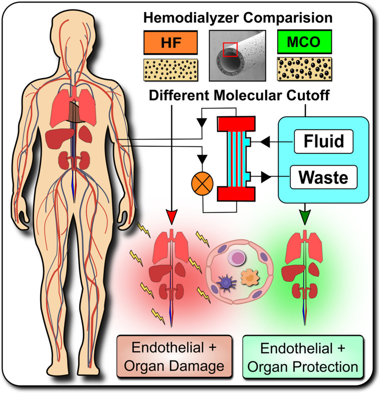
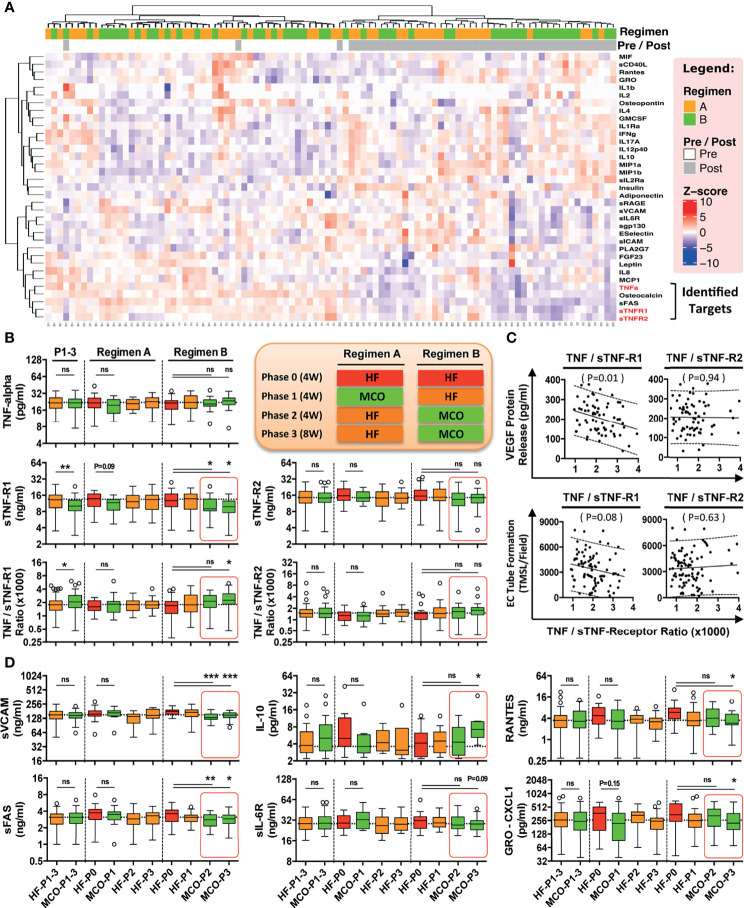
Abstract: Systemic chronic microinflammation and altered cytokine signaling, with adjunct cardiovascular disease (CVD), endothelial maladaptation and dysfunction is common in dialysis patients suffering from end-stage renal disease and associated with increased morbidity and mortality. New hemodialysis filters might offer improvements. We here studied the impact of novel improved molecular cut-off hemodialysis filters on systemic microinflammation, uremia and endothelial dysfunction. Human endothelial cells (ECs) were incubated with uremic serum obtained from patients treated with two different hemodialysis regimens in the Permeability Enhancement to Reduce Chronic Inflammation (PERCI-II) crossover clinical trial, comparing High-Flux (HF) and Medium Cut-Off (MCO) membranes, and then assessed for their vascular endothelial growth factor (VEGF) production and angiogenesis. Compared to HF membranes, dialysis with MCO membranes lead to a reduction in proinflammatory mediators and reduced endothelial VEGF production and angiogenesis. Cytokine multiplex screening identified tumor necrosis factor (TNF) superfamily members as promising targets. The influence of TNF-α and its soluble receptors (sTNF-R1 and sTNF-R2) on endothelial VEGF promoter activation, protein release, and the involved signaling pathways was analyzed, revealing that this detrimental signaling was indeed induced by TNF-α and mediated by AP-1/c-FOS signaling. In conclusion, uremic toxins, in particular TNF-signaling, promote endothelial maladaptation, VEGF expression and aberrant angiogenesis, which can be positively modulated by dialysis with novel MCO membranes.

Translational perspective and graphical abstract: Systemic microinflammation, altered cytokine signaling, cardiovascular disease, and endothelial maladaptation/dysfunction are common clinical complications in dialysis patients suffering from end-stage renal disease. We studied the impact of novel improved medium-cut-off hemodialysis filters on uremia and endothelial dysfunction. We can show that uremic toxins, especially TNF-signaling, promote endothelial maladaptation, VEGF expression and aberrant angiogenesis, which can be positively modulated by dialysis with novel improved medium-cut-off membranes.

Trial registration: ClinicalTrials.gov NCT02084381.

Keywords: cardiovascular disease; chronic kidney disease; end-stage renal disease; endothelial cell (dys)function; expanded hemodialysis therapy; tumor necrosis factor alpha (TNF-alpha); uremic toxins / systemic microinflammation; vascular endothelial growth factor (VEGF).


Conflict of interest statement

The authors declare that the research was conducted in the absence of any commercial or financial relationships that could be construed as a potential conflict of interest.

Figures

Graphical Abstract
Graphical Abstract
Systemic microinflammation, altered cytokine signaling and cardiovascular diseases are common in hemodialysis patients contributing to the highly increased cardiovascular morbidity and mortality. One of the pathological causes is the endothelial maladaptation and dysfunction associated with uremia and chronic systemic microinflammation. We here elucidate the molecular and biological mechanisms how endothelial maladaptation is induced, and most importantly also how it can be reversed, with in vivo validation in a crossover randomized multi-center clinical study comparing novel improved medium-cut-off (MCO) dialyzers to standard-of-care high-flux (HF) dialyzers.
Figure 1
Figure 1
Study design and hypothesis: Expanded Hemodialysis Therapy ameliorates systemic Inflammation and endothelial maladaptation and dysfunction. (A) The medical need for Expanded Hemodialysis Therapy and clinical study design: The hemodialysis field has been shown a near exponential growth in the past decades, with >167.000 publications on PUBMED containing the search-term “Hemodialysis” in 2020. Recently, particular attention has been placed into lowering chronic treatment-associated adverse cardiovascular diseases (CVD) and new optimized treatment concepts, such as “Expanded Hemodialysis Therapy” with improved molecular cut-off hemodialyzers (8, 27). Within the PERCI-II study n=48 hemodialysis patients underwent crossover randomized multi-center comparison employing novel medium-cut-off (MCO; MCOI-Ci400, Gambro) dialyzers in comparison to standard of care high-flux (HF) hemodialyzers (PERCI-II-MCO; ClinicalTrials.gov: NCT02084381) (28). These novel MCO dialyzers have an improved molecular size cut-off, which positively modulates systemic microinflammation (28). (B) Goal of the follow up study: To Elucidate the Molecular and Biological Mechanisms: In the present study, we explore the molecular signaling mechanisms underlying this positive antiinflammatory shift and evaluate promising leads identified during the first screen in 2017. In particular, we study the modulation of TNF-superfamily members in sera of patients undergoing MCO dialysis and how this impacts on uremia- and TNF-α-induced endothelial maladaptation and dysfunction (left panel) and the molecular mechanisms (right panel), resulting in aberrant VEGF induction and angiogenesis. Our VEGF promoter activation studies and adjunct signaling pathway experiments elucidated that this detrimental uremia- and TNF-α-induced signaling is mediated via AP-1/c-FOS signaling and that alterations in the serum ratio between TNF-α and sTNF-R1, but not sTNF-R2, are potential indicators for endothelial maladaptation. These findings provide new avenues for molecular targets and treatment modalities to reduce chronic microinflammation in the context of hemodialysis.
Figure 2
Figure 2
Hemodialysis with improved MCO dialyzers normalizes endothelial VEGF production and maladaptive angiogenesis upon uremic serum exposure in vitro. (A) Schematics of patient serum collection for analysis of endothelial VEGF expression and angiogenesis/endothelial tube formation after stimulation of ECs with respective sera. Within the PERCI-II study, patients underwent alternating hemodialysis (HD; in 4 weeks intervals) with either high-flux (HF) or medium cut-off (MCO) dialyzers (n=23-25 patients). Upon a 4-weeks wash-in phase on HF dialyzers, patients were allocated for 4 weeks to either HF or MCO dialyzers, followed by a 4-weeks wash-out phase on HF dialyzers, followed by a 12-weeks allocation to HF or MCO dialyzer. The top row shows regimen A (HF, MCO, HF, HF) and the bottom row shows regimen B (HF, HF, MCO, MCO). The sera/time points used for analysis in the second part of the figure are indicated with red stars: end of wash-in, end of phase 1, 2, 3; and (B, C) Endothelial VEGF mRNA expression (AU; arbitrary units, 3-hour stimulation; n=23-25), and (D, E) VEGF protein release (pg/ml) upon 24-hour stimulation with either 10% HF-HD or 10% MCO-HD serum (n=23-25), and (F, G) Endothelial tube formation (TMSL/field; n=23-25) upon stimulated with either 10% HF-HD or 10% MCO-HD serum for 16 hours, as compared to healthy serum (HS) controls. ANOVA, Mean ± SEM, with *P < 0.05, **P < 0.01, and ***P < 0.001. ns, not significant.
Figure 3
Figure 3
MCO dialysis alters the systemic TNF-α/s-TNF-R1-ratio correlating with endothelial protection in vitro and a beneficial shift in serum cytokine levels in vivo. The patients underwent the two different hemodialysis (HD) regimes as indicated in (either HF/MCO/MCO or MCO/HF/HF; n=23-25) and the levels of soluble mediators at the different time points were analyzed with multiplex Milliplex and Luminex technology (28). (A) Unsupervised clustering heat-map analysis of biomarkers (rows) and patients (columns); (B) Quantification of TNF-α, and sTNF-R1 and sTNF-R1-R2 and their corresponding ratios; (C) Correlation of the TNF-α/sTNF-R1- and TNF-α/sTNF-R2-ratios with endothelial VEGF release (pg/ml) or endothelial tube formation (TMSL/field) upon stimulation with either 10% HF or 10% MCO patient serum; and (D) Patient serum profiling for biomarkers of endothelial activation and systemic inflammation. [Legend to (B) and (D)] Central legend (simplified depiction corresponding to the clinical trial scheme shown in ) indicates the underlying color code of the samples/time points in the box plots from regimen and the length of their phases (Phase 1 and 2 four weeks and phase 3 eight weeks, abbreviated as P1, P2, and P3 with duration shown in brackets), which show cytokine analysis of serum samples at the end of HF wash-in phase (Phase 0, the standard proinflammatory baseline before start of Regimen A or B which indicated in red), and the end of Phase 1, 2, and 3 (HF shown in orange, and MCO shown in green in legend and corresponding box plots), and the analyzed samples correspond to the end of the phases (Corresponding to the red stars in Figure 2A ). Each box plot is labeled with the corresponding dialysis filter device (HF or MCO) and the trial stage (P0, P1, P2, and P3), as indicate in the central legend. ANOVA, Box plots Tukey with interquartile range, with *P < 0.05, **P < 0.01, and ***P < 0.001. ns, not significant.
Figure 4
Figure 4
Elevated TNF-α and VEGF levels in uremic serum and VEGF induction in ECs by uremic serum, but omission of VEGF induction by TNF-α blockade. (A) Levels of TNF-α (pg/ml), sTNF-R1 (ng/ml) and VEGF (pg/ml), in sera derived from healthy control subjects or uremic hemodialysis patients (n=14), used to generate the healthy and uremic serum pools (HSP and USP, respectively; Mann-Whitney test, Box plots min-max range); (B, C) Kinetics and dose-response of endothelial cell (EC) VEGF mRNA (AU; arbitrary units; n=6) and protein production (pg/ml) in response to incubation with HSP or USP (both 2way-ANOVA); (B) To assess kinetics of VEGF production, the ECs were incubated for different time points (1-24 hours) with 10% serum with the peak of VEGF mRNA expression detected at 3 hours and maximal protein expression at 6-24 hours; (C) To assess the dose-response of VEGF production the ECs were incubated with different concentrations of (1-20% serum) with maximal VEGF mRNA expression and protein secretion being detected in response to 10-20% serum after 3 and 24 hours of incubation respectively; and (D–F) The effect of either: (D) Anti-IL-1 receptor antagonist Anakinra, or (E) Anti-TNF-α blocking antibody Infliximab, on human uremic serum-induced VEGF release in ECs. The cells were pre-treated with or without either Anakinra or Infliximab for 1 hour, followed by stimulation for 24 hours with either 10% USP or 10% HSP (n=7), and ECs were subsequently assessed for VEGF release (both 2way-ANOVA); and (F) Dose-dependent effect of 1-20% USP vs. 1-20% HSP on EC viability, with assessment of EC viability (% viable cells, n = 6) with the WST-8 cell viability assay after 24-hour stimulation (2way-ANOVA). Box plots min-max range with Mann-Whitney-test, other plots 2way-ANOVA-testing with mean ± SEM, with *P <0.05, **P <0.01, and ***P <0.001.
Figure 5
Figure 5
TNF-α concentration-dependent VEGF promoter activation and angiogenesis and VEGF promoter sequences responsive to TNF-α stimulation. (A–C) ECs were stimulated with different concentrations of TNF-α (1 to 1000 pg/ml) to assess: (A) VEGF promoter activation (RLU; relative luciferase activity, 3-hour stimulation with TNF-α, n=4) in cells transfected with the pLuc 2068 full-length luciferase-reporter construct; and (B) VEGF mRNA expression (AU; arbitrary units, 3-hour stimulation with TNF-α, n=4); and (C) Endothelial tube formation (TMSL/field; total master segment length per field averaged of 5 assessed fields per condition, 16-hour stimulation with TNF-α, n=12). (D) To identify VEGF promoter sequences responsive to TNF-α stimulation ECs were transfected with different VEGF promoter constructs subjected to progressive 5’-deletions (full-length -2018 construct and -1286, -267, and -52 deletion) and the relative luciferase activity (RLU, n=4) determined in cells stimulated for 6 hours with TNF-α (1 pg/mL or 1000 pg/ml) compared to unstimulated resting control cells, demonstrating a loss in VEGF promoter activity upon truncation of the promoter region spanning the positions -267 to -52, and simultaneous identification of a potential high affinity AP-1/c-FOS binding site at position -102 with computation analysis. (E) Validation of the AP-1/c-FOS transcription factor-binding site with EMSA (one representative experiment shown) using biotin-labeled double-stranded oligonucleotides targeting to the calculated AP-1/c-FOS positions at -95 to -119 of the corresponding VEGF promoter region. ECs were stimulated for 6 h with TNF-a (1 pg/mL or 1000 pg/ml) and nuclear fractions analyzed for formation of nuclear complexes with the c-FOS probe (labeled as a shift with arrow); and (F) Effect of sTNF-R1 (1, 10, and 100 ng/ml) on the modulation of TNF-α induced c-FOS mRNA-expression (AU; 3-hour stimulation with 1 or 1000 pg/ml TNF-α, n=4) with corresponding TNF-α/sTNF-R1 ratios (0.01, 0.1, 1.0, 10, 100, 1000). 1way-ANOVA with Mean ± SEM, with *P < 0.05, **P < 0.01, and ***P < 0.001. ns, not significant.
Figure 6
Figure 6
Analysis of the molecular signaling pathways underling TNF-α induced endothelial VEGF induction and angiogenesis identifies AP-1/cFOS-signaling. (A, B) Role of the AP-1/c-FOS signaling pathway in low and high TNF-α level-induced endothelial VEGF induction (AU; n=5) shown in (A) and in vitro angiogenesis/endothelial tube formation (TMSL/field; total master segment length per field averaged out of 5 assessed fields per condition, 16-hour stimulation with TNF-α, n=6) shown in (B). Cells were first pre-treated with or without AP-1 blocker SR-11302 (10 nM) for 1 hour, followed by stimulation for 16 hours with or without 1 or 1000 pg/ml TNF-α in the presence or absence TNF-α blocking antibody Infliximab (100 ug/ml). Mann-Whitney-test with mean ± SEM, *P < 0.05 and **P < 0.01.

Similar articles

Cited by

References

    1. Eckardt KU, Coresh J, Devuyst O, Johnson RJ, Kottgen A, Levey AS, et al. . Evolving Importance of Kidney Disease: From Subspecialty to Global Health Burden. Lancet (2013) 382:158–69. doi: 10.1016/S0140-6736(13)60439-0 - DOI - PubMed
    1. Remuzzi G, Benigni A, Finkelstein FO, Grunfeld JP, Joly D, Katz I, et al. . Kidney Failure: Aims for the Next 10 Years and Barriers to Success. Lancet (2013) 382:353–62. doi: 10.1016/S0140-6736(13)60438-9 - DOI - PubMed
    1. Bikbov B, Purcell CA, Levey AS, Smith M, Abdoli A, Abebe M, et al. . Global, Regional, and National Burden of Chronic Kidney Disease, 1990–2017: A Systematic Analysis for the Global Burden of Disease Study 2017. Lancet (2020) 395:709–33. doi: 10.1016/S0140-6736(20)30045-3 - DOI - PMC - PubMed
    1. Nadim MK, Forni LG, Mehta RL, Connor MJ, Liu KD, Ostermann M, et al. . COVID-19-Associated Acute Kidney Injury: Consensus Report of the 25th Acute Disease Quality Initiative (ADQI) Workgroup. Nat Rev Nephrol (2020) 16:747–64. doi: 10.1038/s41581-020-00356-5 - DOI - PMC - PubMed
    1. Perico L, Benigni A, Casiraghi F, Ng LFP, Renia L, Remuzzi G. Immunity, Endothelial Injury and Complement-Induced Coagulopathy in COVID-19. Nat Rev Nephrol (2021) 17:46–64. doi: 10.1038/s41581-020-00357-4 - DOI - PMC - PubMed

Publication types

MeSH terms

Associated data