Review
. 2021 Nov 8:12:775447.
doi: 10.3389/fimmu.2021.775447. eCollection 2021.

β2 Integrin CD11d/CD18: From Expression to an Emerging Role in Staged Leukocyte Migration

Affiliations
Review

β2 Integrin CD11d/CD18: From Expression to an Emerging Role in Staged Leukocyte Migration

Eoin N Blythe et al. Front Immunol. .

Abstract

CD11d/CD18 is the most recently discovered and least understood β2 integrin. Known CD11d adhesive mechanisms contribute to both extravasation and mesenchymal migration - two key aspects for localizing peripheral leukocytes to sites of inflammation. Differential expression of CD11d induces differences in monocyte/macrophage mesenchymal migration including impacts on macrophage sub-set migration. The participation of CD11d/CD18 in leukocyte localization during atherosclerosis and following neurotrauma has sparked interest in the development of CD11d-targeted therapeutic agents. Whereas the adhesive properties of CD11d have undergone investigation, the signalling pathways induced by ligand binding remain largely undefined. Underlining each adhesive and signalling function, CD11d is under unique transcriptional control and expressed on a sub-set of predominately tissue-differentiated innate leukocytes. The following review is the first to capture the nearly three decades of CD11d research and discusses the emerging role of CD11d in leukocyte migration and retention during the progression of a staged immune response.

Keywords: CD11d; CD18; beta 2 integrin; extravasation; inflammation; leukocyte; migration.


Conflict of interest statement

The authors declare that the research was conducted in the absence of any commercial or financial relationships that could be construed as a potential conflict of interest.

Figures
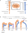
Figure 1
Figure 1
Visual representation of β2 integrin structure and conserved regulatory conformations. (A) Organization of the domains composing the CD11 and CD18 chains (15, 16). The ligand binding α-I domain is highlighted by a hatched pattern. The metal ion-dependant adhesion site (MIDAS) and the socket for isoleucine (SILEN) motifs are located within the α-I domain (16, 17). The SILEN motif interacts with an invariant isoleucine located in the α7 helix to maintain the inactive conformation (17). (B) Representation of the β2 integrin regulatory conformations. The bent-closed inactive conformation predominates under basal conditions (1). Stimulation can activate the integrin and induce the extended-closed conformation. Additional stimulation and the binding of a divalent metal ion to the MIDAS motif, can induce the extended-open conformation. The bent-open conformation is stabilized by binding a cis ligand and may provide an alternative activation pathway to the extended-open conformation (–20). The extended-open conformation is characterized by the separation of the cytosolic tails in additional to local conformational changes within the α-I domain, including shifting of the α7 helix (–16, 21, 22).
Figure 2
Figure 2
Visual representation of a probable CD11d structure including amino acid homolog comparisons of key motifs. (A) Sequence comparison of the CD11d α-I domain major ligand binding region. The ligand binding CD11d α-I domain is highlighted by a hatched pattern. Residues determined to be important in the ligand binding pocket of CD11b are underlined and percent homology to CD11d is in brackets. Alignment and CD11b residue analysis performed in previous study (51). (B) Sequence comparison of the CD11 α7 helix. An invariable isoleucine is highlighted in red and percent homology to CD11d is in brackets. Alignment was performed in previous study (17). Conformational changes to the α7 helix within CD11d have been shown to alter ligand affinities, thus implying the presence of an open and closed α-I domain conformation (51). (C) Sequence comparison of complete CD11 cytoplasmic tails. Yellow denotes potential phosphorylation sites, red denotes conserved residues of interest, and the underlined sequence denotes a potential CK2 site. The GFFKR “hinge” motif is required to maintain the association of the CD11 and CD18 cytoplasmic tails (52). The constitutive phosphorylation of a serine residue is conserved across CD11a (Ser1165) (53), CD11b (Ser1143) (54), and CD11c (Ser1158) (55). CD11d has a putative CK2 site at Ser1148-Cys1154 using the consensus sequence (S-X-X-D/E-X-pS-P) (56). The same sequence would predict Ser1153 to be constitutively phosphorylated as observed in other β2 integrins. The function of the conserved GFFKR +2 tyrosine residue in CD11b-d is largely undefined. The tyrosine appears to be embedded into the membrane during the inactive conformation, while exposed during the active conformation (57). Long isoform CD11a (NP_002200.2), CD11b (NP_001139280.1), CD11c (NP_000878.2), and CD11d (NP_001305114.1) amino acid sequences were acquired from the National Center for Biotechnology Information database (–31).
Figure 3
Figure 3
Diagram of known CD11d/CD18 ligands. Cellular receptors are shaded green, extracellular matrix proteins are blue, and proteins/protein modifications prevalent within the ECM during inflammation are red (51, 68). Shared ligand specificities with CD49d/CD29 is denoted in a red outline (70), while shared ligand specificities with CD11b/CD18 is outlined in black (51).
Figure 4
Figure 4
Representation of the impact CD11d density has on monocyte/macrophage mesenchymal migration. Low density of CD11d expression supports migration, while high densities inhibits migration and promotes tissue retention (8).

References

    1. Fagerholm SC, Guenther C, Llort Asens M, Savinko T, Uotila LM. Beta2-Integrins and Interacting Proteins in Leukocyte Trafficking, Immune Suppression, and Immunodeficiency Disease. Front Immunol (2019) 10:254. doi: 10.3389/fimmu.2019.00254 - DOI - PMC - PubMed
    1. Dib K. BETA 2 Integrin Signaling in Leukocytes. Front Biosci (2000) 5:438–51. doi: 10.2741/pathology - DOI - PubMed
    1. Hu P, Luo B-H. Integrin Bi-Directional Signaling Across the Plasma Membrane. J Cell Physiol (2013) 228:306–12. doi: 10.1002/jcp.24154 - DOI - PubMed
    1. Danilenko DM, Rossitto PV, Vieren MVd, Trong HL, McDonough SP, Affolter VK, et al. . A Novel Canine Leukointegrin, Alpha D Beta 2, Is Expressed by Specific Macrophage Subpopulations in Tissue and a Minor CD8+ Lymphocyte Subpopulation in Peripheral Blood. J Immunol (1995) 155:35–44. - PubMed
    1. Van der Vieren M, Le Trong H, Wood CL, Moore PF, St John T, Staunton DE, et al. . A Novel Leukointegrin, Alpha D Beta 2, Binds Preferentially to ICAM-3. Immunity (1995) 3:683–90. doi: 10.1016/1074-7613(95)90058-6 - DOI - PubMed

Publication types

MeSH terms

Grants and funding

LinkOut - more resources