Review
. 2021 Nov 1:9:739079.
doi: 10.3389/fcell.2021.739079. eCollection 2021.

Navigating the Light-Sheet Image Analysis Software Landscape: Concepts for Driving Cohesion From Data Acquisition to Analysis

Affiliations
Review

Navigating the Light-Sheet Image Analysis Software Landscape: Concepts for Driving Cohesion From Data Acquisition to Analysis

Holly C Gibbs et al. Front Cell Dev Biol. .

Abstract

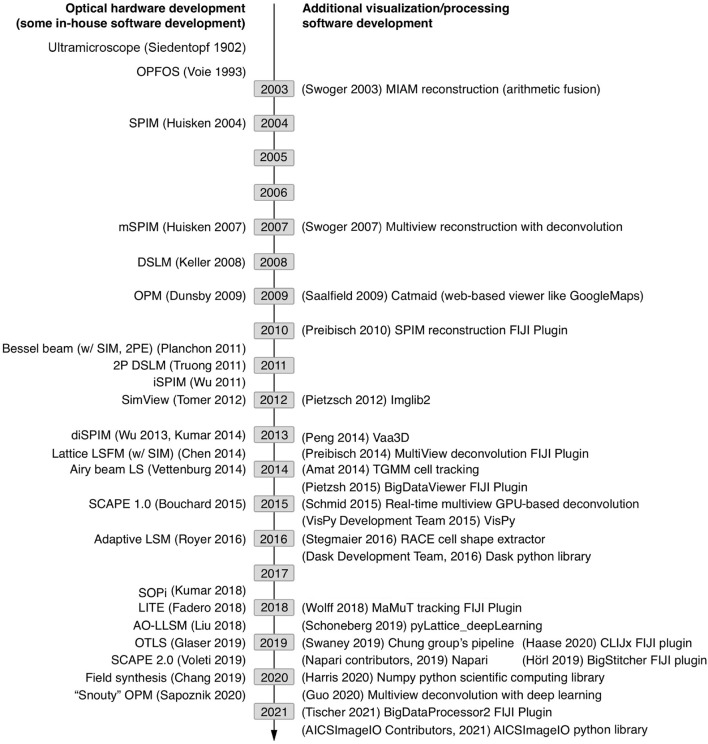
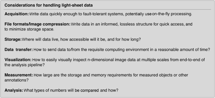
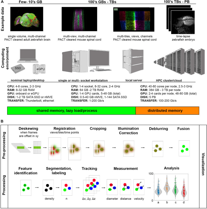
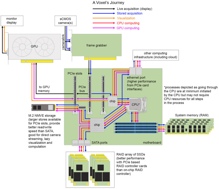
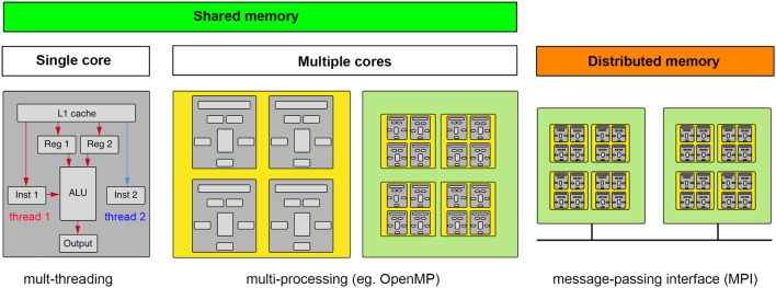
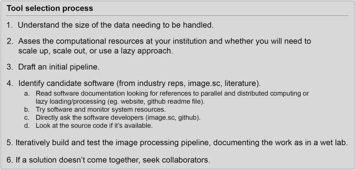
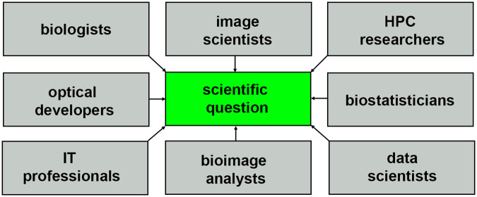
From the combined perspective of biologists, microscope instrumentation developers, imaging core facility scientists, and high performance computing experts, we discuss the challenges faced when selecting imaging and analysis tools in the field of light-sheet microscopy. Our goal is to provide a contextual framework of basic computing concepts that cell and developmental biologists can refer to when mapping the peculiarities of different light-sheet data to specific existing computing environments and image analysis pipelines. We provide our perspective on efficient processes for tool selection and review current hardware and software commonly used in light-sheet image analysis, as well as discuss what ideal tools for the future may look like.

Keywords: image analysis; light-sheet; multiview deconvolution; parallel processing; tool selection.


Conflict of interest statement

The authors declare that the research was conducted in the absence of any commercial or financial relationships that could be construed as a potential conflict of interest.

Figures

FIGURE 1
FIGURE 1
Timeline of sampled light-sheet microscopy optical hardware development and processing software across a variety of applications. As unique species of light-sheet microscopes have been developed, unique analysis solutions have been created.
FIGURE 2
FIGURE 2
Data handling steps that require special attention with light-sheet image data. If possible, it is advantageous to select a compressed file format that can be utilized by the requisite analysis software. Unfortunately, not all analysis software can read all image formats and often data must be resaved or restructured as it travels through an analysis pipeline.
FIGURE 3
FIGURE 3
Range of light-sheet data sizes, computing environments, and processing tasks. (A) As data size increases, increasingly parallel computation helps to prevent bottlenecking during image analysis. (B) Components of a light-sheet image analysis pipeline have highly variable pre-processing steps dependent on the particular type of light-sheet microscope used and more uniform processing steps depending on the biological measurement of interest.
FIGURE 4
FIGURE 4
The journey of a voxel. Hardware components and interfaces for transferring data between them on one example of a typical image analysis workstation.
FIGURE 5
FIGURE 5
Computing concepts for scaling up and scaling out. Computation requires data and instructions to be loaded into registers directly accessible by an arithmetic logic unit (ALU). Multi-threading makes computations parallel by taking advantage of dead time when data are being fetched. Multi-processing is when the computation can be spread to multiple cores (ALUs), whether on a CPU or GPU, that have access to the same memory (GPU’s typically having their own smaller on-board memory). If the data and/or computations do not fit in shared memory, a message-passing interface must help coordinate the broadcasting of data and computations across a distributed memory system. (gray, core; yellow, CPU; green, motherboard).
FIGURE 6
FIGURE 6
Outline of software tool selection process.
FIGURE 7
FIGURE 7
Community of researchers that help to turn light-sheet image data into scientific insight.

References

    1. AICSImageIO Contributors (2021). AICSImageIO: Image Reading, Metadata Conversion, and Image Writing for Microscopy Images in Pure Python [Computer software]. GitHub. Available online at: https://github.com/AllenCellModeling/aicsimageio (accessed October 7, 2021).
    1. Albert-Smet I., Marcos-Vidal A., Vaquero J. J., Desco M., Muñoz-Barrutia A., Ripoll J. (2019). Applications of Light-Sheet Microscopy in Microdevices. Front. Neuroanat. 13:1. - PMC - PubMed
    1. Amat F., Lemon W., Mossing D. P., McDole K., Wan Y., Branson K., et al. (2014). Fast, accurate reconstruction of cell lineages from large-scale fluorescence microscopy data. Nat. Methods 11 951–958. 10.1038/nmeth.3036 - DOI - PubMed
    1. Andreev A., Koo D. E. S. (2020). Practical guide to storing large amounts of microscopy data. Microsc. Today 28 42–45. 10.1017/s1551929520001091 - DOI
    1. Bai C., Liu C., Yu X., Peng T., Min J., Yan S., et al. (2019). Imaging Enhancement of Light-Sheet Fluorescence Microscopy via Deep Learning. IEEE Photon. Technol. Lett. 31 1803–1806. 10.1109/lpt.2019.2948030 - DOI

LinkOut - more resources