Neuroanatomical anomalies associated with rare AP4E1 mutations in people who stutter
- PMID: 34859215
- PMCID: PMC8633735
- DOI: 10.1093/braincomms/fcab266
Neuroanatomical anomalies associated with rare AP4E1 mutations in people who stutter
Abstract
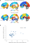
Developmental stuttering is a common speech disorder with strong genetic underpinnings. Recently, stuttering has been associated with mutations in genes involved in lysosomal enzyme trafficking. However, how these mutations affect the brains of people who stutter remains largely unknown. In this study, we compared grey matter volume and white matter fractional anisotropy between a unique group of seven subjects who stutter and carry the same rare heterozygous AP4E1 coding mutations and seven unrelated controls without such variants. The carriers of the AP4E1 mutations are members of a large Cameroonian family in which the association between AP4E1 and persistent stuttering was previously identified. Compared to controls, mutation carriers showed reduced grey matter volume in the thalamus, visual areas and the posterior cingulate cortex. Moreover, reduced fractional anisotropy was observed in the corpus callosum, consistent with the results of previous neuroimaging studies of people who stutter with unknown genetic backgrounds. Analysis of gene expression data showed that these structural differences appeared at the locations in which expression of AP4E1 is relatively high. Moreover, the pattern of grey matter volume differences was significantly associated with AP4E1 expression across the left supratentorial regions. This spatial congruency further supports the connection between AP4E1 mutations and the observed structural differences.
Keywords: corpus callosum; fractional anisotropy; gene expression; thalamus; voxel-based morphometry.
© The Author(s) (2021). Published by Oxford University Press on behalf of the Guarantors of Brain.
Figures


Similar articles
-
White matter neuroanatomical differences in young children who stutter.Brain. 2015 Mar;138(Pt 3):694-711. doi: 10.1093/brain/awu400. Epub 2015 Jan 24. Brain. 2015. PMID: 25619509 Free PMC article.
-
Association between Rare Variants in AP4E1, a Component of Intracellular Trafficking, and Persistent Stuttering.Am J Hum Genet. 2015 Nov 5;97(5):715-25. doi: 10.1016/j.ajhg.2015.10.007. Am J Hum Genet. 2015. PMID: 26544806 Free PMC article.
-
A voxel-based morphometry (VBM) analysis of regional grey and white matter volume abnormalities within the speech production network of children who stutter.Cortex. 2013 Sep;49(8):2151-61. doi: 10.1016/j.cortex.2012.08.013. Epub 2012 Sep 17. Cortex. 2013. PMID: 23140891 Free PMC article.
-
The Role of Basal Ganglia and Its Neuronal Connections in the Development of Stuttering: A Review Article.Cureus. 2022 Aug 31;14(8):e28653. doi: 10.7759/cureus.28653. eCollection 2022 Aug. Cureus. 2022. PMID: 36196326 Free PMC article. Review.
-
Research updates in neuroimaging studies of children who stutter.Semin Speech Lang. 2014 May;35(2):67-79. doi: 10.1055/s-0034-1382151. Epub 2014 May 29. Semin Speech Lang. 2014. PMID: 24875668 Free PMC article. Review.
Cited by
-
Knowns and unknowns about the neurobiology of stuttering.PLoS Biol. 2024 Feb 22;22(2):e3002492. doi: 10.1371/journal.pbio.3002492. eCollection 2024 Feb. PLoS Biol. 2024. PMID: 38386639 Free PMC article.
-
Stuttering: Our Current Knowledge, Research Opportunities, and Ways to Address Critical Gaps.Neurobiol Lang (Camb). 2025 Apr 2;6:nol_a_00162. doi: 10.1162/nol_a_00162. eCollection 2025. Neurobiol Lang (Camb). 2025. PMID: 40201450 Free PMC article. Review.
References
-
- Kornfeld S, Sly W.. I-cell disease and pseudo-Hurler polydystrophy: disorders of lysosomal enzyme phosphorylation and localization. In: Scriver C, Beaudet A, Sly W, Valle D, eds. The Metabolic and molecular bases of inherited disease. New York: McGraw-Hill; 1995:2495–2508.
Grants and funding
LinkOut - more resources
Full Text Sources
