. 2022 Feb 1;205(3):300-312.
doi: 10.1164/rccm.202009-3686OC.

Targeting the P2Y13 Receptor Suppresses IL-33 and HMGB1 Release and Ameliorates Experimental Asthma

Affiliations

Targeting the P2Y13 Receptor Suppresses IL-33 and HMGB1 Release and Ameliorates Experimental Asthma

Rhiannon B Werder et al. Am J Respir Crit Care Med. .

Abstract

Rationale: The alarmins IL-33 and HMGB1 (high mobility group box 1) contribute to type 2 inflammation and asthma pathogenesis. Objectives: To determine whether P2Y13-R (P2Y13 receptor), a purinergic GPCR (G protein-coupled receptor) and risk allele for asthma, regulates the release of IL-33 and HMGB1. Methods: Bronchial biopsy specimens were obtained from healthy subjects and subjects with asthma. Primary human airway epithelial cells (AECs), primary mouse AECs, or C57Bl/6 mice were inoculated with various aeroallergens or respiratory viruses, and the nuclear-to-cytoplasmic translocation and release of alarmins was measured by using immunohistochemistry and an ELISA. The role of P2Y13-R in AEC function and in the onset, progression, and exacerbation of experimental asthma was assessed by using pharmacological antagonists and mice with P2Y13-R gene deletion. Measurements and Main Results: Aeroallergen exposure induced the extracellular release of ADP and ATP, nucleotides that activate P2Y13-R. ATP, ADP, and aeroallergen (house dust mite, cockroach, or Alternaria antigen) or virus exposure induced the nuclear-to-cytoplasmic translocation and subsequent release of IL-33 and HMGB1, and this response was ablated by genetic deletion or pharmacological antagonism of P2Y13. In mice, prophylactic or therapeutic P2Y13-R blockade attenuated asthma onset and, critically, ablated the severity of a rhinovirus-associated exacerbation in a high-fidelity experimental model of chronic asthma. Moreover, P2Y13-R antagonism derepressed antiviral immunity, increasing IFN-λ production and decreasing viral copies in the lung. Conclusions: We identify P2Y13-R as a novel gatekeeper of the nuclear alarmins IL-33 and HMGB1 and demonstrate that the targeting of this GPCR via genetic deletion or treatment with a small-molecule antagonist protects against the onset and exacerbations of experimental asthma.

Keywords: GPCR; alarmin; epithelium; pneumonia virus of mice; purinergic; rhinovirus.


Figures

Figure 1.
Figure 1.
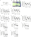
Airway epithelial cells (AECs) express and respond to P2Y13-R (P2Y13 receptor) activation. (A) Representative image of P2Y13-R immunohistochemistry (pink) in a healthy human lung biopsy sample (left) or an asthmatic human lung biopsy sample (center). The antibody was preincubated with a peptide before staining to ensure that immunoreactivity was specific (right). P2Y13-R+ AECs were quantified as a percentage of total, Nuc., or Cyto. expression. Scale bar, 20 μm. (B) P2Y13-R expression on cultured, differentiated human AECs (hAECs) (top) or cultured, undifferentiated hAECs (P2Y13-R, red; nuclei, blue). Scale bar, 10 μm. (C) Multiplex immunohistochemistry of a human lung biopsy sample stained for P2Y13-R (red), IL-33 (green), and HMGB1 (high mobility group box 1) (yellow) expression. Arrows denote P2Y13-R+ (red +), HMGB1+ (yellow +) and/or IL-33+ (green +) AECs. Scale bar, 10 μm. (D) P2Y13-R expression in lung sections of WT and P2Y13-R−/− mice. Scale bars, 20 μm (left 2 panels) or 50 μm (right 2 panels). (E) hAECs were stimulated with 100 μM of ADP or ATP for 2 hours, fixed, and stained for IL-33 or HMGB1 (red) and DAPI (blue). Scale bar, 10 μm. (F) Healthy hAECs were stimulated with house dust mite extract (HDM) (50 μg/ml), and supernatants were collected to probe for ADP (top) and ATP (bottom). (G) Healthy (black) and asthmatic (red) hAECs cultured at the air–liquid interface were stimulated with HDM (50 μg/ml), and basolateral supernatants were collected to probe for ADP (top) and ATP (bottom) at 8 hours. Representative images from three independent experiments (n = 4 subjects per group) are shown in BE. In F and G, **P < 0.01 is compared with Time 0 or unstimulated samples. Cyto. = cytoplasmic; Expt = experimental; Nuc. = nuclear; WT = wild type.
Figure 2.
Figure 2.
P2Y13-R (P2Y13 receptor) activation mediates alarmin release in vitro. (A and B) Human AECs (hAECs) and (C) mouse AECs (mAECs) were preincubated with 1 μM of MRS2211 before incubation with house dust mite extract (HDM) (50 μg/ml). Cells were fixed and stained for IL-33 and HMGB1 (high mobility group box 1). Translocation of alarmins from the nucleus to cytoplasm was quantified (cyto–IL-33 [cytoplasmic IL-33] or cyto-HMGB1) and expressed as a percentage of total cells. Scale bar, 10 μm. (D) HMGB1 release into the supernatant was measured by using an ELISA in HDM-stimulated (50 μg/ml) hAECs (left) and mAECs (right). (E) hAECs from healthy subjects or subjects with asthma were preincubated with 1 μM of MRS2211 and then stimulated with ADP. Translocation of alarmins from the nucleus to cytoplasm was quantified (cyto–IL-33 or cyto-HMGB1) and expressed as a percentage of total cells. (F) hAECs were infected with RV, and the cytoplasmic translocation of IL-33 and HMGB1 was quantified. Data are presented as the mean ± SEM and are representative of three independent experiments (n = 3–4 subjects per group or 3–5 mice per group). For F, cells from six donors were used. *P < 0.05, **P < 0.01, and ***P < 0.001 are compared with Time 0 and # P < 0.05, ## P < 0.01, and ### P < 0.001 are compared with HDM-treated samples at each time point or as specified. AEC = airway epithelial cell; ind. = induced; RV = rhinovirus; V = vehicle.
Figure 3.
Figure 3.
P2Y1-R (P2Y1 receptor), P2Y2-R, and P2Y12-R do not contribute to house dust mite extract (HDM)-induced alarmin translocation and release in vitro. (A) hAECs and (B) mAECs were preincubated with MRS2500 (P2Y1-R antagonist), AR-C118925XX (P2Y2-R antagonist), ticagrelor (P2Y12-R antagonist), or MRS2211 (P2Y13-R antagonist) before incubation with HDM. Nuclear-to-cytoplasmic translocation of IL-33 and HMGB1 (high mobility group box 1) was quantified. (C and D) mAECs obtained from wild-type (+/+) or P2Y13-R−/− (−/−) mice were stimulated with HDM, the nuclear-to-cytoplasmic translocation of IL-33 and HMGB1 was quantified (C), and the HMGB1 release into the supernatant was measured by using an ELISA (D). Data are presented as mean ± SEM or as box-and-whisker plots showing quartiles (boxes) and ranges (whiskers) and are representative of two independent experiments (n = 3–4 subjects per group or 4–6 mice per group). *P < 0.05, **P < 0.01, and ***P < 0.001 are compared with HDM-treated cells or Time 0, and ## P < 0.01 and ### P < 0.001 are compared with HDM-treated samples at each time. AEC = airway epithelial cell; Cyto-HMGB1 = cytoplasmic HMGB1; Cyto–IL-33 = cytoplasmic IL-33; hAEC = human AEC; mAEC = mouse AEC.
Figure 4.
Figure 4.
P2Y13-R (P2Y13 receptor) promotes allergen-induced alarmin release and type 2 inflammation in vivo. (A) Study design. Wild-type mice were injected intraperitoneally with MRS2211 (1 mg/kg) 1 hour before inoculation with 100 μg of house dust mite extract (HDM), 25 μg of Alt, or 100 μg of CRE (i.n.). Mice were killed 2 or 72 hours later. (B) HMGB1 (high mobility group box 1) immunostaining on lung tissues. Arrows indicate HMGB1 nuclear-to-cytoplasmic translocation, and translocation was quantified as a percentage of AECs. Scale bar, 100 μm. (C) HMGB1 and (D) IL-33 release in the BAL fluid (BALF). (E) Type-2 innate lymphoid cells (ILC2s) in the lung. (F and G) Eosinophils and neutrophils in the BALF. (HL) Wild-type (+/+), P2Y13-R−/+, and P2Y13-R−/− mice were intranasally inoculated with 100 μg of HDM, and endpoints were analyzed 72 hours later. (H) HMGB1 and (I) IL-33 release in the BALF. (J) Number of ILC2s in the lung. (K and L) Number of eosinophils and neutrophils in the BALF. Data are presented as box-and-whisker plots showing quartiles (boxes) and ranges (whiskers) and are representative of two independent experiments (n = 4–7 mice per group). *P < 0.05, **P < 0.01, ***P < 0.001, and ****P < 0.0001. Alt = Alternaria alternata extract; AEC = airway epithelial cell; CRE = cockroach extract; Cyto-HMGB1 = cytoplasmic HMGB1; ns = not significant; Veh = vehicle.
Figure 5.
Figure 5.
P2Y13-R (P2Y13 receptor) antagonism attenuates house dust mite extract (HDM)-induced experimental asthma. (A) Study design. Wild-type mice were given MRS2211 (1 mg/kg, i.p.) 1 hour before inoculation with 100 μg of HDM at Day 0. Mice were challenged at Days 14, 15, 16, and 17 with 10 μg of HDM, and endpoints were measured 3 hours after the final challenge. (B and C) Number of eosinophils and neutrophils in BAL fluid (BALF). (D and E) Number of ILC2s and Th2 cells in the lung. (F and G) Concentration of IL-4 and IL-5 in the BALF. (H) Representative images of Muc5a immunostaining on lung tissue. Muc5a expression was quantified as a percentage of AECs. Scale bars, 100 μm. Data are presented as box-and-whisker plots showing quartiles (boxes) and ranges (whiskers) and are representative of two independent experiments (n = 4–8 mice per group). *P < 0.05, **P < 0.01, and ***P < 0.001. AEC = airway epithelial cell; ILC2 = type 2 innate lymphoid cell; PBS = phosphate-buffered saline; Th2 = T-helper cell type 2; Veh. = vehicle.
Figure 6.
Figure 6.
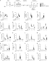
P2Y13-R (P2Y13 receptor) antagonism ameliorates the severity of a rhinovirus (RV)-induced asthma exacerbation. (A) Study design. Mice were inoculated with pneumonia virus of mice (PVM) (1 plaque-forming unit [pfu], i.n.) at 7 days old and with cockroach extract (CRE) (1 μg, i.n.) 3 days later. Six weeks later, mice were reinfected with PVM (100 pfu, i.n.) and exposed weekly to CRE (1 μg, i.n.). Mice were inoculated with RV-1B (5 × 106 virions median tissue culture infectious dose) at 94 days after the primary infection. In the treatment group, mice were injected intraperitoneally with MRS2211 (1 mg/kg) 1 hour before RV-1B and were then injected daily. Mice were killed at 1 and 3 dpi. (B) IL-33 and (C) HMGB1 (high mobility group box 1) release in the BAL fluid (BALF). (D and E) RV-1B viral copies were determined by using quantitative PCR (D) analysis and IFN-λ protein expression (E) in the BALF. (F and G) Number of eosinophils and neutrophils in the BALF. (H and I) Number of type 2 innate lymphoid cells (ILC2s) and T-helper cell type 2 (Th2) cells in the lung. (JL) Concentration of IL-4, IL-5, and IL-13 in the BALF. (M) Muc5a expression was quantified as a percentage of airway epithelial cells (AECs). (NU) Wild-type (WT) or P2Y13-R−/− mice were treated as per study design in A. (N and O) Number of eosinophils and neutrophils in the BALF. (P and Q) Number of ILC2s and Th2 cells in the lung. (RT) Concentration of IL-4, IL-5, and IL-13 in the BALF. (U) Muc5a expression was quantified as a percentage of AECs. Data are presented as box-and-whisker plots showing quartiles (boxes) and ranges (whiskers) and are representative of two independent experiments (n = 4–8 mice per group). *P < 0.05, **P < 0.01, and ***P < 0.001 are compared with the V-treated group and # P < 0.05, ## P < 0.01, and ### P < 0.001 are compared with RV-infected group or WT group at each time point. dpi = days postinfection; HPRT = hypoxanthine guanine phosphoribosyltransferase; PND = postnatal day; V = vehicle.

Comment in

Similar articles

Cited by

References

    1. Schmitz J, Owyang A, Oldham E, Song Y, Murphy E, McClanahan TK, et al. IL-33, an interleukin-1-like cytokine that signals via the IL-1 receptor-related protein ST2 and induces T helper type 2-associated cytokines. Immunity . 2005;23:479–490. - PubMed
    1. Neill DR, Wong SH, Bellosi A, Flynn RJ, Daly M, Langford TK, et al. Nuocytes represent a new innate effector leukocyte that mediates type-2 immunity. Nature . 2010;464:1367–1370. - PMC - PubMed
    1. Shim EJ, Chun E, Lee HS, Bang BR, Kim TW, Cho SH, et al. The role of high-mobility group box-1 (HMGB1) in the pathogenesis of asthma. Clin Exp Allergy . 2012;42:958–965. - PubMed
    1. Zhou B, Comeau MR, De Smedt T, Liggitt HD, Dahl ME, Lewis DB, et al. Thymic stromal lymphopoietin as a key initiator of allergic airway inflammation in mice. Nat Immunol . 2005;6:1047–1053. - PubMed
    1. Werder RB, Lynch JP, Simpson JC, Zhang V, Hodge NH, Poh M, et al. PGD2/DP2 receptor activation promotes severe viral bronchiolitis by suppressing IFN-λ production. Sci Transl Med . 2018;10:eaao0052. - PubMed

Publication types