. 2022 Jan 7;21(1):243-249.
doi: 10.1021/acs.jproteome.1c00822. Epub 2021 Dec 3.

Mass Spectrometry Imaging of N-Linked Glycans in a Formalin-Fixed Paraffin-Embedded Human Prostate by Infrared Matrix-Assisted Laser Desorption Electrospray Ionization

Affiliations

Mass Spectrometry Imaging of N-Linked Glycans in a Formalin-Fixed Paraffin-Embedded Human Prostate by Infrared Matrix-Assisted Laser Desorption Electrospray Ionization

Crystal L Pace et al. J Proteome Res. .

Abstract

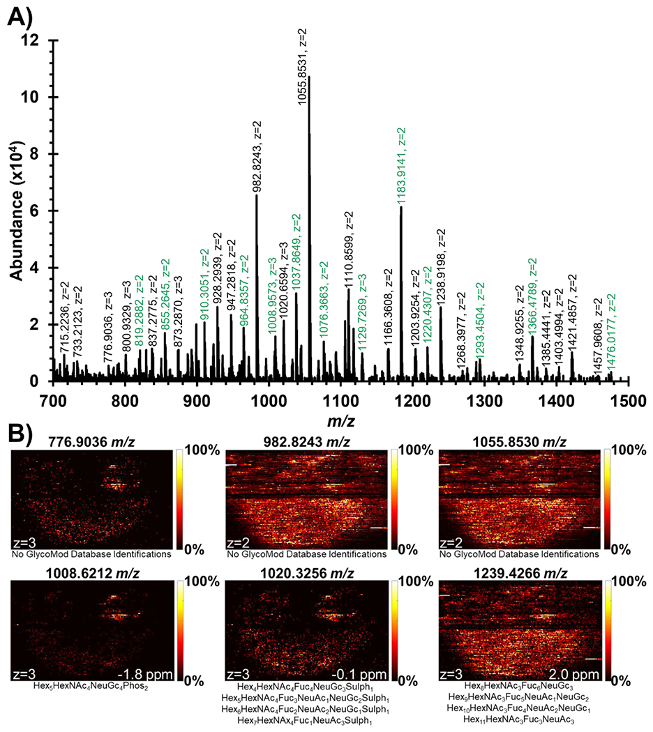
N-Linked glycans are structurally diverse polysaccharides that represent significant biological relevance due to their involvement in disease progression and cancer. Due to their complex nature, N-linked glycans pose many analytical challenges requiring the continued development of analytical technologies. Infrared matrix-assisted laser desorption electrospray ionization (IR-MALDESI) is a hybrid ionization technique commonly used for mass spectrometry imaging (MSI) applications. Previous work demonstrated IR-MALDESI to significantly preserve sialic acid containing N-linked glycans that otherwise require chemical derivatization prior to detection. Here, we demonstrate the first analysis of N-linked glycans in situ by IR-MALDESI MSI. A formalin-fixed paraffin-embedded human prostate tissue was analyzed in negative ionization mode after tissue washing, antigen retrieval, and pneumatic application of PNGase F for enzymatic digestion of N-linked glycans. Fifty-three N-linked glycans were confidently identified in the prostate sample where more than 60% contained sialic acid residues. This work demonstrates the first steps in N-linked glycan imaging of biological tissues by IR-MALDESI MSI. Raw data files are available in MassIVE (identifier: MSV000088414).

Keywords: IR-MALDESI; N-linked glycans; mass spectrometry imaging; prostate cancer.


Figures

Figure 1.
Figure 1.
Optical image of FFPE human prostate tissue showing multiple large morphological features. The tumorous region as determined by H&E staining is roughly outlined. A few examples of glandular and stromal regions are highlighted and will be discussed later with ion image distributions.
Figure 2.
Figure 2.
Experimental workflow implemented in this study for the analysis of N-linked glycans. Tissue washing was utilized for dewaxing and delipidating the tissue. Antigen retrieval was required to reduce protein crosslinking prior to enzymatic digestion by pneumatically applied PNGase F. IR-MALDESI MSI was used to spatially detect N-Linked glycans in negative ionization mode. Data analysis included spatial interpretation in MSiReader and peak picking of multiply-charged species via GlycoHunter.
Figure 3.
Figure 3.
Colocalization with spatial features of human prostate sample. Ion images of three putatively identified N-linked glycans are shown for reference (left). The optical image of the prostate sample is overlayed on top of each ion image (middle) to show colocalization with morphological features of the tissue with smaller regions of interest zoomed-in to exhibit close alignment (right). The black line seen halfway through the ion images was added by MSiReader to separate two regions of interest collected for this prostate sample.
Figure 4.
Figure 4.
A) Mass spectrum #7719 showing significant number of multiply-charged peaks. Labels highlighted in green were already annotated by GlyConnect database searching (Table 1). This scan was searched for peak pairs via GlycoHunter with a 0.5017 Da or 0.3345 Da m/z offset to find multiply-charged monoisotopic peaks to search within GlycoMod database. B) Ion images of multiply-charged peaks with no GlycoMod database identifications and potential sulfate/phosphate modifications represent significant spatially informative distributions that could be of biological importance to the human prostate sample. The black line seen halfway through the ion images was added by MSiReader to separate two regions of interest collected for this prostate sample.
Figure 5.
Figure 5.
Fragmentation spectra for a putatively identified N-linked glycan and an unidentified multiply-charged peak. A) The glycosidic cleavages shown for the 1037 m/z peak confirm the putative identification of Hex5HexNAc4Fuc1NeuAc1. B) The unknown peak at 1055 m/z showed extremely similar fragmentation peaks to the 1037 m/z peak. Additionally, the singly charged form of Hex5HexNAc4Fuc1NeuAc1 was also detected indicating that this is likely the core structure of the 1055 m/z peak with an additional unknown modification.
Figure 6.
Figure 6.
Isotopic distributions of two detected N-linked glycans with theoretical distributions of chlorine and deprotonated adducts overlayed. The [M-2H+]2+ isotopic distribution would be observed at a lower m/z value but has been shifted to compare probabilities of abundance. Chi-squared values testing a goodness of fit indicate that the N-linked glycans were detected with chlorine adducts.

References

    1. Hart GW; Copeland RJ Glycomics Hits the Big Time. Cell 2010, 143 (5), 672–676. - PMC - PubMed
    1. Abdul Rahman S; Bergström E; Watson CJ; Wilson KM; Ashford DA; Thomas JR; Ungar D; Thomas-Oates JE Filter-Aided N-Glycan Separation (FANGS): A Convenient Sample Preparation Method for Mass Spectrometric N-Glycan Profiling. J. Proteome Res 2014, 13 (3), 1167–1176. - PMC - PubMed
    1. Hecht ES; McCord JP; Muddiman DC A Quantitative Glycomics and Proteomics Combined Purification Strategy. J. Vis. Exp 2016, 2016 (109), 1–13. - PMC - PubMed
    1. Powers TW; Jones EE; Betesh LR; Romano PR; Gao P; Copland JA; Mehta AS; Drake RR Matrix Assisted Laser Desorption Ionization Imaging Mass Spectrometry Workflow for Spatial Profiling Analysis of N-Linked Glycan Expression in Tissues. Anal. Chem 2013, 85 (20), 9799–9806. - PMC - PubMed
    1. Eshghi ST; Yang S; Wang X; Shah P; Li X; Zhang H Imaging of N-Linked Glycans from Formalin-Fixed Paraffin-Embedded Tissue Sections Using MALDI Mass Spectrometry. ACS Chem. Biol 2014, 9 (9), 2149–2156. - PMC - PubMed

Publication types