. 2021 Dec 3;6(66):eabf1152.
doi: 10.1126/sciimmunol.abf1152. Epub 2021 Dec 3.

A particulate saponin/TLR agonist vaccine adjuvant alters lymph flow and modulates adaptive immunity

Affiliations

A particulate saponin/TLR agonist vaccine adjuvant alters lymph flow and modulates adaptive immunity

Murillo Silva et al. Sci Immunol. .

Abstract

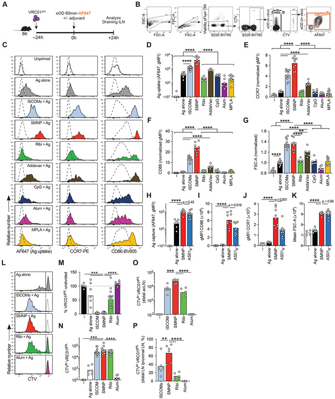
Saponins are potent and safe vaccine adjuvants, but their mechanisms of action remain incompletely understood. Here, we explored the properties of several saponin formulations, including immune-stimulatory complexes (ISCOMs) formed by the self-assembly of saponin and phospholipids in the absence or presence of the Toll-like receptor 4 agonist monophosphoryl lipid A (MPLA). We found that MPLA self-assembles with saponins to form particles physically resembling ISCOMs, which we termed saponin/MPLA nanoparticles (SMNP). Saponin-containing adjuvants exhibited distinctive mechanisms of action, altering lymph flow in a mast cell–dependent manner and promoting antigen entry into draining lymph nodes. SMNP was particularly effective, exhibiting even greater potency than the compositionally related adjuvant AS01B in mice, and primed robust germinal center B cell, TFH, and HIV tier 2 neutralizing antibodies in nonhuman primates. Together, these findings shed new light on mechanisms by which saponin adjuvants act to promote the immune response and suggest that SMNP may be a promising adjuvant in the setting of HIV, SARS-CoV-2, and other pathogens.


Figures

Figure 1:
Figure 1:. Characterization of saponin-based nanoparticle adjuvants.
(A) Dynamic Light Scattering (DLS) size measurements of ISCOMs and SMNP. (B) Size exclusion chromatogram of SMNP particles. (C) Representative transmission electron microscope (TEM) images of ISCOM and SMNP particles. Scale bars, 50 nm. (D) Activation analysis of Raw-Blue TLR reporter cell line after 18hr incubation with ISCOMS, SMNP, or free MPLA. ISCOMs were added at molar equivalent concentrations to SMNP as a control. Symbols represent mean values from duplicate wells. (E) Analysis of cytokine secretion by bone marrow-derived DCs 6hrs after incubation with 1 μg/mL SMNP or ISCOMs. Each symbol represents a replicate well (n = 3) and the bar represents the mean. (F) SMNP was added at indicated concentrations to human PBMCs in vitro for 24 hr and then cytokines and chemokines in the supernatant were analyzed by multiplexed ELISA. Shown are means ± SEM. (G-H) Analysis of proinflammatory cytokine secretion in draining inguinal LNs of mice at 3, 6, and 18 hrs after adjuvant injection. Adjuvant was injected subcutaneously at the base of the tail in Balb/C mice. Representative data from 2 independent experiments (n=3 mice/group). Shown are the experiment timeline (G) and mean cytokine concentrations at each timepoint (H). Symbols represent mean values. Error bars are standard error of the mean (SEM). Statistical analysis for (E) was performed by one-way ANOVA, followed by Tukey’s post-test. Statistical analysis for (H) is comparing responses to SMNP using two-way ANOVA, followed by Dunnett’s post test. *, p < 0.05; **, p < 0.01; ***, p < 0.001; ****, p < 0.0001.
Figure 2.
Figure 2.. SMNP is a potent adjuvant for humoral immunity in mice.
(A) Schematic of the experiment for (A-F). Balb/C mice were immunized s.c. with specified adjuvants and 2 μg of MD39 HIV Env trimer, and were boosted with identical formulations 4 weeks later. Germinal center B cell (BGC) and T follicular helper (TFH) responses were analyzed 12 days post prime immunization and antibody titers were measured 2, 4, 5, and 7 weeks post prime. Pooled data from 2 independent experiments. Each symbol represents a mouse and the bar represents the mean. n = 9 or 10 mice per group. (B) Representative flow plots and quantification of BGC cells in draining iLN on day 12. Live, single cells were gated on B220+, CD4, CD38lo, GL7hi (C) Representative flow plots and quantification of TFH cells in draining iLN on day 12. Live, single cells were gated on B220, CD4+, CXCR5hi, PD1hi. (D) Enumeration of BGC and TFH cells in axillary LNs on day 12. (E) Total HIV MD39 Env-specific IgG Ab titers. (F) HIV MD39 Env-specific IgG1 Ab titers. (G) HIV MD39 Env-specific IgG2a Ab titers. Statistical analysis (B-D) was performed by One-way Anova, followed by Dunnett’s post-test. Statistical analysis (E-G) was performed by Two-way Anova, followed by Benjamin, Krieger and Yekitieli’s post-test. Log-distributed datasets (E-G) were log-transformed before statistical analysis. *, p < 0.05; **, p < 0.01; ***, p < 0.001; ****, p < 0.0001.
Figure 3.
Figure 3.. SMNP enhances antigen uptake, activation, and proliferation of B cells in draining LNs.
(A-G) 106 CTV+ VRC01gHL B cells were adoptively transferred into B6 mice prior to vaccination with eOD-60mer-AF647 with various adjuvants. Antigen uptake and activation of VRC01gHL B cells in draining iLNs was assessed at 24h post-immunization. (A) Schematic of the experiment. (B) Gating strategies to identify endogenous B cells (Endo B) and VRC01gHL B cells. Cells were stained in vitro using AF488-labeled eOD-60mer to identify VRC01gHL B cells. AF647 fluorescence indicates in vivo antigen uptake. Representative plots from B6 mice immunized with ISCOMs + eOD-60mer-AF647 are shown. (C) Representative histograms showing VRC01gHL B cell phenotype in LN. The uptake of eOD-60mer was inferred from AF647 fluorescence (left column). Surface expression of early activation markers, CCR7 (middle) and CD86 (right), was assessed. Dotted line represents endogenous B cells. (D) Quantification of AF647 uptake (geometric MFI) in LN. (E) Quantification of CCR7 expression (geometric MFI normalized to Ag alone group) in iLNs. (F) Quantification of CD86 expression (geometric MFI normalized to Ag alone group) in iLNs. (G) Quantification of cell size (mean FSC-A normalized to Ag alone group) in iLNs. (D-G) Each symbol represents a mouse and the bar represents the mean (e-g) or geometric mean (d). Pooled data from 2-4 independent experiments. n = 8 mice (unprimed; 4 experiments), n = 12 mice (Ag alone, ISCOM; 4 experiments), n = 9 mice (SMNP, MPLA; 3 experiments), n = 6 mice (AddaVax, Ribi, amph-CpG, Alum; 2 experiments). (H-K) Antigen uptake and activation of VRC01gHL B cells in draining iLNs was assessed 24h after immunization with eOD-60mer-AF647 and SMNP, AS01B or no adjuvant. (H) Quantification of AF647 uptake (geometric MFI). (I) Quantification of CCR7 expression (geometric MFI). (J) Quantification of CD86 expression (geometric MFI). (K) Quantification of cell size (mean FSC-A). (H-K) Each symbol represents a mouse and the bar represents the mean (K) or geometric mean (H-J). Pooled data from 2 independent experiments. N = 4 mice (unprimed), n = 7 mice (Ag alone), n = 9 mice (SMNP, AS01B). (L-N) 106 CTV+ VRC01gHL B cells were adoptively transferred into B6 mice prior to vaccination with eOD-GT5 60mer with various adjuvants. VRC01gHL B cell proliferative responses in draining iLNs were assessed at d3. (L) Representative histograms (tinted) showing VRC01gHL B cell proliferation following different immunizations. Unprimed VRC01gHL cells (dotted histograms) are overlaid. The vertical line divides CTVhi (undivided) and CTVlo (divided) populations. (M) Normalized frequency of undivided VRC01gHL B cells in proximal draining (inguinal) LNs. Frequency of undivided VRC01gHL B cells of the total B cells were calculated for each mouse. Normalization was performed for each experiment by dividing the frequencies with the average (mean) in unimmunized mice, and the values were expressed as percentages. (N) Quantification of divided VRC01gHL B cell numbers in draining inguinal LNs. (O) Quantification of divided VRC01gHL B cell numbers in distal draining axillary LNs. (P) Ratio of divided cells in the distal axillary LN vs the proximal inguinal LN. (L-P) Each symbol represents a mouse and the bar represents the mean. (L-N) Pooled data from 2 independent experiments. n = 6 mice (unprimed; 4 experiments), n = 6 mice (Ag alone, alum; 2 experiments), n = 12 mice (ISCOM, SMNP; 4 experiments). (O, P) Pooled data from 2 independent experiments, n = 6 mice per group. Statistical analysis was performed by One-way Anova, followed by Tukey’s post-test (D, H) or Dunnett’s post-test (E-G, I-P). **, p < 0.01; ***, p < 0.001; ****, p < 0.0001. Log-distributed datasets (D, H) were log-transformed before statistical analysis.
Figure 4.
Figure 4.. SMNP enhances TFH differentiation and IL-21 production by CD4+ T cells in draining LN.
(A–C) 106 CTV+ SMARTA CD4+ T cells were adoptively transferred into B6 mice prior to vaccination with eODgp61-60mer with various adjuvants. Activation of SMARTA CD4+ T cells in draining iLN was assessed at 24h post-immunization. (A) Schematic of the experiment. (B) Representative histograms showing surface expression of ICOS (left) and CD25 (right) by SMARTA (solid line) and endogenous CD4+ T cells (dotted line). (C) Quantification (geometric MFI) of ICOS (left) and CD25 (right). Each symbol represents a mouse, bars indicate the mean. (B, C) Pooled data from two independent experiments. n = 3 mice (unimmunized) or n = 6 mice (no adjuvant, ISCOM, SMNP, Ribi). (D–J) 2×10⁴ SMARTA CD4+ T cells were adoptively transferred into B6 mice prior to vaccination with eODgp61-60mer with various adjuvants. SMARTA responses in iLNs were analyzed at d4 post-immunization. (D) Schematic of the experiment. (E) Quantification of total SMARTA CD4+ T cells. (F) Quantification of CXCR5hi PD1hi SMARTA TFH. (G–J) Cells were re-stimulated ex vivo with LCMV gp66–77 or irrelevant OVA323–339 to assess cytokine responses. (G) SMARTA CD4+ T cells stimulated with LCMV gp66–77. Numbers in quadrants indicate the percentages. (H) Quantification of IL-21+ cells as a percentage of SMARTA CD4+ T cells. (I) Quantification of IFNγ + cells as a percentage of SMARTA CD4+ T cells. (J) Quantification of IL-21+ cells as a percentage of SMARTA TFH. (K) Quantification of IFNγ + cells as a percentage of SMARTA TFH. (E-K) Pooled data from 3-5 independent experiments. Ag alone, n = 6 (Ag alone; 2 experiments), n = 15 (ISCOMs, SMNP; 5 experiments), n = 11 (Ribi; 4 experiments), n = 9 (AS01B; 3 experiments). Each symbol represents a mouse, bars represent the geometric mean (C, E, F) or the mean (H-K). **, p < 0.01; ***, p < 0.001; ****, p < 0.0001 by One-way Anova with Tukey’s test. Log-distributed data sets (E-F) were log-transformed before statistical analysis.
Figure 5.
Figure 5.. SMNP adjuvant enhances lymph flow and promotes antigen delivery to draining lymph nodes.
(A) Histological images of fluorescent 70kDa dextran drainage to iLN 4h after subcutaneous base-of-tail injection in balb/C mice. Scale bar = 200 um. (B) Quantification of fluorescent 70kDa dextran diffusion into the LN parenchyma 4h after injection in balb/C mice. Dextran intensity values were measured every 10um from LN surface. n = 6 mice per group. Each symbol represents an analyzed LN section. 9 LN sections were evaluated for Naïve, 7 for SMNP, and 13 for AddaVax. Data representative of 2 independent experiments. (C) Area under curve (AUC) analysis for (B). (D-E) C57Bl/6 mice were subcutaneously injected with Cy-5 labeled SMNP (SMNP-Cy5). Cellular uptake of Cy5 was assessed at 2h post-injection. (D) Representative flow cytometry histograms of uptake of Cy5-labeled SMNP by the indicated cell type (tinted red histogram) in the draining iLN 2h after subcutaneous injection. The vertical line divides Cy5+ and Cy5 cells. The number indicates the frequency (percentage) of Cy5+ cells from a representative sample. The gate was set based on background signals from mice immunized with non-fluorescent SMNP (dotted histogram). Subcapsular sinus macrophage (SSM), medullary macrophage (MSM). (E) Quantification of percent Cy5+ cells for each indicated cell type. Each symbol represents a mouse and bars indicate the mean. n = 6 mice for SMNP-Cy5, n = 3 mice for SMNP. Pooled data from two independent experiments. Gating strategy is depicted in fig. S5B. (F) Flow cytometry analysis of CD169+ SSM remaining in the draining iLN 4hrs after subcutaneous adjuvant injection. n = 6 mice per group. Live single cells were gated on B220, CD3, Ly6G, Ly6C, CD11b+, CD169hi. Pooled data from 2 independent experiment. Each symbol represents a mouse and the bar represents the mean. (G) Representative confocal images of CD169+ SSM in the draining LN at 24h after injection of adjuvants with eOD-GT5 60mer. CD169 staining is shown in red, IgD staining is shown in blue. Scale bar = 100μm. (H) Quantification of loss of SSM from (G). Each symbol represents a mouse and bars represent the mean. n = 6 mice per group. Pooled data from two independent experiments. (I) Casp-1/Casp-11 KO, NLRP3 KO, or WT C57Bl/6 mice were injected with 5ug of SMNP. 24hrs later, the draining LNs were harvested for flow cytometry analysis of SSMs. Each symbol represents a mouse. Bars represent the mean. n = 3 or 4 mice per group. (J-K) B6 mice were pre-treated with clodronate liposome (CLL) to deplete CD169+ macrophages or with control liposome. 106 CTV+ VRC01gHL B cells were then adoptively transferred one day before immunization with eOD-60mer:AF647 alone or with SMNP. (J) Quantification of antigen uptake by VRC01gHL B cells in draining iLNs at 24h post-immunization. Each symbol represents a mouse. Bars represent the geometric mean. Results from two independent experiments, n = 6 mice per group. (K) Activation of VRC01gHL B cells as assessed by CCR7 expression (geometric MFI). Each symbol represents a mouse. Bars represent the mean. Results from two independent experiments, n = 6 per group. (L-P) B6 mice were pre-treated with CLL or control liposome. 2×10⁴ SMARTA CD4+ T cells were then adoptively transferred prior to vaccination with eOD-GT5gp61 60mer with SMNP. (L) SMARTA CD4+ T cell numbers in iLNs at d4 post-vaccination. (M) CXCR5hi PD1hi SMARTA TFH cell numbers in iLNs at d4 post-vaccination. (N) Percentage of IL-21+ SMARTA CD4+ T cells upon ex vivo gp66-77 re-stimulation. (O) Percentage of IFNγ + SMARTA CD4+ T cells upon ex vivo gp66-77 re-stimulation. (P) Percentage of IL-21+ SMARTA TFH cells upon ex vivo gp66-77 re-stimulation. For (C, E, F, H, I, J and K) statistical analysis was performed by One-way Anova, followed by Tukey’s post-test. Datasets in (K) were log-transformed before statistical analysis. For (L-P), statistical analysis was performed using Mann Whitney U test. *, p < 0.05; **, p < 0.01; ***, p < 0.001; ****, p < 0.0001.
Figure 6.
Figure 6.. SMNP adjuvant enhances lymph flow in a mast cell-dependent manner.
(A) Fluorescent antigen (DTx) remaining at injection site 24hrs after injection. Wistar rats were injected with 20 μg of fluorescent antigen intradermally in 20 or 40 μl of PBS. Representative data from 2 independent experiments. (n=4 animals, 4 separate injection sites were evaluated in each animal per group). (B-C). Balb/C mice were injected with 5 μg Indocyanine Green (ICG) dye in the footpad and the draining popliteal LN (pLN) was monitored. Shown are intravital images showing ICG drainage to popliteal lymph-node in mice 1hr following injection (B) and quantification of ICG in pLN (C). Representative data from 2 separate experiments. n=4 mice (no adj) or n=6 mice (adjuvant groups). Each symbol represents a mouse and the bar represents the mean. (D) Quantification of DTx trafficking to iLN in balb/C mice 4hr after subcutaneous base-of-tail injection (one on each side) with 20ug of fluorescent DTx. Representative data from 2 separate experiments. n = 4 mice per group. Each symbol represents an individual iLN measurement (2 per mouse) and the bar represents the mean. (E) Representative images of lymphatic vessels 1hr after subcutaneous injection. Scale bar = 50 μm. (F) Quantification of lymphatic vessel diameter in (E). n = 6 mice per group. Each symbol represents one mouse and the bar represents the mean. Shown are results from 2 independent experiments. (G) Quantification of ICG drainage into inguinal LNs of Balb/C mice 4 hrs after base of tail injection. A subset of mice were treated with antihistamine (diphenhydramine) just prior to ICG injection. Representative data from 2 separate experiments. n=4 mice (naive) or n=10 mice (ICG groups). Each symbol represents a mouse and the bar represents the mean. (H) Quantification of ICG drainage into inguinal LNs of C57/b6 or cKitw-sh (mast cell-deficient) mice 4 hrs after base of tail injection. Representative data from 2 separate experiments. n=10 mice (C57b/6) or n=8 mice (cKitw-sh). Each symbol represents a mouse and the bar represents the mean. (J-L) 106 CTV+ VRC01gHL B cells were adoptively transferred into B6 or cKitw-sh mice prior to vaccination with eOD-60mer-AF488 with SMNP. Antigen uptake and activation of VRC01gHL B cells in draining iLNs was assessed at 24h post-immunization. (I) Quantification of eOD-60mer AF488 uptake (geometric MFI). (J) Quantification of CCR7 expression (geometric MFI). (K) Quantification of CD86 expression (geometric MFI normalized). (I-K) Pooled data from 2 independent experiments. n = 7 (B6) or n = 8 (cKitw-sh). Each symbol represents a mouse, bars represent the geometric mean (I) or the mean (J and K). (L-Q) 2×10⁴ SMARTA CD4+ T cells were adoptively transferred into B6 or cKitw-sh mice prior to vaccination with eODgp61-60mer with SMNP. SMARTA responses in iLNs were analyzed at d4 post-immunization. (L) Quantification of total SMARTA CD4+ T cells. (M) Quantification of CXCR5hi PD1hi SMARTA TFH (N–Q) Cells were re-stimulated ex vivo with LCMV gp66–77 or irrelevant OVA323–339 to assess cytokine responses. (N) Quantification of IL-21+ cells as a percentage of SMARTA CD4+ T cells. (O) Quantification of IFNγ + cells as a percentage of SMARTA CD4+ T cells. (P) Quantification of IL-21+ cells as a percentage of SMARTA TFH cells. (Q) Quantification of IFNγ + cells as a percentage of SMARTA TFH cells. (N-Q) Pooled data from 2 independent experiments. n = 8 per group. Each symbol represents a mouse, bars represent the geometric mean (L, M) or the mean (N-Q). For (A, H-Q) statistical analysis performed by Mann-Whitney test. For (C -D, F-G) statistical analysis was One-way Anova, followed by Dunnett’s post-test. *, p < 0.05; **, p < 0.01; ***, p < 0.001; ****, p < 0.0001.
Figure 7.
Figure 7.. SMNP is a potent adjuvant in non-human primates.
(A–D) RMs were subcutaneously immunized in the deltoid area with soluble native-like Env trimer BG505 MD39 with SMNP at week 0, 10 and 24. LN necropsies were performed at week 30. (A) Autologous BG505 pseudovirus ID50 neutralization titers assessed at week 26. Each symbol represents an animal, bars indicate the geometric mean. Previously published data (ref. (10)) of RMs subcutaneously immunized in the mid-thighs with soluble native-like Env trimer BG505 Olio6CD4ko with ISCOMs in the same schedule are shown as a reference. n = 6 animals per group. Data from one experiment. (B) Schematic showing the cluster of axillary LNs (lateral, central, pectoral and apical LNs) draining the deltoid region (triangle) in NHPs with approximate distances from the injection site. (C) Top panel, Representative FACS plots pre-gated on B cells, showing CD38 CD71+ total BGC responses. Bottom panel, MD39 Env-binding cells within the BGC gate are identified. Numbers indicate percentages. (D) Quantification of MD39 Env+ BGC cells in draining LNs identified as in (C), expressed as number per 106 lymphocytes. n = 6 animals. Left and right LNs were separately examined. Each symbol represents one side of an animal. Data from one experiment. (E) Schematic of experimental design for (F-M) to compare the adjuvanticity of CDN, amph-CpG, and SMNP in NHPs. RMs were immunized subcutaneously in each inner thigh with MD39-ferritin NPs along with the specified adjuvant at week 0, 10 and 24. n = 5 (no antigen), n = 6 (adjuvant groups). (F) HIV MD39 Env binding titers at various time points post prime and boost immunization. Symbols represent mean values. Error bars are SEM. (G) Antibody dependent cellular phagocytosis, neutrophil mediated phagocytosis, and complement deposition from purified Ab 25 wks post immunization. Data is displayed in arbitrary units (AU). (H) ID50 autologous BG505 pseudovirus neutralizing titers analyzed 25wks post immunization. (I, J) BGC responses of biopsied dLNs at (I) 6 and (J) 14 wks post immunization. Data is shown as percent of total B cells. (K) HIV MD39 Env binding BGC cells 6 wks post immunization. Data is shown as percent of total BGC cells. (L-M) TFH responses of biopsied dLNs at 6 and 14 wks post immunization. Data is shown as percent of total CD4+ T cells. For (A, G-M), each symbol represents one animal and the bar represents the mean. Statistical analysis was performed by Mann-Whitney test (A), or One-way Anova, followed by Dunnett’s post-test (G-M). *, p < 0.05; **, p < 0.01; ***, p < 0.001; ****, p < 0.0001.

References

    1. Rao M, Alving CR, Adjuvants for HIV vaccines. Current Opinion in HIV and AIDS 11, 585–592 (2016). - PubMed
    1. Noe AR, Kotraiah V, Gutierrez GM, Enabling Vaccine Delivery Platforms and Adjuvants for Malaria. Current Topics in Malaria, 387 (2016).
    1. Arunachalam PS, Walls AC, Golden N, Atyeo C, Fischinger S, Li C, Aye P, Navarro MJ, Lai L, Edara VV, Roltgen K, Rogers K, Shirreff L, Ferrell DE, Wrenn S, Pettie D, Kraft JC, Miranda MC, Kepl E, Sydeman C, Brunette N, Murphy M, Fiala B, Carter L, White AG, Trisal M, Hsieh CL, Russell-Lodrigue K, Monjure C, Dufour J, Spencer S, Doyle-Meyers L, Bohm RP, Maness NJ, Roy C, Plante JA, Plante KS, Zhu A, Gorman MJ, Shin S, Shen X, Fontenot J, Gupta S, O'Hagan DT, Van Der Most R, Rappuoli R, Coffman RL, Novack D, McLellan JS, Subramaniam S, Montefiori D, Boyd SD, Flynn JL, Alter G, Villinger F, Kleanthous H, Rappaport J, Suthar MS, King NP, Veesler D, Pulendran B, Adjuvanting a subunit COVID-19 vaccine to induce protective immunity. Nature 594, 253–258 (2021). - PubMed
    1. Lvgren Bengtsson K, Morein B, Osterhaus ADME, ISCOM technology-based Matrix M\texttrademark adjuvant: success in future vaccines relies on formulation. Expert review of vaccines 10, 401–403 (2011). - PubMed
    1. Ragupathi G, Gardner JR, Livingston PO, Gin DY, Natural and synthetic saponin adjuvant QS-21 for vaccines against cancer. Expert Rev Vaccines 10, 463–470 (2011). - PMC - PubMed

Publication types