. 2022 Feb 1;61(6):e202111855.
doi: 10.1002/anie.202111855. Epub 2021 Dec 27.

Bioorthogonal Ligation-Activated Fluorogenic FRET Dyads

Affiliations

Bioorthogonal Ligation-Activated Fluorogenic FRET Dyads

Evelin Albitz et al. Angew Chem Int Ed Engl. .

Abstract

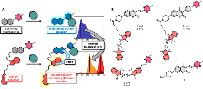
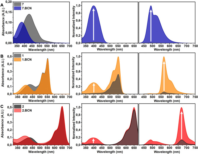
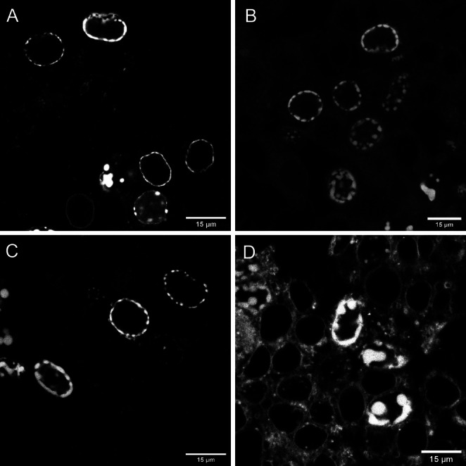
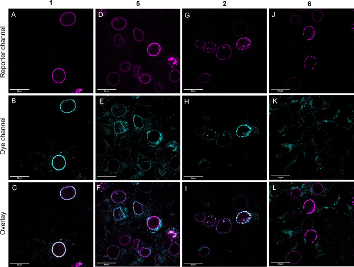
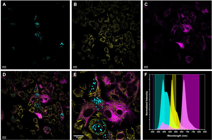
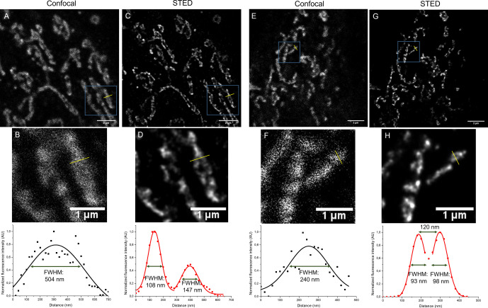
An energy transfer-based signal amplification relay concept enabling transmission of bioorthogonally activatable fluorogenicity of blue-excitable coumarins to yellow/red emitting cyanine frames is presented. Such relay mechanism resulted in improved cyanine fluorogenicities together with increased photostabilities and large apparent Stokes-shifts allowing lower background fluorescence even in no-wash bioorthogonal fluorogenic labeling schemes of intracellular structures in live cells. These energy transfer dyads sharing the same donor moiety together with their parent donor molecule allowed three-color imaging of intracellular targets using one single excitation source with separate emission windows. Sub-diffraction imaging of intracellular structures using the bioorthogonally activatable FRET dyads by STED microscopy is also presented.

Keywords: FRET dyad; bioorthogonal; fluorogenic; multicolor; single excitation.


Conflict of interest statement

The authors declare no conflict of interest.

Figures

Figure 1
Figure 1
A) Representation of the bioorthogonally controlled fluorogenicity relay concept, B) Structures of bioorthogonal fluorogenic dyads 14 and further probes (57) used in this study.
Figure 2
Figure 2
Absorption (5 μM), excitation and emission spectra (1 μM) of (A) 7, λem: 560 nm, λex: 405 nm; B) 1, λem: 620 nm, λex: 405 nm; C) 2, λem: 690 nm, λex: 405 nm and their BCN conjugates in PBS‐SDS; fluorescence spectra were normalized for each BCN conjugates.
Figure 3
Figure 3
Confocal microscopy images of Lamin‐HaloTag expressing HEK293T cells treated with Halo‐BCN (3 μM) and probes 1 (A), 2 (B) and 5 (C), 6 (D) (1 μM) following washing. Spectral detection: A) (dye 1): λexc: 405 nm/ λem: 560–800 nm; B) (dye 2): λexc: 405 nm/ λem: 560–800 nm; C) (5): λexc: 543 nm/ λem: 560–800 nm; D) (6): λexc: 633 nm/ λem: 650–800 nm.
Figure 4
Figure 4
Confocal microscopy images of Lamin‐LTAG‐miRFP (A–F) or Lamin‐GFPTAG (G–L) expressing HEK293T cells treated with Lys(BCN) and probes 1 (A–C), 2 (G–I) or 5 (D–F), 6 (J–L). Scale bar: 20 μm. Spectral detection: A,D) (miRFP/reporter channel): λexc: 633 nm/ λem: 645–800 nm; G,J) (GFP/reporter channel): λexc: 488 nm/ λem: 495–600 nm; B) (1): λexc: 405 nm/ λem: 560–600 nm; E) (5): λexc: 543 nm/ λem: 560–600 nm; H) (2): λexc: 405 nm/ λem: 620–800 nm; K) (6): λexc: 633 nm/ λem: 645–800 nm. Overlay pictures demonstrate the merged fluorescent signals of the reporter and dye channels.
Figure 5
Figure 5
Multicolor imaging of HepG2 cells. A) Lamin‐HaloTag treated with 7 tagged Halo‐substrate (Halo‐7), λexc: 405 nm/ λem: 420–500 nm); B) TOMM20 stained with 1‐tagged antibody, λexc: 405 nm/ λem: 570–600 nm); C) CK‐19 stained with 2‐tagged antibody, λexc: 405 nm/ λem: 660–800 nm; D) Overlay of A, B and C; E) magnification of D, Scale bar: 20 μm; F) Emission spectra of dyes 7 (cyan), 1 (yellow) and 2 (magenta) together with excitation wavelength (405 nm) and detection windows.
Figure 6
Figure 6
Confocal (A,B,E,F) and STED (C,D,G,H) images of COS‐7 cells immunostained for TOMM20 with secondary antibodies labeled with probe 1 (A–D) and probe 2 (E–H) (λexc: 405 nm/ λem: 560–755 nm; λSTED=775 nm (pulsed)). Scale bar: 5 μm (A,C,E,G) and in magnified pictures (B,D,F,H): 1 μm. FWHM: full width at half maxima.

References

    1. Sigal Y. M., Zhou R., Zhuang X., Science 2018, 361, 880–887. - PMC - PubMed
    1. Wang L., Frei M. S., Salim A., Johnsson K., J. Am. Chem. Soc. 2019, 141, 2770–2781. - PubMed
    1. Grimm J. B., English B. P., Choi H., Muthusamy A. K., Mehl B. P., Dong P., Brown T. A., Lippincott-Schwartz J., Liu Z., Lionett T., Lavis L. D., Nat. Methods 2016, 13, 985–988. - PubMed
    1. Grimm J. B., English B. P., Chen J., Slaughter J. P., Zhang Z., Revyakin A., Patel R., Macklin J. J., Normanno D., Singer R. H., Lionnet T., Lavis L. D., Nat. Methods 2015, 12, 244–250. - PMC - PubMed
    1. Wang L., Tran M., D′Este E., Roberti J., Koch B., Xue L., Johnsson K., Nat. Chem. 2020, 12, 165–172. - PubMed

Publication types

Substances

LinkOut - more resources