. 2021:50:249-300.
doi: 10.1016/bs.enz.2021.06.007. Epub 2021 Aug 23.

Retroviral integrase: Structure, mechanism, and inhibition

Affiliations

Retroviral integrase: Structure, mechanism, and inhibition

Dario Oliveira Passos et al. Enzymes. 2021.

Abstract

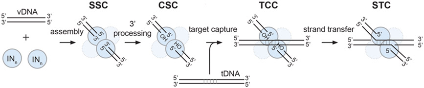
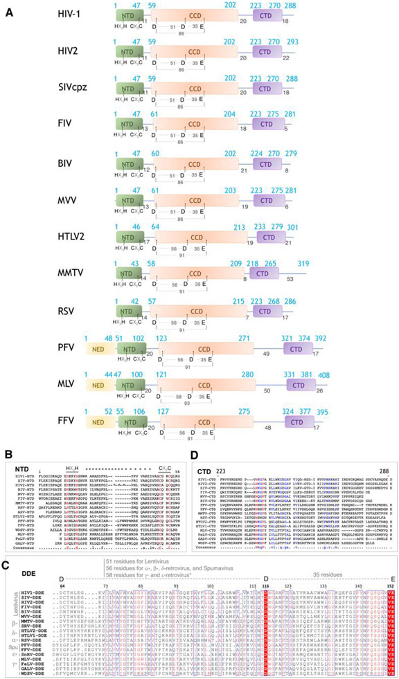
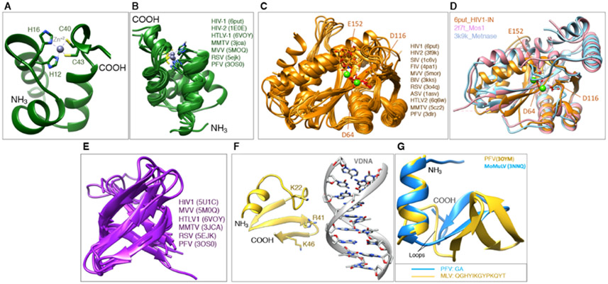
The retroviral protein Integrase (IN) catalyzes concerted integration of viral DNA into host chromatin to establish a permanent infection in the target cell. We learned a great deal about the mechanism of catalytic integration through structure/function studies over the previous four decades of IN research. As one of three essential retroviral enzymes, IN has also been targeted by antiretroviral drugs to treat HIV-infected individuals. Inhibitors blocking the catalytic integration reaction are now state-of-the-art drugs within the antiretroviral therapy toolkit. HIV-1 IN also performs intriguing non-catalytic functions that are relevant to the late stages of the viral replication cycle, yet this aspect remains poorly understood. There are also novel allosteric inhibitors targeting non-enzymatic functions of IN that induce a block in the late stages of the viral replication cycle. In this chapter, we will discuss the function, structure, and inhibition of retroviral IN proteins, highlighting remaining challenges and outstanding questions.

Keywords: Antiretroviral therapy; Drug design; Enzyme; Hydrolase; Retrovirus.


Figures

Figure 1.
Figure 1.
Phylogenetic tree of retroviral INs.
Figure 2.
Figure 2.
Schematic mechanism of intasome assembly. Under the right biochemical conditions, an oligomer of IN proteins assembles on the ends of vDNA to form the stable synaptic complex (SSC). IN then cleaves two nucleotides from the 3′ ends of vDNA, forming the cleaved synaptic complex (CSC) that exposes the conserved free 3′-OH groups of the catalytically competent CA dinucleotides. CSC intasomes can capture tDNA to form the target capture complex (TCC), which will rapidly catalyze strand transfer to form the post-catalytic strand transfer complex (STC) in which the tDNA and the integrated vDNA are still bound to the intasome. In this schematic, there are two oligonucleotides that mimic the two long-terminal repeats of vDNA ends, which facilitates in vitro assembly and biochemical/structural studies. However, in a cell, these would be replaced by a single vDNA genome containing long-terminal repeats on each end, respectively.
Figure 3.
Figure 3.
Domain organization and conservation of retroviral IN proteins. (A) Schematic representation of different retroviral INs. The distinct domains (NED, NTD, CCD, and CTD) are indicated, along with amino acid positions. (B-D) sequence conservation among retroviral IN domains, including (B) the NTD, (C) the CCD, and (D) the CTD.
Figure 4.
Figure 4.
Structures of individual IN domains. (A) The NTD from HIV-1 IN first solved by NMR, showing the basic organization and the Zn-binding HHCC motif. (B) Overlay of NTDs from different retroviral INs in the public domain. (C) Overlay of CCDs from different retroviral INs in the public domain. (D) Overlay of the CCD from HIV-1 IN with the catalytic domain from transposases, Metnase and Mos1. (E) Overlay of CTDs from different retroviral INs in the public domain. (F) The NED from PFV shown in its configuration interacting with vDNA. (G) Overlay of the NED from PFV and from MoMuLV.
Figure 5.
Figure 5.
Structures of oligomeric and multi-domain constructs of IN. (A-C) Individual domains of HIV-1 IN form homodimers. Each domain is colored accordingly to the domain representation on the top left of the panel. (D-F) Structures of the SIV, RSV and HIV-1 INs comprising the CCD-CTD domains and organized into dimers. (G) Structure of the NTD-CCD multi-domain construct from HIV-1 IN in a tetrameric configuration. The tetramer, or dimer of dimers, is maintained by CCD-CCD dimers that interact through the loop comprising residues ~186-195, as well as interactions involving NTD dimers. (H) Structure of the NTD-CCD multi-domain construct from MVV IN, also in a tetrameric configuration. This complex also had the IN-binding domain (IBD) of the protein LEDGF/p75 (discussed later) bound to each CCD dimer. This structure differs slightly by the manner in which the CCD-CCD dimers interact, as well as by the configuration of the NTDs. (I) Structure of the NTD-CCD multi-domain construct from HIV-2 IN, packed as a trimer of dimers in a distinct configuration from the other NTD-CCD constructs. This structure also had the IBD domain bound. (J) Structure of the NTD-CCD multi-domain construct from HIV-1 IN carrying a Phenylalanine at the N-terminus.
Figure 6.
Figure 6.
Structures of intasomes from different retroviruses. Each IN protomer is colored with a different color. The oligomeric forms are tetramer (PFV and HTLV1), octamer (MMTV and RSV), hexadecamer (MVV) and both tetramer and dodecamer (HIV). The SIV structural form depicted here is the base unit of an intasome stack, which is a run-on oligomer of incompletely formed “proto-intasome” octamers containing 2 vDNAs per octameric unit. They are not biologically relevant as an oligomeric species, but should contain an identical active site and can therefore be successfully used to study INSTIs.
Figure 7.
Figure 7.. Structural basis of integrase 3′ processing and strand transfer activities.
(A) Structure of the manganese-bound SSC (PDB 4E7I). The water labeled WNuc performs a nucleophilic attack on scissile phosphodiester bond of vDNA (bold red arrow), displacing two or three nucleotides from the end. The DNA and IN backbones are colored khaki and blue, respectively; red and orange sticks are oxygen and phosphorus, respectively. Purple and red spheres are manganese ions and water molecules, respectively, with the nucleophilic water labeled WNuc. Purple dashed lines indicate metal ion interactions. (B) Overlay of metal ion-bound TCC (PDB 4E7K); DNA and protein in green) and STC (PDB 4E7L; DNA and protein in pink) structures. The 3´OH performs a nucleophilic attack on the scissile phosphodiester bond of tDNA (bold red arrow) [80]. Both sets of metal ions are shown; the 3.8 Å spacing between ions in the TCC contracts to 3.2 Å in the STC. The curved black line indicates the displacement of the viral-target DNA phosphodiester bond after strand transfer relative to the scissile bond in tDNA. Other labeling is the same as in panel A.
Figure 8.
Figure 8.
Structure of the PFV intasome bound to the nucleosome core particle. The intasome binds to superhelical location at position +/−3.5 on the nucleosome
Figure 9.
Figure 9.
Clinically approved drugs used to treat HIV-infected individuals. The first-generation drugs include Raltegravir and Elvitegravir, and the second generation drugs include Dolutegravir, Bictegravir, and Cabotegravir.
Figure 10.
Figure 10.
Binding modes of second generation and developmental naphthyridine-scaffold INSTIs to lentiviral intasomes. (A) Superposition of second generation INSTIs bound to SIVrcm and HIV-1 intasomes. (B) Superimposed binding modes of the second generation INSTI BIC and the developmental naphthyridine-scaffold compound 4d, bound to the HIV-1 intasome, are displayed. The compound 4d binds closer to the Mg2+ ions because the electronegative atoms are embedded into the core of the scaffold. The terminal adenine base of vDNA and all water molecules are omitted for clarity.
Figure 11.
Figure 11.
INSTIs can bind differently to PFV and HIV intasomes. (A and B) Compound 4f bound to the (A) HIV (pink) and (B) PFV (gray) intasome. (C) Overlay of compound 4f binding modes. (D and E) Compound 4c, containing a 6-pentanol substituent, bound to the (D) HIV (green) and (E) PFV (gray, PDB 5FRN) intasome. (F) Overlay of compound 4c binding modes. Compound 4d, containing a 6-hexanol substituent, is also shown in its binding mode to the HIV (light blue) intasome. In (A), (B), (D), and (E), intasome active sites are shown as surface views, with labeled residues. R231 is poorly ordered in the map and is, therefore, displayed as an Ala stub. The terminal adenine is removed for clarity.
Figure 12.
Figure 12.
Schematic representation that recapitulates the receptor molecular environment and the water (W) networks with which the naphthyridine scaffold ligands interact when coordinating the Mg2+ ions. The scheme summarizes interactions by their locations with respect to the metal coordination plane of the naphthyridine scaffold (above, in-plane, or below). For clarity, the two water molecules coordinating the Mg2+ ions from above are not shown.
Figure 13.
Figure 13.
Binding mode of ALLINIs to HIV IN. (A) The structure of the IBD of LEDGF/p75 (green) bound to a dimer of CCDs of HIV-1 IN (brown) (PDB 2B4J). Residues in contact distance are depicted. (B) The structure of compound KF116 (yellow surface) bound to the CCD dimer interface of HIV-1 IN (PDB 4O55). The chemical structure of KF116 is on the top left of the panel. (C) The structure of compound BI224436 (gray) bound to HIV-1 IN (PDB 4NYF). The chemical structure of BI224436 is on the top left of the panel. Green circles depict two critical regions of ALLINIs potency, the carboxylic acid, and tert-butoxy, that mimic the key residues Ile-365 and Asp-366 of LEDGF/p75, respectively. (D) The structure of the compound GSK1264 bound to the CCD:CTD dimer of HIV-1 IN (PDB 5HOT). Both the CCD and the CTD form part of the ALLINI interface. (E) Depiction of the CCD dimer interface where GSK1264 binds for comparison with panels A-C (here, the CTD has been omitted for clarity).

References

    1. Grandgenett DP, Vora AC, Schiff RD, A 32,000-Dalton nucleic acid-binding protein from avian retravirus cores possesses DNA endonuclease activity, Virology. 89 (1978) 119–132. 10.1016/0042-6822(78)90046-6. - DOI - PubMed
    1. Engelman A, Mizuuchi K, Craigie R, HIV-1 DNA integration: Mechanism of viral DNA cleavage and DNA strand transfer, Cell. 67 (1991) 1211–1221. 10.1016/0092-8674(91)90297-c. - DOI - PubMed
    1. Randolph CA, Champoux JJ, The Majority of Simian Immunodeficiency Virus/Mne Circle Junctions Result from Ligation of Unintegrated Viral DNA Ends That Are Aberrant for Integration, Virology. 194 (1993) 851–854. 10.1006/viro.1993.1329. - DOI - PubMed
    1. Brown PO, Bowerman B, Varmus HE, Bishop JM, Retroviral integration: structure of the initial covalent product and its precursor, and a role for the viral IN protein, Proc National Acad Sci. 86 (1989) 2525–2529. 10.1073/pnas.86.8.2525. - DOI - PMC - PubMed
    1. Du Z, Ilyinskii PO, Lally K, Desrosiers RC, Engelman A, A mutation in integrase can compensate for mutations in the simian immunodeficiency virus att site., J Virol. 71 (1997) 8124–8132. 10.1128/jvi.71.11.8124-8132.1997. - DOI - PMC - PubMed

LinkOut - more resources