. 2022 Jan 26;42(4):692-701.
doi: 10.1523/JNEUROSCI.0636-21.2021. Epub 2021 Dec 3.

Feeling of Ownership over an Embodied Avatar's Hand Brings About Fast Changes of Fronto-Parietal Cortical Dynamics

Affiliations

Feeling of Ownership over an Embodied Avatar's Hand Brings About Fast Changes of Fronto-Parietal Cortical Dynamics

Elias Paolo Casula et al. J Neurosci. .

Abstract

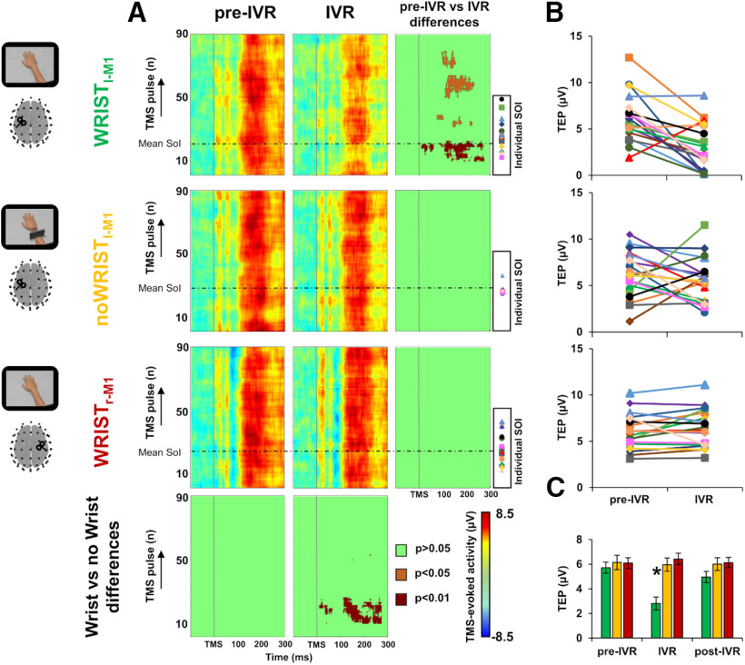
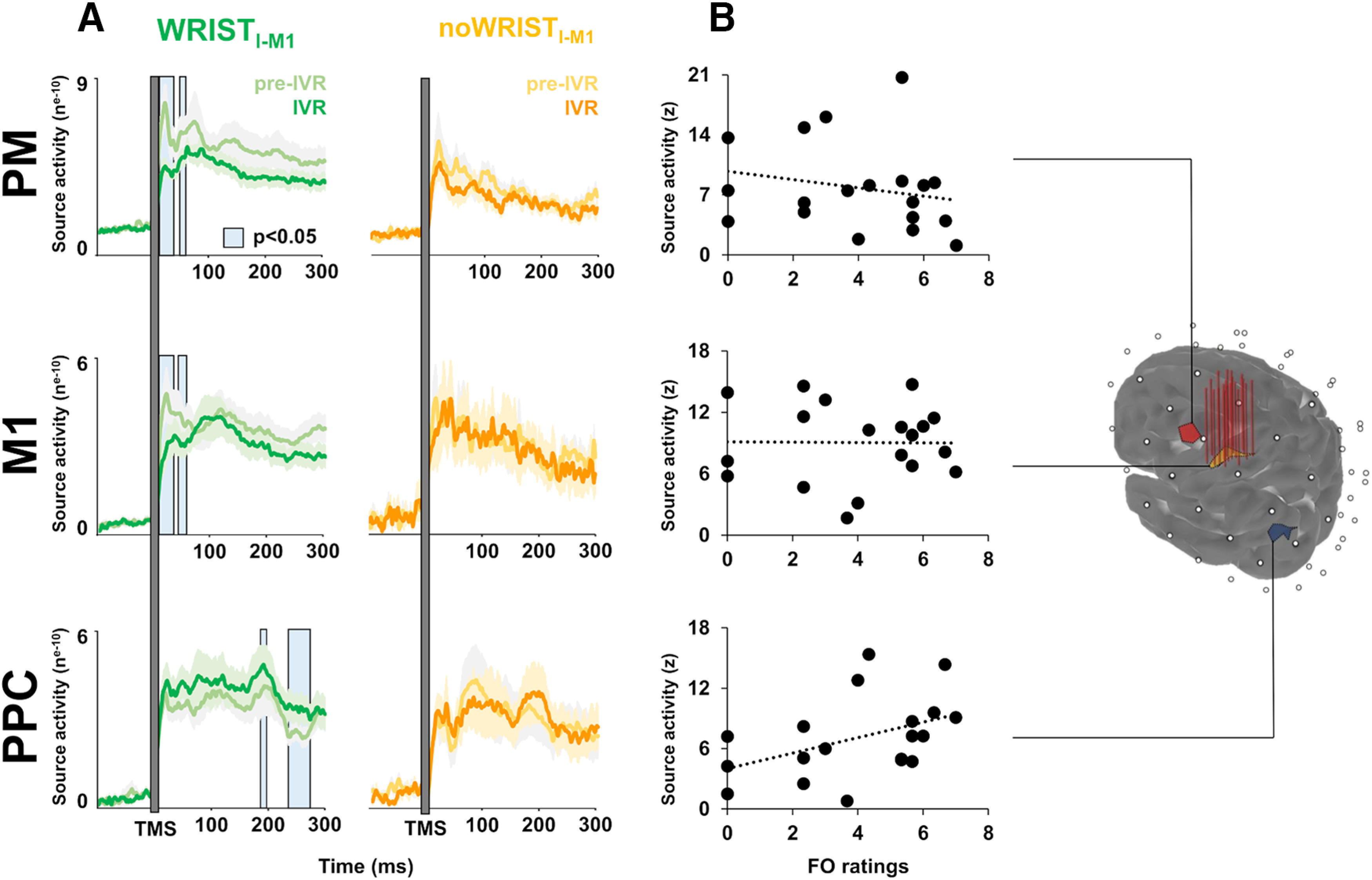
When we look at our body parts, we are immediately aware that they belong to us and we rarely doubt about the integrity, continuity, and sense of ownership of our body. Despite this certainty, immersive virtual reality (IVR) may lead to a strong feeling of embodiment over an artificial body part seen from a first-person perspective (1PP). Although such feeling of ownership (FO) has been described in different situations, it is not yet understood how this phenomenon is generated at neural level. To track the real-time brain dynamics associated with FO, we delivered transcranial magnetic stimuli over the hand region in the primary motor cortex (M1) and simultaneously recorded electroencephalography (EEG) in 19 healthy volunteers (11 male/8 female) watching IVR renderings of anatomically plausible (full-limb) versus implausible (hand disconnected from the forearm) virtual limbs. Our data show that embodying a virtual hand is temporally associated with a rapid drop of cortical activity of the onlookers' hand region in the M1 contralateral to the observed hand. Spatiotemporal analysis shows that embodying the avatar's hand is also associated with fast changes of activity within an interconnected fronto-parietal circuit ipsilateral to the brain stimulation. Specifically, an immediate reduction of connectivity with the premotor area is paralleled by an enhancement in the connectivity with the posterior parietal cortex (PPC) which is related to the strength of ownership illusion ratings and thus likely reflects conscious feelings of embodiment. Our results suggest that changes of bodily representations are underpinned by a dynamic cross talk within a highly-plastic, fronto-parietal network.SIGNIFICANCE STATEMENT Observing an avatar's body part from a first-person perspective (1PP) induces an illusory embodiment over it. What remains unknown are the cortical dynamics underpinning the embodiment of artificial agents. To shed light on the physiological mechanisms of embodiment we used a novel approach that combines noninvasive stimulation of the cortical motor-hand area and whole-scalp electroencephalographic (EEG) recordings in people observing an embodied artificial limb. We found that just before the illusion started, there is a decrease of activity of the motor-hand area accompanied by an increase of connectivity with the parietal region ipsilateral to the stimulation that reflects the ratings of the embodiment illusion. Our results suggest that changes of bodily representations are underpinned by a dynamic cross talk within a fronto-parietal circuit.

Keywords: EEG; TMS; brain dynamics; embodiment.


Figures

Figure 1.
Figure 1.
The TMS-EEG-IVR setup. During IVR, each participant was instructed to observe a virtual right hand while TMS was applied over (1) the left (Wristl-M1 condition) or (2) the right (Wristr-M1) hand M1 representation (M1hand) or (3) a right disconnected hand while stimulated over the left M1hand (noWristl-M1). TMS was constantly monitored through stereotaxic neuronavigation, and cortical activation was continuously recorded with EEG. During IVR, participants were asked to refer whether they start to feel the ownership illusion over the virtual right hand, i.e., SoI. TMS, transcranial magnetic stimulation; EEG, electroencephalography; IVR, immersive virtual reality; M1, primary motor cortex; SoI, start of illusion.
Figure 2.
Figure 2.
Schematic representation of the experimental procedure. Each participant underwent three TMS-EEG-IVR sessions (one for each condition, i.e., Wristl-M1, noWristl-M1, Wristr-M1), each consisting of an IVR block (160 pulses) preceded by a pre-IVR (120 pulses) and followed by a post-IVR (120 pulses) block. The order of observation conditions was counterbalanced across participants. At the end of IVR block, a black screen covered the whole virtual scenario, and the participants verbally rated the strength of FO and VA over the virtual hand by answering to the embodiment questionnaires. IVR, immersive virtual reality; TMS, transcranial magnetic stimulation; EEG, electroencephalography; M1, primary motor cortex; FO, feeling of ownership; VA, vicarious agency.
Figure 3.
Figure 3.
Embodiment measures. Mean ratings of FO (panel A) and VA (panel B) for the Wristl-M1 (green bars), noWristl-M1 (yellow bars), and Wristr-M1 (red bars) condition. Striped bars depict the control items; *p < 0.05. Error bars indicate SE. FO, feeling of ownership; VA, vicarious agency; M1, primary motor cortex.
Figure 4.
Figure 4.
Spatiotemporal analysis of TEPs. TEPs (rectified) from left M1hand for the three sessions (Wristl-M1, noWristl-M1, and Wristr-M1) in the three blocks: pre-IVR, IVR, and post-IVR. (Panel A) Topography of voltage distribution represent TEPs from 40 to 230 ms for all the conditions and in the three blocks. (Panel B) Significant electrodes (p < 0.05) from the cluster-based comparison between pre-IVR and IVR block within the Wristl-M1 session are represented by gray and white circles; significant differences (p < 0.05) between the two IVR blocks of the Wristl-M1 and the noWristl-M1 conditions are represented by only gray circles. IVR, immersive virtual reality; TMS, transcranial magnetic stimulation; TEP, TMS-evoked potential; M1, primary motor cortex.
Figure 5.
Figure 5.
Trial-by-trial analysis. Panel A, Trial-by-trial plots of TMS-evoked activity representing variations in EEG amplitude for the Wristl-M1, noWristl-M1, and the Wristr-M1 condition, in the pre-IVR (left column) and IVR block (central column). The horizontal dotted line represents the average time at which participants started to perceive the illusory ownership of the virtual arm (SoI). Significant differences between blocks (right column) and between conditions (lower row) are depicted in dark red (p < 0.01), orange (p < 0.05), and green (nonsignificant, p > 0.05). Individual SoI are depicted for each participant with a different marker. Panel B, TEP amplitude for each participant in the SoI time window for the Wristl-M1 condition (upper plot), the noWristl-M1 (central plot), and the Wristr-M1 (lower plot). Panel C, ANOVA conducted on the TEP amplitude among the three conditions (green bars, Wristl-M1; yellow bars, nowristl-M1; red bars, Wristr-M1) and the three blocks (pre-IVR, IVR, post-IVR). Error bars indicate SEM; *p < 0.05. IVR, immersive virtual reality; TMS, transcranial magnetic stimulation; M1, primary motor cortex; SoI, start of illusion; EoI, end of illusion.
Figure 6.
Figure 6.
Cortical dynamics in the space domain. Source reconstruction of TMS-evoked activity at ROI level: the stimulated hand area in the M1 (M1hand), the PM, and the PPC. Vertical red lines over the M1hand ROI indicate the stimulated hotspot of the 19 participants. Panel A: timing of the source activity evoked for the Wristl-M1 condition (pre-IVR block, light green line; IVR block, dark green line) and for the noWristl-M1 condition (pre-IVR block, light yellow line; IVR block, dark yellow line). Panel B: the correlation analysis between source activity of the three ROIs and FO rating. IVR, immersive virtual reality; TMS, transcranial magnetic stimulation; M1, primary motor cortex; PM, premotor cortex; PPC, posterior parietal cortex; FO, feeling of ownership.

References

    1. Bäumer T, Schippling S, Kroeger J, Zittel S, Koch G, Thomalla G, Rothwell JC, Siebner HR, Orth M, Münchau A (2009) Inhibitory and facilitatory connectivity from ventral premotor to primary motor cortex in healthy humans at rest–a bifocal TMS study. Clin Neurophysiol 120:1724–1731. 10.1016/j.clinph.2009.07.035 - DOI - PubMed
    1. Blanke O, Slater M, Serino A (2015) Behavioral, neural, and computational principles of bodily self-consciousness. Neuron 88:145–166. 10.1016/j.neuron.2015.09.029 - DOI - PubMed
    1. Botvinick M, Cohen J (1998) Rubber hands 'feel' touch that eyes see. Nature 391:756. 10.1038/35784 - DOI - PubMed
    1. Buch ER, Mars RB, Boorman ED, Rushworth MF (2010) A network centered on ventral premotor cortex exerts both facilitatory and inhibitory control over primary motor cortex during action reprogramming. J Neurosci 30:1395–1401. 10.1523/JNEUROSCI.4882-09.2010 - DOI - PMC - PubMed
    1. Cash RF, Noda Y, Zomorrodi R, Radhu N, Farzan F, Rajji TK, Fitzgerald PB, Chen R, Daskalakis ZJ, Blumberger DM (2017) Characterization of glutamatergic and GABA A-mediated neurotransmission in motor and dorsolateral prefrontal cortex using paired-pulse TMS–EEG. Neuropsychopharmacology 42:502–511. 10.1038/npp.2016.133 - DOI - PMC - PubMed

Publication types

LinkOut - more resources