. 2022 Dec;71(12):2414-2429.
doi: 10.1136/gutjnl-2021-324725. Epub 2021 Dec 3.

Neutrophils prevent rectal bleeding in ulcerative colitis by peptidyl-arginine deiminase-4-dependent immunothrombosis

Affiliations

Neutrophils prevent rectal bleeding in ulcerative colitis by peptidyl-arginine deiminase-4-dependent immunothrombosis

Moritz Leppkes et al. Gut. 2022 Dec.

Abstract

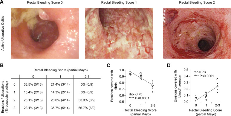
Objective: Bleeding ulcers and erosions are hallmarks of active ulcerative colitis (UC). However, the mechanisms controlling bleeding and mucosal haemostasis remain elusive.

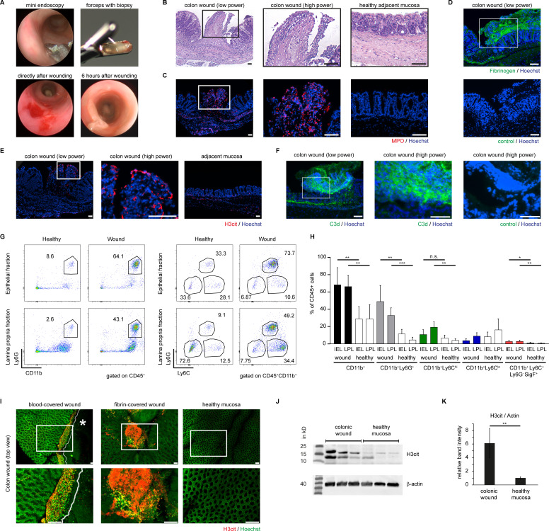
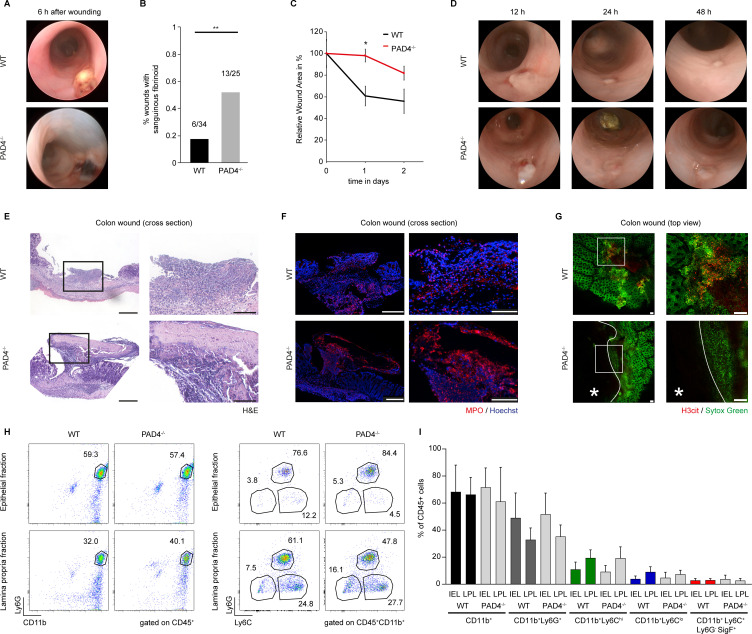
Design: We used high-resolution endoscopy and colon tissue samples of active UC (n = 36) as well as experimental models of physical and chemical mucosal damage in mice deficient for peptidyl-arginine deiminase-4 (PAD4), gnotobiotic mice and controls. We employed endoscopy, histochemistry, live-cell microscopy and flow cytometry to study eroded mucosal surfaces during mucosal haemostasis.

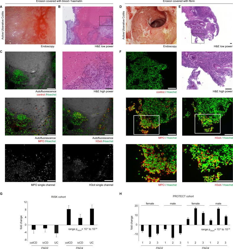
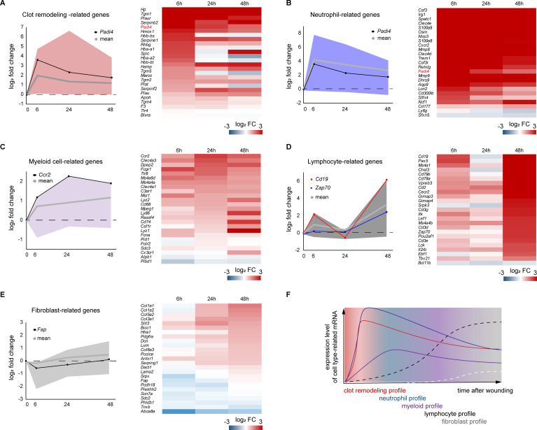
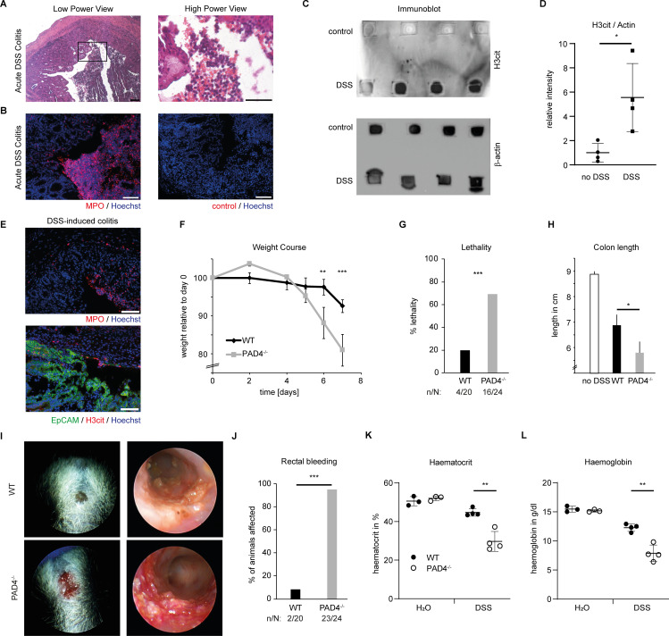
Results: Erosions and ulcerations in UC were covered by fresh blood, haematin or fibrin visible by endoscopy. Fibrin layers rather than fresh blood or haematin on erosions were inversely correlated with rectal bleeding in UC. Fibrin layers contained ample amounts of neutrophils coaggregated with neutrophil extracellular traps (NETs) with detectable activity of PAD. Transcriptome analyses showed significantly elevated PAD4 expression in active UC. In experimentally inflicted wounds, we found that neutrophils underwent NET formation in a PAD4-dependent manner hours after formation of primary blood clots, and remodelled clots to immunothrombi containing citrullinated histones, even in the absence of microbiota. PAD4-deficient mice experienced an exacerbated course of dextrane sodium sulfate-induced colitis with markedly increased rectal bleeding (96 % vs 10 %) as compared with controls. PAD4-deficient mice failed to remodel blood clots on mucosal wounds eliciting impaired healing. Thus, NET-associated immunothrombi are protective in acute colitis, while insufficient immunothrombosis is associated with rectal bleeding.

Conclusion: Our findings uncover that neutrophils induce secondary immunothrombosis by PAD4-dependent mechanisms. Insufficient immunothrombosis may favour rectal bleeding in UC.

Keywords: IBD; bleeding; leukocytes; mucosal injury; ulcerative colitis.


Conflict of interest statement

Competing interests: None declared.

Figures

Figure 1
Figure 1
Bleeding in active UC is controlled by successful formation of fibrin on mucosal erosions. (A) Patients experiencing flares of UC often suffer from rectal bleeding, typically assessed by the partial Mayo score (0: none, 1: visible blood with stool (< 50 %), 2: visible blood with stool (> 50 %), 3: passing blood alone). Patients suffering from flares of UC underwent routine endoscopy. Representative endoscopic images of patients with various degrees of rectal bleeding are depicted. (B) Clinically observed rectal bleeding coincided with the enhanced presence of mucosal erosions on endoscopy as graded by an experienced endoscopist in a blinded fashion inspired by the Blackstone score (0: no visible erosions, 1: less than 10 erosions (< 5 mm in size) per 10 cm section, 2: more than 10 erosions (< 5 mm in size) per 10 cm section to 3: more than 10 erosions (< 5 mm in size) and ulcerations (> 5 mm in size) per 10 cm section). The section most strongly affected determined the grading result (Spearman’s rho: 0.38, * p < 0.05). additionally, the morphology of mucosal erosions was analysed and the frequency of either fibrin coverage or persistence of fresh blood or haematin was assessed in these images supported by digital image analysis. (C) Presence of erosions did not necessarily prompt rectal bleeding, rather complete coverage by fibrin on all mucosal erosions achieved haemostasis. Less efficient fibrin coverage was observed in patients suffering from rectal bleeding as assessed by partial Mayo score (spearman Rho: 0.73, **** p < 0.0001). (D) Persistent presence of blood or haematin on erosions was associated with clinical rectal bleeding (spearman Rho: 0.73, **** p < 0.0001). The study cohort included 36 patients suffering from active UC with varying degrees of rectal bleeding. UC, ulcerative colitis.
Figure 2
Figure 2
Eroded colonic mucosa features blood clots which are remodelled to a fibrin layer rich in aggregated granulocytes and NETs. During colonoscopy of active UC, biopsies were taken specifically from either (A–C) blood clot-covered or (D–F) fibrin-covered erosions and analysed by endoscopy and microscopy as indicated. Representative endoscopic images of (A) blood/haematin-covered and (D) fibrin-covered colon erosions of patients suffering from active UC are shown. (B) Representative low-power and high-power images are provided (scale bars: 100 µm). We identified marked infiltration of neutrophils into the blood clot and adjacent amorphous haematoxyline-affine material. Samples from (E) fibrin-covered erosions showed a strong abundance of aggregated neutrophils (scale bars = 100 µm). (C, F) Immunofluorescence of such biopsies is shown using either MPO or H3cit-directed primary antibodies. (C) Aggregates of MPO+ neutrophils were present at the edge of the autofluorescent primary blood clot (488/525 nm). In this area, decondensed chromatin and H3cit was detected. Combined and single channel analyses are presented (scale bars = 100 µm). (F) Fibrin visible on the endoscopic level is rich in H3cit and aggregated MPO+ neutrophils. As depicted, H3cit is rather restricted to the fibrin layer (representative of n = 22 biopsies studied). (G–H) Publicly available datasets of RNAseq-based transcriptomics of large cohorts of patients suffering from icCD (1), colCD (2) or UC (3) were reanalysed for expression of PAD2 and PAD4, respectively. Fold change as compared with non-diseased controls is depicted. colCD, colonic Crohn’s disease; H3cit, citrullinated histone H3; icCD, ileocolonic Crohn’s disease; MPO, myeloperoxidase; NETs, neutrophil extracellular traps; PAD, peptidyl-arginine deiminase; UC, ulcerative colitis.
Figure 3
Figure 3
Mucosal damage leads to the formation of red blood clots subject to remodelling to neutrophil-rich fibrin layers characterised by marked PAD-activity. Colonoscopy was performed in mice and mucosal wounds were induced using an endoscopic forceps. (A) Top Left: forceps during endoscopy; top right: forceps with biopsy; bottom right: red blood clot on the mucosal wound directly after wounding; bottom left: whitish fibrin covers the mucosal wound 6 hours after wounding. The red blood clot was remodelled. (B) H&E staining of sections of mucosal ulcers (18 hours after injury) display an amorphous layer rich in granulocytes at the surface of the mucosal ulceration. An overview of a cross-section (left) and a high-power magnification (centre) is presented, as well as an image of healthy mucosa (right). (C) The remodelled layer covering the wound bed is positive for MPO as evidenced by immunofluorescence. MPO staining shows fiber-like constitution, indicative of a partially extracellular localisation, colocalised to DNA. (D) The remodelled layer is also characterised by fibrin deposition, as evidenced by fibrinogen immunofluorescence (top) as compared with control (bottom) (scale bars = 100 µm). (E) H3cit is preferentially detected in the wound surface, also featuring (F) cleaved complement C3d. (G) Flow cytometric analysis of healthy and wound tissue 18 hours after wounding. Both the LPL fractions and the IEL fractions of wounds were analysed. In wound tissues, CD11b+Ly6G+ neutrophils are the dominant cell type in the IEL fraction, whereas the lamina propria features large populations of both CD11b+Ly6G+ neutrophils and CD11b+Ly6Chi monocytes. (H) Quantification of the cellular composition of colon wounds and adjacent healthy colon tissue (n = 18 wounds studied, *** p < 0.001, Student’s t-test, ** p < 0.01, Student’s t-test, * p < 0.05, Student’s t-test). (I) Colonic wounds were mounted on glass slides as a whole and subjected to epifluorescence analysis after immunostaining of H3cit (in red) and Hoechst (in green) staining. The wound surface of two different wounds both derived from wild-type mice is presented. Left: a wound with persistent presence of the primary blood clot. A cell-rich layer in proximity to the blood clot characterised by H3cit-positive chromatin threads separates the blood clot from the adjacent mucosa. Centre: a remodelled colonic ulcer surface, covered by H3cit+ chromatin. Right: no H3cit is detectable on healthy mucosa. Images are representative of > 10 colonic wounds studied. (J) Western blot analyses of colonic wounds and healthy mucosa collected 18 hours after wounding were performed. (K) Densitometry of Western blots as in (J) shows increased presence of H3cit in the wound bed as compared with healthy control tissues (** p < 0.001, Student’s t-test, 3 independent experiments performed). Loading control: β-Actin. All scale bars = 100 µm. H3cit, citrullinated histone H3; IEL, intraepithelial lymphocyte; LPL, lamina propria leucocyte; MPO, myeloperoxidase; PAD, peptidyl-arginine deiminase.
Figure 4
Figure 4
An increased neutrophil-related transcriptional signature coincides with the increase of clot remodelling-associated transcripts. RNA-sequencing was performed studying the transcriptome of healthy colon and wound tissue from three defined time points after wounding (6 hours, 24 hours, 48 hours after wounding, n = 3 wounds per time point). Expression was analysed as a fold-change in relation to the healthy state. An expression time course for each gene set (clot remodelling-related, neutrophil-related, myeloid cell-related, lymphocyte-related, fibroblast-related) is provided. The area between the minimal and maximal differentially expressed genes of each gene set is coloured. Additionally, the mean of each gene set and exemplary single gene time courses are depicted for the genes indicated. A heat map of the entire topic-related gene set studied is provided visualising the log2 fold change in colour (increase in red, decrease in blue). (A) Expression of clot remodelling-related genes is most strongly increased 6 hours after wound infliction. (B) Neutrophil-related genes – among these Padi4 – are also most strongly increased 6 hours after wound infliction. (C) Expression of myeloid cell-related genes show a slower increase within the first day of healing peaking at 24–48 hours. (D) The profile of lymphocyte-related genes peaks at 48 hours after damage. (E) Expression of fibroblast-related genes shows only a moderate increase over time. (F) Summarised depiction of the expression profiles shown in (A)–(E) over time. Please appreciate the sequential dynamics of the designated profiles.
Figure 5
Figure 5
Deficiency of PAD4 disturbs blood clot remodelling on mucosal ulcerations and delays mucosal wound healing. Mini-colonoscopy was repetitively performed after physical wounding of the colon mucosa. (A) Representative endoscopic imaging of colon wounds 6 hours after wounding performed in PAD4−/− and wild-type (WT) mice display the disturbed remodelling of the blood clot on the ulcer surface in PAD4−/− mice. Please appreciate the persistent sanguinous appearance of wounds in PAD4−/− mice. (B) Quantification of the fraction of wounds displaying a sanguinous morphology covering the wounds (** p < 0.05, Fisher’s exact test for 2×2 contingency tables). (C) Wound bed size was measured on repeated endoscopic examinations. Wounds in PAD4-deficient mice had a delayed kinetic of wound healing (* p < 0.05, Student’s t-test). (D) Time course of mucosal healing as evidenced by representative endoscopic images of colon wounds performed in PAD4−/− and WT mice (n = 24 wounds per group (* p < 0.05, Student’s t-test)). (E) H&E staining of colonic wound sections from WT and PAD4−/− mice after 18 hours shows a remodelled blood clot in the WT colon, characterised by a high abundance of invading granulocytes throughout the fibrin layer. In contrast, the PAD4−/− wound clot still contains the primary blood clot with granulocytes only present at the clot edge (scale bars: low power = 200 µm, high power = 100 µm). The same wound as in (E) was subjected to immunofluorescence. (F) Staining of MPO shows neutrophil infiltration throughout the clot in WT mice. The wound from PAD4−/− mice features MPO+ cells surrounding the unstained sanguinous core (scale bars: low power = 200 µm, high power = 100 µm). (G) Epifluorescence analyses of the colon ulcer surface of PAD4−/− and WT mice displayed the reduced presence of H3cit in PAD4−/− mice as well as the persistence of the primary blood clot (marked by the asterisk). High-power insets are presented on the right. Counterstaining with sytox green. (H) Flow cytometric analyses of cellular infiltrates of wound tissues from WT and PAD4−/− mice 18 hours after wounding are presented. Both genotypes exhibit a marked infiltration of CD11b+Ly6G+ neutrophils to the wound site. Especially in the epithelial fraction representing the ulcer surface, CD11b+Ly6G+ neutrophils are strongly enriched in both groups. (I) Comparative quantification of the cellular composition of WT and PAD4−/− wound tissue as assessed in (H) shows no significant differences in the cellular composition with regard to the examined leucocyte populations (n = 18 wounds from two independent experiments evaluated). H3cit, citrullinated histone H3; MPO, myeloperoxidase; PAD, peptidyl-arginine deiminase; WT, wild-type.
Figure 6
Figure 6
Immunothrombosis occurs on mucosal erosions even in the absence of bacteria. Colonoscopy was performed in mice and mucosal wounds were induced using an endoscopic forceps system in SPF and gnotobiotic wild-type C57BL/6 mice under sterile conditions. (A) Faecal bacterial load was assessed by 24 h-culture of faecal homogenates on lb agar plates. Gnotobiotic mice remained sterile throughout the experiment. (B) Mini-colonoscopy was performed 6 and 18 hours after wounding. Representative endoscopic images of colon wounds show that remodelling of the wound bed surface occurs in a similar fashion independently of the colonic microbiota. (C) H&E staining of wounded colon tissue sections shows infiltration of leucocytes to the wounded colon and submucosal oedema in both SPF and gnotobiotic mice. (D) Submucosal oedema thickness was quantified in cross sections in SPF and gnotobiotic mice (N: 11–14 wounds per group, *** p < 0.001, Student’s t-test, bars represent the means) (E) scatter plots of leucocyte numbers infiltrating the submucosa quantified in cross sections in SPF and gnotobiotic mice (** p < 0.01, Student’s t-test, bars represent the means). (F) Marked infiltration of MPO+ neutrophils to both wound bed and submucosa in SPF and gnotobiotic mice was observed by immunofluorescence, (G) H3cit was preferentially detected in wound beds of SPF and gnotobiotic mice as compared with the adjacent mucosa as studied by immunofluorescence. Dashed lines indicate the ROI of the immunothrombus area. (H) Complement activation as studied by C3d staining in both SPF and gnotobiotic mice was prominent in the wound bed area in both groups. (I) MPO+ cells in the submucosa were quantified in images as in (F) (N: 8–10 wounds per group, * p < 0.05, Student’s t-test, mean ± SD are depicted). Dashed lines indicate the ROI of the immunothrombus area. The solid line indicates the intact border of the wound (J) H3cit+ speckles per ROI in the wound bed and adjacent mucosa were quantified in images as in (G) (N: 3–7 wounds per group, *** p < 0.001, Student’s t-test, mean ± SD are depicted) (K) C3d relative fluorescence intensity per ROI in the wound bed and adjacent mucosa was quantified in images as in (H) (N: 5–10 wounds per group, *** p < 0.001, Student’s t-test, mean ± SD are depicted, scale bars = 100 µm). H3cit, citrullinated histone H3; MPO, myeloperoxidase; ROI, region of interest; SPF, specific pathogen-free.
Figure 7
Figure 7
PAD4-mediated immunothrombosis controls mucosal haemostasis in acute DSS-induced colitis. Acute colitis was induced in mice by 4 % DSS in the drinking water for 7 days. (A) H&E staining and (B) immunofluorescence of MPO of murine colonic wounds at day 9 shows a massive destruction of the epithelial cell layer with abundant neutrophils in the colon lumen (scale bars = 100 µm). (C) Colon tissue lysates from day 9 of acute DSS-induced colitis and untreated controls were analysed by an immuno-dot blot technique. The amount of citrullinated histone H3 and hence PAD activity is markedly increased under inflammatory conditions. β−actin immunoblot serves as a loading control (representative of 3 independent experiments with a total of n = 12 samples). (D) Semiquantitative image density analyses of immuno-dot blots as in (C) reveal a significantly higher H3cit expression in DSS-colitis tissue compared with healthy tissue (* p < 0.05, Student’s t test). (E) Immunofluorescence of MPO (top), EpCAM and H3cit (bottom) performed on cryosections of colonic tissue samples derived from day 9 of acute DSS-induced colitis (4 %) shows the increased presence of MPO in the inflamed mucosa with H3cit restricted to the eroded area with a breached epithelial lining (as seen by lack of EpCAM staining (top part of the lower micrograph)) (representative of n > 10 samples, scale bar = 100 µm). (F) Weight (in %) of mice subjected to 4 % DSS in the drinking water was measured during the course of the experiments comparing PAD4-deficient (PAD4−/−) mice and PAD4-proficient littermates (WT). Please appreciate the accelerated weight loss in PAD4−/− mice. (G) Lethality of mice, interpreted by the number of mice suffering a weight loss of > 20 %, was strongly increased in the PAD4−/− group (*** p < 0.001, Fisher’s two-tailed exact test). (H) At the time of sacrifice, colon length of mice was measured. A reduced length is typical of increased inflammation. A reduced colon length was observed in PAD4−/− mice as compared with WT littermates after DSS treatment. (I) Rectal bleeding is a typical hallmark of severe acute DSS-induced colitis, detectable on both clinical assessment (left) and on analyses by endoscopy of affected mice (right). This was evident more often in PAD4−/− mice. (J) The number of mice suffering from persistent rectal bleeding was strongly increased in PAD4−/− mice subjected to DSS as compared with wild-type littermates (*** p < 0.001, Fisher’s two tailed exact test, n: number of mice suffering from rectal bleeding, N: total number of mice studied). Complete blood count was performed on day 10 of DSS-induced colitis. (K) Haematocrit and (L) haemoglobin were measured in the blood of WT and PAD4−/− mice subjected to drinking water containing DSS or not (H2O). when subjected to DSS, PAD4−/− mice exhibited significantly more blood loss than WT mice, whereas untreated mice showed no differences (** p < 0.01, Student’s t test). DSS, dextrane sodium sulfate; MPO, myeloperoxidase; PAD, peptidyl-arginine deiminase; WT, wild-type.

Comment in

  • What good can neutrophils do in UC?
    Salas A. Salas A. Gut. 2022 Dec;71(12):2375-2376. doi: 10.1136/gutjnl-2021-326484. Epub 2022 Jan 24. Gut. 2022. PMID: 35074905 No abstract available.

References

    1. Truelove SC, Witts LJ. Cortisone in ulcerative colitis; preliminary report on a therapeutic trial. Br Med J 1954;2:375–8. 10.1136/bmj.2.4884.375 - DOI - PMC - PubMed
    1. Samuel S, Ingle SB, Dhillon S, et al. . Cumulative incidence and risk factors for hospitalization and surgery in a population-based cohort of ulcerative colitis. Inflamm Bowel Dis 2013;19:1858–66. 10.1097/MIB.0b013e31828c84c5 - DOI - PMC - PubMed
    1. Robert JH, Sachar DB, Aufses AH, et al. . Management of severe hemorrhage in ulcerative colitis. Am J Surg 1990;159:550–5. 10.1016/s0002-9610(06)80064-4 - DOI - PubMed
    1. Kaser A, Zeissig S, Blumberg RS. Inflammatory bowel disease. Annu Rev Immunol 2010;28:573–621. 10.1146/annurev-immunol-030409-101225 - DOI - PMC - PubMed
    1. Neurath MF, Leppkes M. Resolution of ulcerative colitis. Semin Immunopathol 2019;41:747–56. 10.1007/s00281-019-00751-6 - DOI - PubMed

Publication types