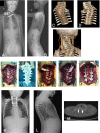
3D printed templates improve the accuracy and safety of pedicle screw placement in the treatment of pediatric congenital scoliosis
- PMID: 34863150
- PMCID: PMC8645104
- DOI: 10.1186/s12891-021-04892-4
3D printed templates improve the accuracy and safety of pedicle screw placement in the treatment of pediatric congenital scoliosis
Abstract
Background: Three-dimensional (3-D) printed guidance templates are being increasingly used in spine surgery. The purpose of this study was to determine if 3D printed navigation templates can improve the accuracy of pedicle screw placement and decrease the complication rate compared to freehand screw placement in the treatment of children with congenital scoliosis.
Methods: The records of pediatric patients with congenital scoliosis treated at our hospital from January 2017 to January 2019 were retrospectively reviewed. Patients were divided into those where a 3D printed guidance templated was used and those in which the freehand method was used for pedicle screw placement. The accuracy rate of pedicle screw placement, surgical outcomes, and complications were compared between groups.
Results: A total of 67 children with congenital scoliosis were included (43 males and 24 females; mean age of 4.13 ± 2.66 years; range, 2-15 years). There were 34 children in the template-assisted group and 33 in the freehand group. The excellent accuracy rate of pedicle screw placement was significantly higher in the template-assisted group (96.10% vs. 88.64%, P = 0.007). The main Cobb angle and kyphosis angle were similar between the 2 groups preoperatively and postoperatively (all, P > 0.05), and in both groups both angles were significantly decreased after surgery as compared to the preoperative values (all, P < 0.001). The degree of change of the Cobb angle of the main curve and kyphosis angle were not significantly different between the 2 groups. There were no postoperative complications in the template group and 4 in the freehand group (0% vs. 12.12%; P = 0.009). All 4 patients with complications required revision surgery.
Keywords: 3D printed template; Computer-assisted surgery; Pediatric congenital scoliosis; Screw placement.
© 2021. The Author(s).
Conflict of interest statement
The authors declare that they have no competing interests.
Figures


Similar articles
-
Comparison of 3D-printed Navigation Template-assisted Pedicle Screws versus Freehand Screws for Scoliosis in Children and Adolescents: A Systematic Review and Meta-analysis.J Neurol Surg A Cent Eur Neurosurg. 2023 Mar;84(2):188-197. doi: 10.1055/a-1938-0254. Epub 2022 Sep 7. J Neurol Surg A Cent Eur Neurosurg. 2023. PMID: 36070792
-
3D-printed guides versus computer navigation for pedicle screw placement in the surgical treatment of congenital scoliosis deformities.J Orthop Surg (Hong Kong). 2024 Jan-Apr;32(1):10225536241233785. doi: 10.1177/10225536241233785. J Orthop Surg (Hong Kong). 2024. PMID: 38378476
-
Does Three-dimensional Printing Plus Pedicle Guider Technology in Severe Congenital Scoliosis Facilitate Accurate and Efficient Pedicle Screw Placement?Clin Orthop Relat Res. 2019 Aug;477(8):1904-1912. doi: 10.1097/CORR.0000000000000739. Clin Orthop Relat Res. 2019. PMID: 31107327 Free PMC article. Clinical Trial.
-
3D printed pedicle screw guides reduce the rate of intraoperative screw revision in adolescent idiopathic scoliosis surgery.Spine J. 2023 Dec;23(12):1894-1899. doi: 10.1016/j.spinee.2023.08.001. Epub 2023 Aug 6. Spine J. 2023. PMID: 37553024
-
Accuracy and postoperative assessment of robot-assisted placement of pedicle screws during scoliosis surgery compared with conventional freehand technique: a systematic review and meta-analysis.J Orthop Surg Res. 2024 Jun 20;19(1):365. doi: 10.1186/s13018-024-04848-z. J Orthop Surg Res. 2024. PMID: 38902785 Free PMC article.
Cited by
-
Let's think beyond the pedicle: A biomechanical study of a new conceptual extra pedicular screw and hook construct.J Clin Orthop Trauma. 2023 Jun 4;41:102173. doi: 10.1016/j.jcot.2023.102173. eCollection 2023 Jun. J Clin Orthop Trauma. 2023. PMID: 37483911 Free PMC article.
-
Clinical applications of 3D printing in spine surgery: a systematic review.Eur Spine J. 2025 Feb;34(2):454-471. doi: 10.1007/s00586-024-08594-y. Epub 2025 Jan 7. Eur Spine J. 2025. PMID: 39774918
-
The expanding use of three-dimensional printing in orthopaedic and spine surgery.J Spine Surg. 2022 Sep;8(3):300-303. doi: 10.21037/jss-22-63. J Spine Surg. 2022. PMID: 36285096 Free PMC article. No abstract available.
-
3D printing applications in spine surgery: an evidence-based assessment toward personalized patient care.Eur Spine J. 2022 Jul;31(7):1682-1690. doi: 10.1007/s00586-022-07250-7. Epub 2022 May 19. Eur Spine J. 2022. PMID: 35590016 Review.
-
Three-Dimensional Printing Applications in Pediatric Spinal Surgery: A Systematic Review.Global Spine J. 2024 Mar;14(2):718-730. doi: 10.1177/21925682231182341. Epub 2023 Jun 5. Global Spine J. 2024. PMID: 37278022 Free PMC article. Review.
References
-
- Farley FA. Etiology of congenital scoliosis. Semin Spine Surg. 2010;22:110–112. doi: 10.1053/j.semss.2010.03.001. - DOI
MeSH terms
LinkOut - more resources
Full Text Sources
Medical

