. 2021 Dec 4;18(1):54.
doi: 10.1186/s12987-021-00289-6.

Elevated CSF inflammatory markers in patients with idiopathic normal pressure hydrocephalus do not promote NKCC1 hyperactivity in rat choroid plexus

Affiliations

Elevated CSF inflammatory markers in patients with idiopathic normal pressure hydrocephalus do not promote NKCC1 hyperactivity in rat choroid plexus

Sara Diana Lolansen et al. Fluids Barriers CNS. .

Abstract

Background: Idiopathic normal pressure hydrocephalus (iNPH) is a potentially reversible neurological condition of unresolved etiology characterized by a clinical triad of symptoms; gait disturbances, urinary incontinence, and cognitive deterioration. In the present study, we aimed to elucidate the molecular coupling between inflammatory markers and development of iNPH and determine whether inflammation-induced hyperactivity of the choroidal Na+/K+/2Cl- cotransporter (NKCC1) that is involved in cerebrospinal fluid (CSF) secretion could contribute to the iNPH pathogenesis.

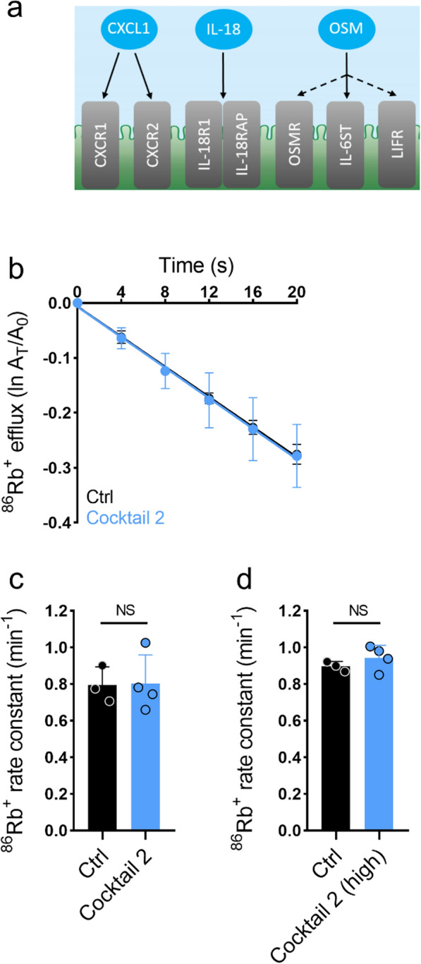
Methods: Lumbar CSF samples from 20 iNPH patients (10 with clinical improvement upon CSF shunting, 10 without clinical improvement) and 20 elderly control subjects were analyzed with the novel proximity extension assay technique for presence of 92 different inflammatory markers. RNA-sequencing was employed to delineate choroidal abundance of the receptors for the inflammatory markers found elevated in the CSF from iNPH patients. The ability of the elevated inflammatory markers to modulate choroidal NKCC1 activity was determined by addition of combinations of rat version of these in ex vivo experiments on rat choroid plexus.

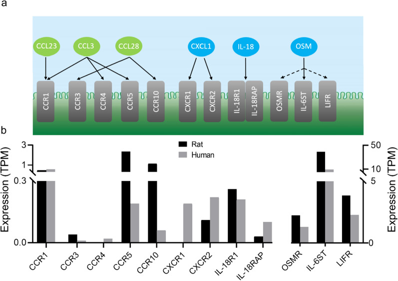
Results: 11 inflammatory markers were significantly elevated in the CSF from iNPH patients compared to elderly control subjects: CCL28, CCL23, CCL3, OPG, CXCL1, IL-18, IL-8, OSM, 4E-BP1, CXCL6, and Flt3L. One inflammatory marker, CDCP1, was significantly decreased in iNPH patients compared to control subjects. None of the inflammatory markers differed significantly when comparing iNPH patients with and without clinical improvement upon CSF shunting. All receptors for the elevated inflammatory markers were expressed in the rat and human choroid plexus, except CCR4 and CXCR1, which were absent from the rat choroid plexus. None of the elevated inflammatory markers found in the CSF from iNPH patients modulated the choroidal NKCC1 activity in ex vivo experiments on rat choroid plexus.

Conclusion: The CSF from iNPH patients contains elevated levels of a subset of inflammatory markers. Although the corresponding inflammatory receptors are, in general, expressed in the choroid plexus of rats and humans, their activation did not modulate the NKCC1-mediated fraction of choroidal CSF secretion ex vivo. The molecular mechanisms underlying ventriculomegaly in iNPH, and the possible connection to inflammation, therefore remains to be elucidated.

Keywords: Biomarkers; Cerebrospinal fluid; Choroid plexus; Inflammatory marker; Normal pressure hydrocephalus.


Conflict of interest statement

The authors declare that they have no competing interests.

Figures

Fig. 1
Fig. 1
Inflammatory receptors are present in the choroid plexus. a Schematic illustration of the inflammatory markers and their corresponding receptors. The inflammatory markers (green and blue circles) and their corresponding receptors (grey bars) are indicated by the black arrows. For OSM, the dotted black arrows indicate binding to IL-6ST in combination with either OSMR or LIFR. The illustration is based on the information provided in [67, 88, 102, 106]. b Expression of the inflammatory receptors in the rat and human choroid plexus. The expression levels were evaluated with RNA-seq and given in transcript per million (TPM). The human choroid plexus data was obtained from the GEO database, accession number GSE137619 [59]
Fig. 2
Fig. 2
The choroid plexus remains viable ex vivo. a Calcein-AM staining of choroid plexus directly after isolation (0 h). b Calcein-AM staining of choroid plexus after 16 h of incubation in cell culture medium (16 h). c Calcein-AM staining of choroid plexus after 16 h of incubation in sterile water (16 h H2O), scale bars 500 μm. Inserts in a-c contain representative regions of the choroid plexus (white boxes) upon ×5 magnification. d 86Rb+ efflux from the choroid plexus as a function of time in control solution (ctrl, n = 5) or in presence of 20 µM bumetanide (bum, n = 5). The y-axis represents the natural logarithm of the 86Rb+ amount left in the choroid plexus at time T (AT) divided by the initial amount at time 0 (A0). Insert: 86Rb+ efflux rate constant in control solution (0.50 ± 0.10 min−1, n = 5) or in presence of 20 µM bumetanide (0.22 ± 0.06 min−1, n = 5), P = 0.0008. Error bars in d represent standard deviation and statistical significance was tested with an unpaired two-tailed t-test. ***P < 0.001
Fig. 3
Fig. 3
The inflammatory markers CCL28, CCL23, and CCL3 do not modulate the NKCC1 activity. a Schematic illustration of the inflammatory markers in cocktail 1 (CCL28, CCL23, and CCL3) and their corresponding receptors. b 86Rb+ efflux from the rat choroid plexus as a function of time in the control setting (black, n = 4) or with exposure to cocktail 1 (green, n = 4) containing CCL28 (100 ng/ml), CCL23 (200 ng/ml), and CCL3 (100 ng/ml). The y-axis represents the natural logarithm of the 86Rb+ amount left in the choroid plexus at time T (AT) divided by the initial amount at time 0 (A0). c 86Rb+ efflux rate constant in the control setting (0.80 ± 0.09 min−1, n = 4) or with exposure to cocktail 1 (0.70 ± 0.12 min−1, n = 4, CCL28 (100 ng/ml), CCL23 (200 ng/ml), and CCL3 (100 ng/ml). P = 0.21. d 86Rb+ efflux rate constant in the control setting (0.82 ± 0.11 min−1, n = 4) or with exposure to high concentrations of cocktail 1 (0.80 ± 0.19 min−1, n = 4, CCL28 (500 ng/ml), CCL23 (500 ng/ml), and CCL3 (500 ng/ml), P = 0.86. Error bars in b, c and d represent standard deviation and statistical significance was tested with an unpaired two-tailed t-test. NS, not significant
Fig. 4
Fig. 4
The inflammatory markers IL-18, CXCL1, and OSM do not modulate the NKCC1 activity. a Schematic illustration of the inflammatory markers in cocktail 2 (IL-18, CXCL1, and OSM) and their corresponding receptors. b 86Rb+ efflux from the rat choroid plexus as a function of time in the control setting (black, n = 3) or with exposure to cocktail 1 (blue, n = 4) containing IL-18 (200 ng/ml), CXCL1 (100 ng/ml), and OSM (200 ng/ml). The y-axis represents the natural logarithm of the 86Rb+ amount left in the choroid plexus at time T (AT) divided by the initial amount at time 0 (A0). c 86Rb+ efflux rate constant in the control setting (0.79 ± 0.10 min−1, n = 3) or with exposure to cocktail 2 (0.80 ± 0.16 min−1, n = 4) containing IL-18 (200 ng/ml), CXCL1 (100 ng/ml), and OSM (200 ng/ml). P = 0.94. d 86Rb+ efflux rate constant in the control setting (0.90 ± 0.03 min−1, n = 3) or with exposure to high concentrations of cocktail 2 (0.94 ± 0.07 min−1, n = 4, IL-18 (500 ng/ml), CXCL1 (500 ng/ml), and OSM (500 ng/ml)), P = 0.32. Error bars in bd represent standard deviation and statistical significance was tested with an unpaired two-tailed t-test. NS, not significant

References

    1. Hakim S, Adams RD. The special clinical problem of symptomatic hydrocephalus with normal cerebrospinal fluid pressure. J Neurol Sci. 1965;2:307–327. - PubMed
    1. Jaraj D, Rabiei K, Marlow T, Jensen C, Skoog I, Wikkelso C. Prevalence of idiopathic normal-pressure hydrocephalus. Neurology. 2014;82:1449–1454. - PMC - PubMed
    1. Andersson J, Rosell M, Kockum K, Lilja-Lund O, Söderström L, Laurell K. Prevalence of idiopathic normal pressure hydrocephalus: a prospective, population based study. PLoS ONE. 2019;14:e0217705. - PMC - PubMed
    1. Relkin N, Marmarou A, Klinge P, Bergsneider M, Black PM. Diagnosing idiopathic normal-pressure hydrocephalus. Neurosurgery. 2005;57:S4–16. - PubMed
    1. Hebb AO, Cusimano MD. Idiopathic normal pressure hydrocephalus: a systematic review of diagnosis and outcome. Neurosurgery. 2001;49:1166–1186. - PubMed

MeSH terms

Substances

LinkOut - more resources