. 2022 Jan:105:53-71.
doi: 10.1016/j.matbio.2021.11.004. Epub 2021 Dec 1.

Novel regulatory roles of small leucine-rich proteoglycans in remodeling of the uterine cervix in pregnancy

Affiliations

Novel regulatory roles of small leucine-rich proteoglycans in remodeling of the uterine cervix in pregnancy

Mariano Colon-Caraballo et al. Matrix Biol. 2022 Jan.

Retraction in

Abstract

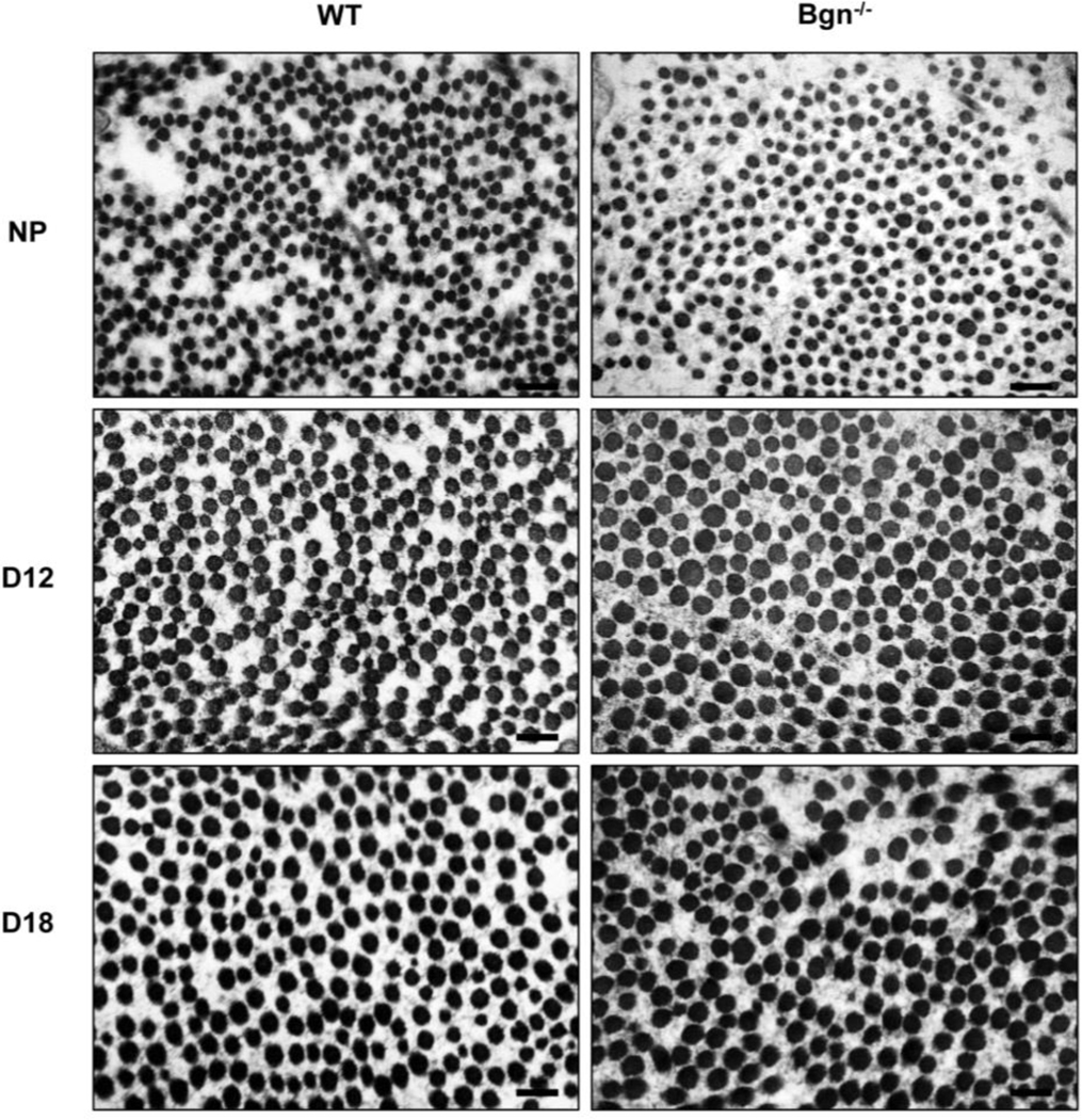
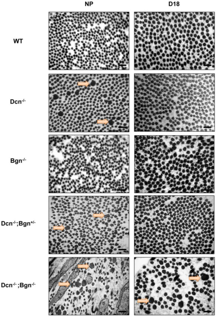
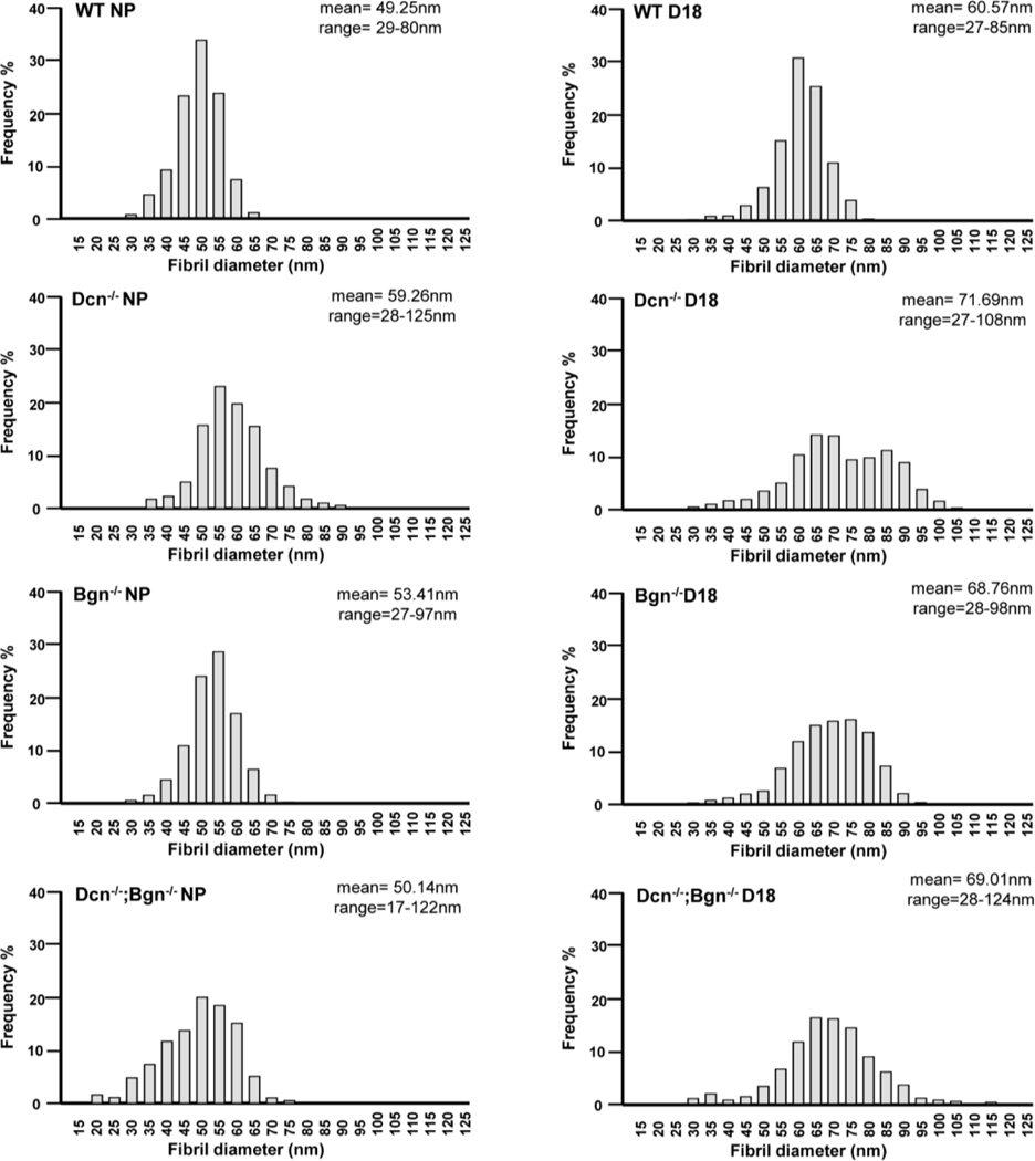
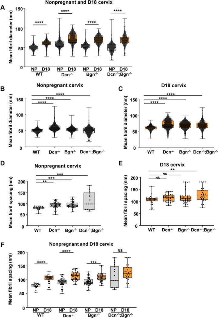
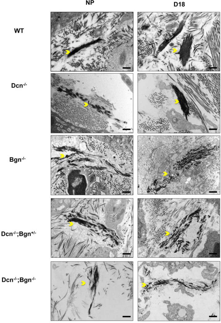
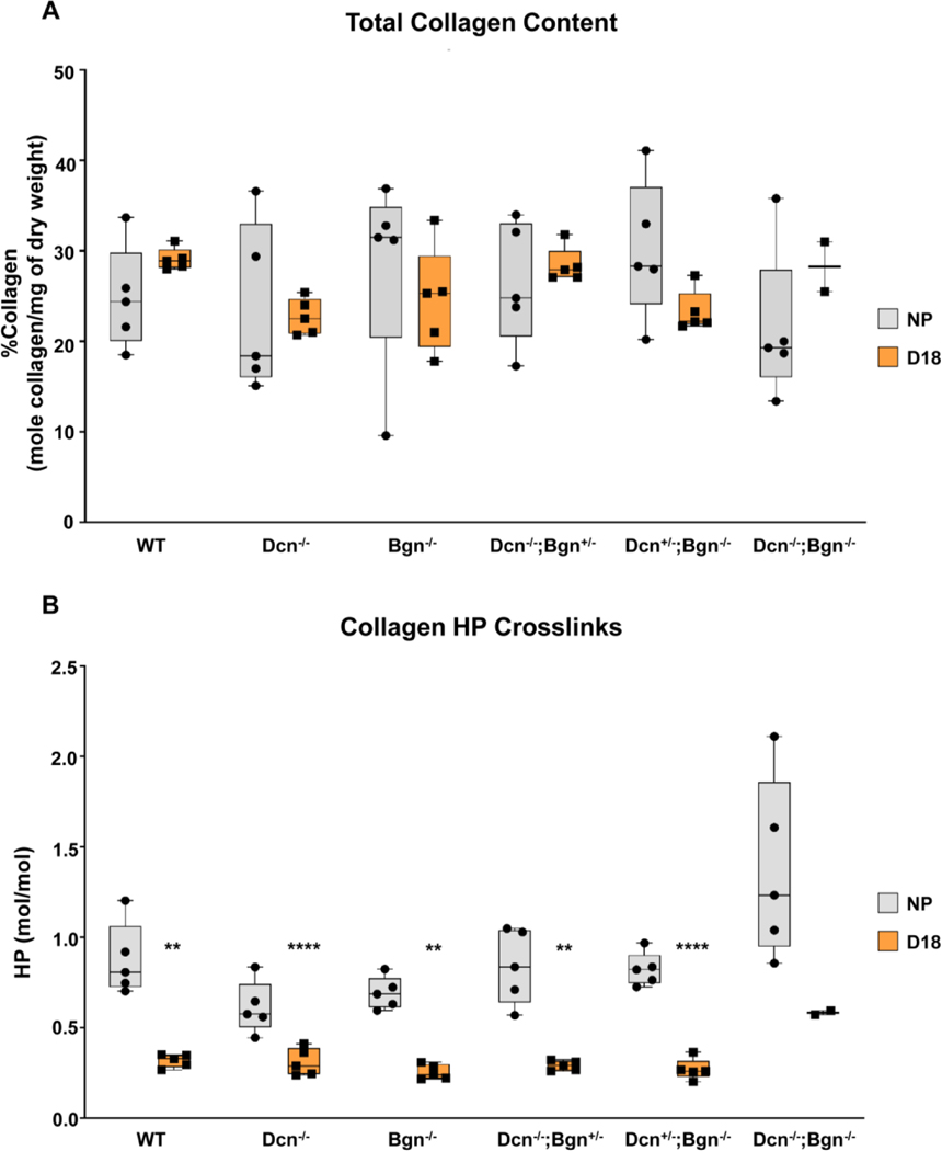
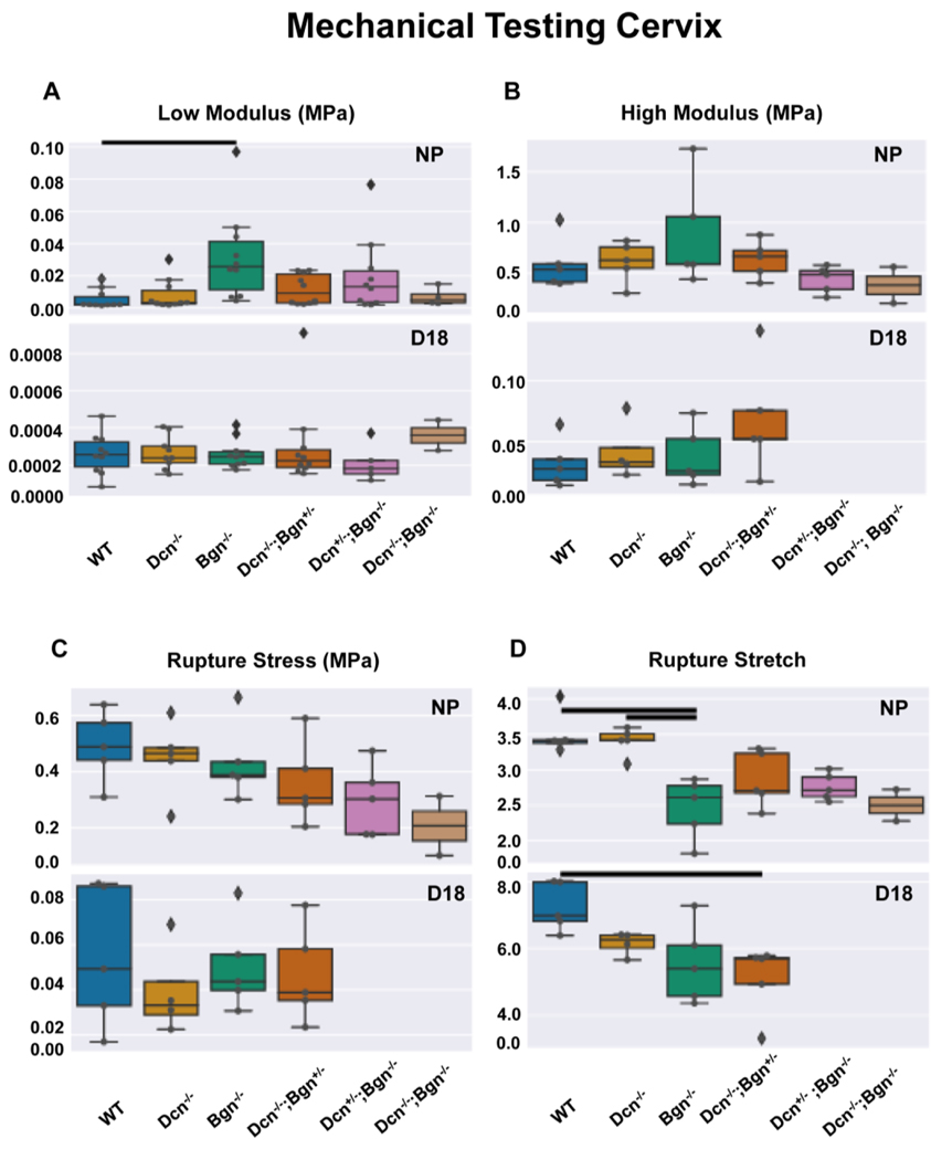
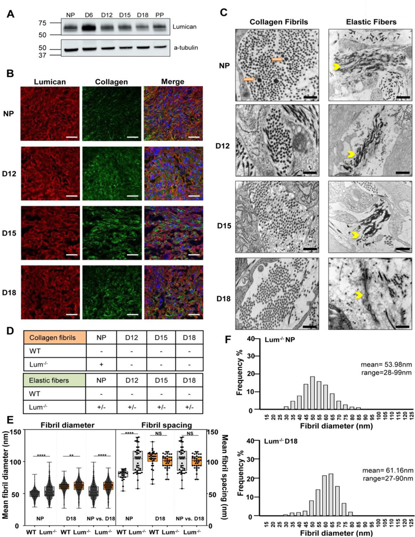
The cervix undergoes rapid and dramatic shifts in collagen and elastic fiber structure to achieve its disparate physiological roles of competence during pregnancy and compliance during birth. An understanding of the structure-function relationships of collagen and elastic fibers to maintain extracellular matrix (ECM) homeostasis requires an understanding of the mechanisms executed by non-structural ECM molecules. Small-leucine rich proteoglycans (SLRPs) play key functions in biology by affecting collagen fibrillogenesis and regulating enzyme and growth factor bioactivities. In the current study, we evaluated collagen and elastic fiber structure-function relationships in mouse cervices using mice with genetic ablation of decorin and/or biglycan genes as representative of Class I SLRPs, and lumican gene representative of Class II SLRP. We identified structural defects in collagen fibril and elastic fiber organization in nonpregnant mice lacking decorin, or biglycan or lumican with variable resolution of defects noted during pregnancy. The severity of collagen and elastic fiber defects was greater in nonpregnant mice lacking both decorin and biglycan and defects were maintained throughout pregnancy. Loss of biglycan alone reduced tissue extensibility in nonpregnant mice while loss of both decorin and biglycan manifested in decreased rupture stretch in late pregnancy. Collagen cross-link density was similar in the Class I SLRP null mice as compared to wild-type nonpregnant and pregnant controls. A broader range in collagen fibril diameter along with an increase in mean fibril spacing was observed in the mutant mice compared to wild-type controls. Collectively, these findings uncover functional redundancy and hierarchical roles of Class I and Class II SLRPs as key regulators of cervical ECM remodeling in pregnancy. These results expand our understating of the critical role SLRPs play to maintain ECM homeostasis in the cervix.

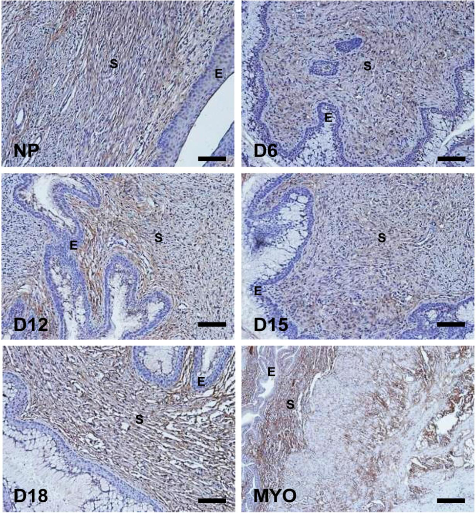
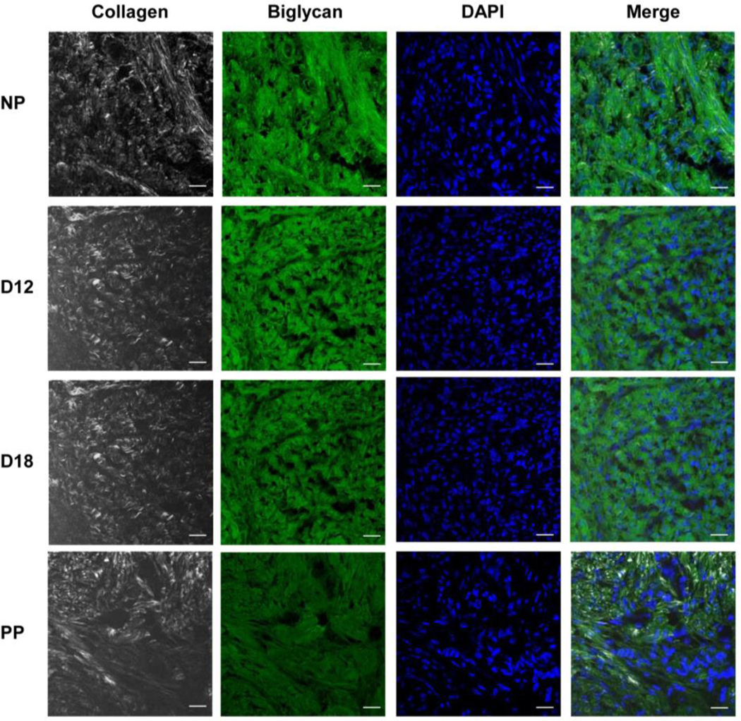
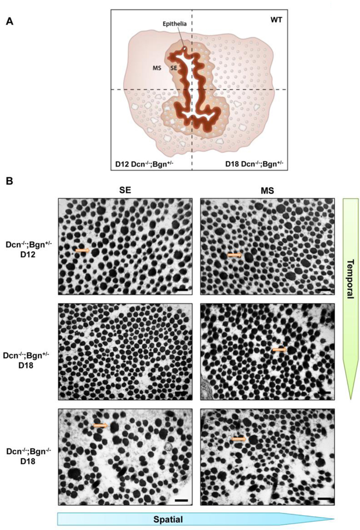
Keywords: Bgn= biglycan; Biomechanics; Cervix; Collagen; D12= gestational day 12; D15= gestational day 15; D18= gestational day 18; D6= gestational day 6; DKO= double knockout; Dcn= decorin; ECM= extracellular matrix; EDS= Ehler-Danlos syndrome; Elastic fibers; Fmod= fibromodulin; HP= hydroxylysylpyridinolines cross-links; LP= lysylpyridinolines cross-links; Lum= lumican; MS= midstroma; Myo= myometrial tissue; NP= non pregnant; PPROM= preterm premature rupture of fetal membranes; PTB= preterm birth; Pregnancy; Proteoglycan; SE= subepithelia; SHG= second harmonic generation imaging; SLRPs= small leucine rich proteoglycans; TEM= tissue electron microscopy; WT= wild-type.


Conflict of interest statement

Declaration of competing interests The authors declare that they have no conflicts of interest with the contents of this article.

Figures

Figure 1:
Figure 1:. Biglycan (Bgn) immunostaining in the cervix during pregnancy.
Cervical sections from three animals per time point were stained with antibody against biglycan and representative images for NP and gestation days 6, 12, 15, 18 are depicted. Myometrial tissue (MYO) was used as a positive control. Immunoreactivity for biglycan was observed as brown staining in the stromal region. S-stroma and E-epithelium. Scale bars, 100μm
Figure 2:
Figure 2:
Biglycan co-aligns with collagen fibers in the cervical extracellular matrix (ECM). Dual imaging of biglycan protein by immunofluorescence (green), collagen fibers by SHG (white), nuclei by DAPI (blue) in cervical sections from NP, day 12 (D12), day 18 (D18), and 1-day PP (PP) mice. Scale bars, 20μm
Figure 3:
Figure 3:. Collagen fibril morphology is normal in the Bgn−/− cervix.
TEM analysis of collagen structure in WT and Bgn−/− cervical sections from NP, gestation days 12 and 18 mice. n= 3 animals per genotype and time point. Scale bars, 500 nm.
Figure 4:
Figure 4:. Severity of defects in collagen fibril morphology progressively increases in the cervix of Dcn−/− with loss of one or both biglycan alleles.
TEM analysis of collagen structure in the subepithelial region in NP cervices reveals large, abnormal collagen fibrils (depicted by the orange arrow) in the Dcn single KO (Dcn−/−) and the mixed compound genotypes (Dcn−/−;Bgn+/− and Dcn−/−;Bgn−/−) but not in the WT or the Bgn single KO (Bgn−/−) (left panel). Defects in collagen fibrils were sustained only in the late pregnant (gestation day 18) Dcn−/−;Bgn−/− (right panel). n= 3 animals per genotype and time point. Scale bars, 500 nm.
Figure 5:
Figure 5:. Spatial pattern of recovery in collagen morphology occurs in the Dcn−/−;Bgn+/− cervix during pregnancy.
Cervix cross-sectional schematic illustrating the subepithelial (SE) and midstromal (MS) regions and the presence of uniform fibrils in both regions of WT mice. Temporal recovery of collagen fibril structure (compare d12 versus day 18) in cervices of Dcn−/−; Bgn+/− uncovers a spatial pattern of recovery (panel A). TEM images from Dcn−/− ;Bgn+/− identify abnormal fibrils in the SE and MS region on day 12 (top panel B) in contrast to day 18 when defects are detected only in the MS region (middle panel B). n= 3 animals per genotype and time point. Scale bars, 500 nm.
Figure 6:
Figure 6:. Loss of decorin and biglycan results in a broader size distribution of collagen fibrils in the cervix.
Tissue electron micrographs were taken at a magnification of 8000× of NP and D18 cervices for each genotype (n= 3 animals per genotype and time point). Collagen fibrils were measured (n=1668–3054 fibrils). Analysis of frequency in fibril diameter shows a heterogeneous distribution in the mutant mice compared to wild-type. A shift towards large diameter fibrils (above 95nm; 95–125nm range) was observed only in the Dcn−/− (NP and day18), and Dcn−/−;Bgn−/− (NP and day18) mice. In contrast, a shift towards smaller diameter fibrils below 26nm (17–26nm range) was detected only the Dcn−/−; Bgn−/− NP.
Figure 7:
Figure 7:. Decorin and biglycan regulate collagen fibril diameter and spacing in the cervical ECM.
Tissue electron micrographs were taken at a magnification of 8000× (n= 3 animals per genotype and time point). Mean fibril diameter distributions between the NP and gestational d18 cervix in wild-type, single KO, and double KO mice (panel A). An increase in mean fibril diameters is observed in late pregnancy (p<0.0001). Mean fibril diameter increases in the NP cervix (panel B; p<0.0001) and d18 cervix (panel C; p<0.0001) in the absence of one or both class I SLRPs. Mean fibril spacing increases in the NP mice lacking decorin, biglycan, or both SLRPs (panel D; p=0.0011 for Dcn−/− p=0.008 for Bgn−/−, and p=0.0002 for Dcn−/−;Bgn−/−). Mean fibril spacing was significantly higher in D18 cervix only in the Dcn−/−;Bgn−/− mice (panel E; p= 0.005). Mean fibril spacing distributions between the NP and gestational d18 cervix in wild-type, single KO, and double KO mice (panel F). An increase in mean fibril spacing is observed between NP and D18 in the Dcn−/− and Bgn−/− mice (p<0.0001; WT and Dcn−/−; p=0.0004 for Bgn−/−).
Figure 8:
Figure 8:. Biglycan plays a dominant role in elastic fiber assembly.
Elastic fiber structure was analyzed by TEM in cervical sections of NP and gestation d18. Defects in elastic fiber structure (yellow arrowheads) were observed in NP single KO (Dcn−/− and Bgn−/−) and the mixed genotypes (Dcn−/− ;Bgn+/− and Dcn−/−;Bgn−/−) but not in the WT mice. On gestation day 18, the abnormalities in elastic fibers were completely resolved in the Dcn−/− but remains evident in the Bgn−/−, mixed genotype and Dcn−/−;Bgn−/− cervices. n= 3 animals per genotype and time point. Scale bars, 500 nm.
Figure 9:
Figure 9:. Collagen content and HP cross-link measurement in non-pregnant and gestation d18 time point within each genotype.
** and **** indicates statistically significant difference from WT (n=5 for all groups except for DKO, n=2), p<0.01 and p<0.001, respectively). Error bars represent SEM.
Figure 10:
Figure 10:. Stiffness and rupture behavior of the cervix described by the load-to-failure mechanical test.
Low modulus calculated in the low stretch region (panel A) and high modulus calculated close to rupture (panel B). Rupture stretch (panel C) and Rupture stress calculated using the image immediately before rupture (panel D). Overlaid dots indicate each sample. Diamonds indicate samples outside 1.5 interquartile range (IQR). Solid lines indicate statistically significant differences between two groups (p<0.05). For low modulus, n=10 animals per genotype and time point except for NP Dcn−/−;Bgn−/− (n=4) and d18 Dcn−/− (n=9), Dcn+/;Bgn−/− (n=5), and Dcn−/−;Bgn−/− (n=2). For all other parameters, n=5 per genotype per time point except NP Dcn−/−;Bgn−/− (n=2) and d18 Dcn−/− (n=4). Dcn+/−;Bgn−/− and Dcn−/−;Bgn−/− were not tested for d18 using the load-to-failure protocol described here.
Figure 11:
Figure 11:. Lumican also plays a role in collagen and elastic fiber formation in the cervical extracellular matrix.
Lumican is expressed in the non-pregnant and pregnant cervix (panel A). Dual imaging of lumican protein by immunofluorescence (red), collagen fibers by SHG (green), nuclei by DAPI (blue) in cervical sections from NP, day 12 (D12), day 15 (D15), and day 18 (D18) mice. Merged images show alignment of lumican with collagen fibers in the cervical stromal region in the NP and pregnant cervix. Scale bar, 20 μm (panel B). Collagen and elastic ultrastructure in non-pregnant and pregnant cervix by TEM. Abnormal collagen fibrils (orange arrows) were noted only in the NP cervix. Defects in elastic fiber structure (yellow arrowhead) was observed in the NP and pregnant cervix (D12-D18). Scale bars 500 nm (panel C). Summary of defects in collagen fibrils and elastic fibers ultrastructure in the cervix. “+” and “−” indicate presence or absence of abnormal fibers, respectively. “+/−” is a mix of normal and abnormal fibers. For each study n= 3 animals per genotype and time point (panel D). Mean collagen fibril diameter (panel E) was significantly greater in NP and d18 Lum−/− cervix (n=2500–2740 fibrils). Collagen fibril spacing was greater in the NP but not gestation day18 Lum−/− cervix compared to WT (panel F).

References

    1. Shynlova O, Mitchell JA, Tsampalieros A, Langille BL, Lye SJ, Progesterone and gravidity differentially regulate expression of extracellular matrix components in the pregnant rat myometrium, Biol Reprod 70(4) (2004) 986–92. - PubMed
    1. Mead TJ, Du Y, Nelson CM, Gueye NA, Drazba J, Dancevic CM, Vankemmelbeke M, Buttle DJ, Apte SS, ADAMTS9-Regulated Pericellular Matrix Dynamics Governs Focal Adhesion-Dependent Smooth Muscle Differentiation, Cell Rep 23(2) (2018) 485–498. - PMC - PubMed
    1. Nallasamy S, Yoshida K, Akins M, Myers K, Iozzo R, Mahendroo M, Steroid Hormones Are Key Modulators of Tissue Mechanical Function via Regulation of Collagen and Elastic Fibers, Endocrinology 158(4) (2017) 950–962. - PMC - PubMed
    1. Byers PH, Murray ML, Ehlers-Danlos syndrome: a showcase of conditions that lead to understanding matrix biology, Matrix Biol 33 (2014) 10–5. - PubMed
    1. Martin JA, Hamilton BE, Osterman MJK, Driscoll AK, Births: Final Data for 2019, Natl Vital Stat Rep 70(2) (2021) 1–51. - PubMed

Publication types