. 2021 Dec 4;12(12):1128.
doi: 10.1038/s41419-021-04412-1.

Enriched environment and visual stimuli protect the retinal pigment epithelium and photoreceptors in a mouse model of non-exudative age-related macular degeneration

Affiliations

Enriched environment and visual stimuli protect the retinal pigment epithelium and photoreceptors in a mouse model of non-exudative age-related macular degeneration

Hernán H Dieguez et al. Cell Death Dis. .

Abstract

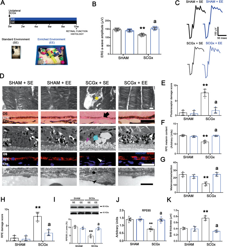
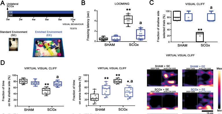
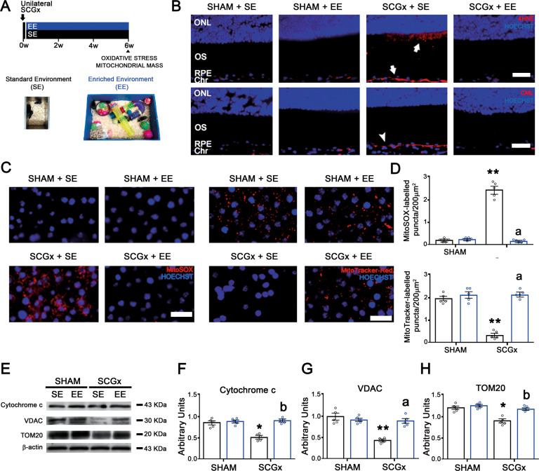
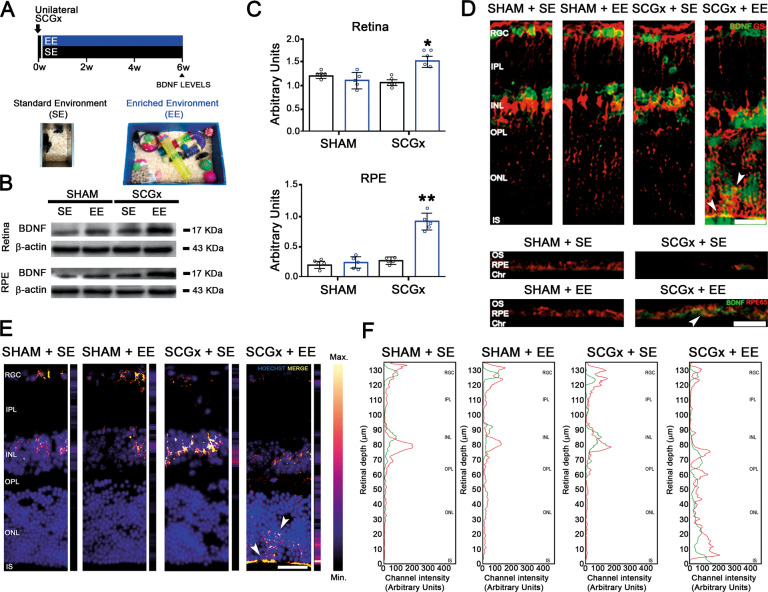
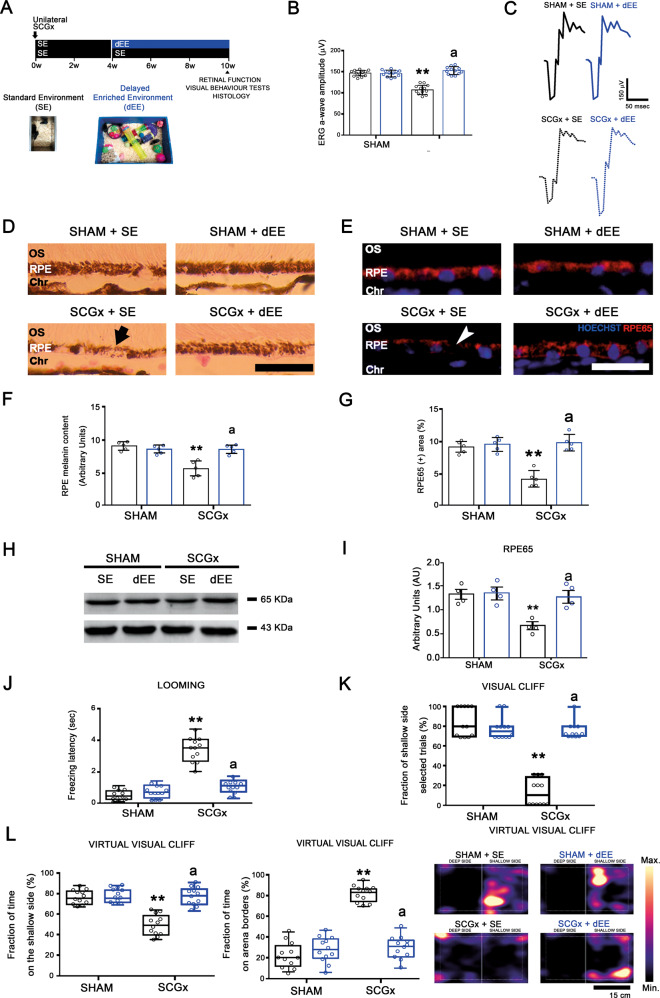
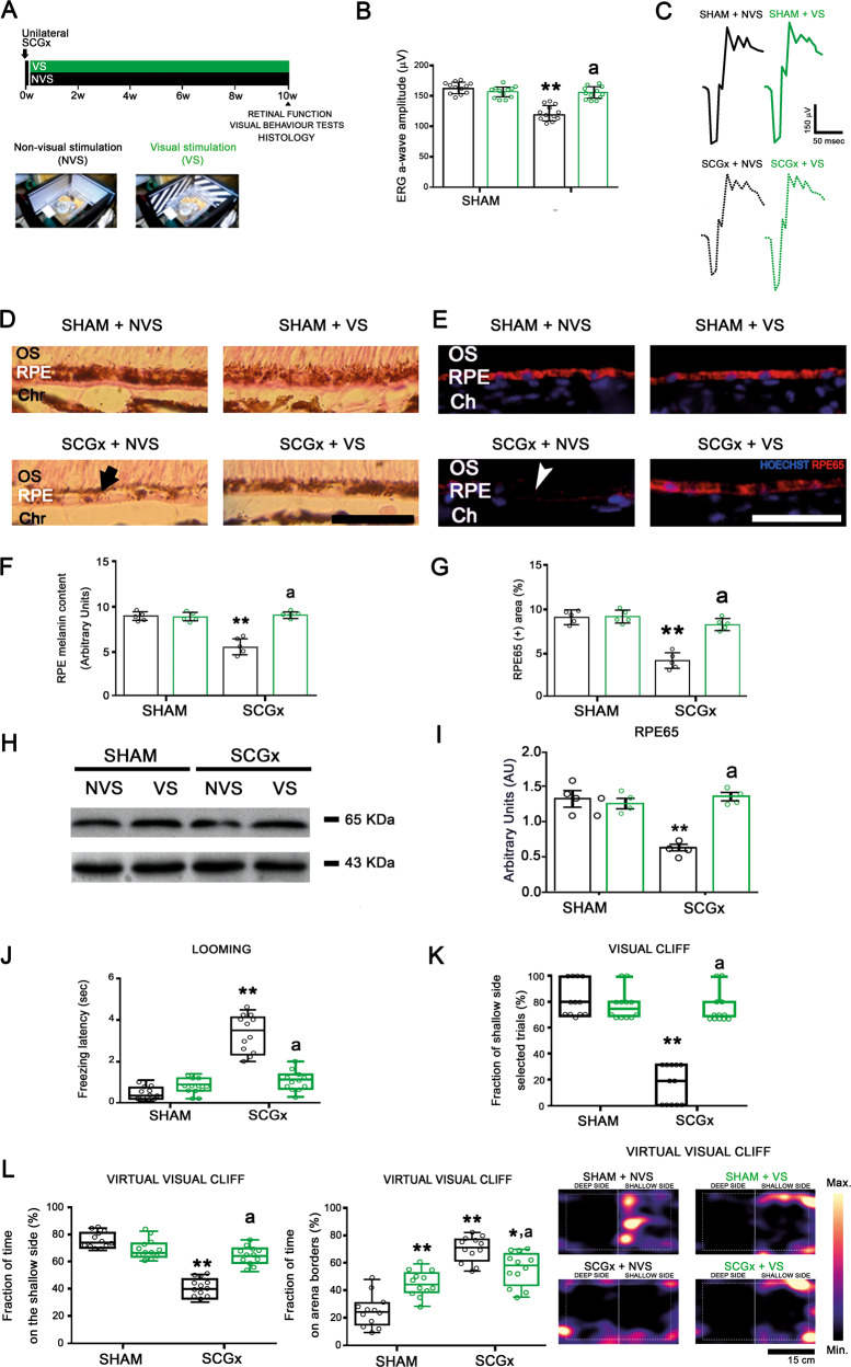
Non-exudative age-related macular degeneration (NE-AMD), the main cause of blindness in people above 50 years old, lacks effective treatments at the moment. We have developed a new NE-AMD model through unilateral superior cervical ganglionectomy (SCGx), which elicits the disease main features in C57Bl/6J mice. The involvement of oxidative stress in the damage induced by NE-AMD to the retinal pigment epithelium (RPE) and outer retina has been strongly supported by evidence. We analysed the effect of enriched environment (EE) and visual stimulation (VS) in the RPE/outer retina damage within experimental NE-AMD. Exposure to EE starting 48 h post-SCGx, which had no effect on the choriocapillaris ubiquitous thickness increase, protected visual functions, prevented the thickness increase of the Bruch's membrane, and the loss of the melanin of the RPE, number of melanosomes, and retinoid isomerohydrolase (RPE65) immunoreactivity, as well as the ultrastructural damage of the RPE and photoreceptors, exclusively circumscribed to the central temporal (but not nasal) region, induced by experimental NE-AMD. EE also prevented the increase in outer retina/RPE oxidative stress markers and decrease in mitochondrial mass at 6 weeks post-SCGx. Moreover, EE increased RPE and retinal brain-derived neurotrophic factor (BDNF) levels, particularly in Müller cells. When EE exposure was delayed (dEE), starting at 4 weeks post-SCGx, it restored visual functions, reversed the RPE melanin content and RPE65-immunoreactivity decrease. Exposing animals to VS protected visual functions and prevented the decrease in RPE melanin content and RPE65 immunoreactivity. These findings suggest that EE housing and VS could become an NE-AMD promising therapeutic strategy.


Conflict of interest statement

The authors declare no competing interests.

Figures

Fig. 1
Fig. 1. Effect of EE on the retinal dysfunction and histological alterations at 10 weeks post-SCGx.
A Experimental protocols. B SCGx induced a significant decrease in ERG a-wave amplitude in animals housed in SE, whereas EE starting 48 h post-SCGx, which had no effect per se, completely prevented the decrease in this parameter (representative traces shown in C). Data are mean ± SEM (n: 12 animals per group), **P < 0.01 vs. sham-treated eyes from SE animals; a: P < 0.01 vs. SCGx-treated eyes from SE animals, by Tukey’s test. D In animals housed in SE, SCGx induced focal losses of PR discs (yellow asterisk) and blebs (quantified in E), and a decrease in RPE melanin content (black arrow) and melanosome number at the central temporal RPE, which were completely prevented by EE (quantified in F and G). EE also prevented RPE vacuolization (cyan arrow) and basal infolding thickening (magenta arrowhead) (quantified as RPE damage score in H), as well as the decrease in RPE65-immunostaining (white arrowhead) and protein levels at the temporal RPE at 10 weeks post-SCGx (quantified in I and J). SCGx induced BrM thickening (quantified in K), a clear loss of its pentalaminar structure (asterisk) and thickening of the endothelial cell basal membrane (black arrowhead), which were prevented by EE. Shown are representative photomicrographs from 5 (for optic microscopy) and 4 (for electronic microscopy) eyes/group, at 800 μm temporally from the ONH. OS photoreceptor outer segments, RPE retinal pigment epithelium, BI RPE basal infoldings, BrM Bruch’s membrane, Ch choriocapillaris, Chr choroid. Scale bars = 500, 20, 25, 1.5, 300 nm. Data are mean ± SEM (n: 5 (for optic microscopy) and 4 (for electronic microscopy) eyes per group), **P < 0.01 vs. sham-treated eyes from SE animals; a: P < 0.01 vs. SCGx-treated eyes from SE animals, by Tukey’s test. Data are mean ± SEM (n: 5 homogenates per group), **P < 0.01 vs. sham-treated eyes from SE animals, by Tukey’s test.
Fig. 2
Fig. 2. Effect of EE on the visual behaviour tests at 10 weeks post-SCGx.
A Experimental protocols. BD In SE animals, SCGx induced a significant increase in the freezing latency of the looming test, and a significant decrease both in the fraction of shallow-side selected trials in the visual cliff test, and the percentage of time spent on the shallow side in the virtual visual cliff test, which were prevented by EE. The time spent at the borders of the virtual visual cliff arena significantly increased in sham-treated animals housed in EE and SGCx-treated animals housed in SE and EE. Heatmaps of the tracked position of sham- and SCGx-treated eyes from SE and EE animals are shown. **P < 0.01 vs. SE animals with sham-treated eyes; a: P < 0.01 vs. SE animals with SCGx-treated eyes, by Tukey’s test (n: 12 animals per group).
Fig. 3
Fig. 3. Effect of EE on the temporal outer retina/RPE oxidative damage, and RPE mitochondria mass at 6 weeks post-SCGx.
A Experimental protocols. B EE prevented the SGCx-induced increase in 4HNE- and CML-immunoreactivity at the outer retina/RPE (arrow), and RPE (arrowhead), respectively. Shown are representative photomicrographs from 5 eyes/group, at 800 μm temporally from the ONH. ONL outer nuclear layer, OS photoreceptor outer segments, RPE retinal pigment epithelium, Chr choroid. Scale bars: 30 μm. C EE significantly prevented both the SCGx-induced increase in MitoSox-Red-labelled mitochondria and decrease in Mitotracker-Red(+) puncta at the RPE (quantified in D). Shown are representative photomicrographs from five eyes/group, at 800 μm temporally from the ONH. Scale bars = 20 μm. Data are mean ± SEM (n: 5 eyes per group), **P < 0.01 vs. sham-treated eyes from SE animals; a: P < 0.01 vs. SCGx-treated eyes from SE animals, by Tukey’s test. EH SCGx induced a decrease in the levels of cytochrome c, VDAC, and TOM20 at the temporal RPE, which were prevented by EE. Data are mean ± SEM (n: 5 homogenates per group), **P < 0.01, *P < 0.05 vs. sham-treated eyes from SE animals; a: P < 0.01, b: P < 0.05 vs. SCGx-treated eyes from SE animals, by Tukey’s test.
Fig. 4
Fig. 4. Effect of EE on the temporal retina/RPE BDNF-immunoreactivity and protein levels, and temporal retina BDNF/GS co-localization at 6 weeks post-SGCx.
A Experimental protocols. B, C BDNF levels were significantly increased in SCGx-treated eyes from animals housed in EE both at the retina and RPE. Data are mean ± SEM (n: 5 homogenates per group), **P < 0.01, *P < 0.05 vs. sham-treated eyes from SE animals, by Tukey’s test. D EE, which had no effect on BDNF-immunoreactivity in sham-treated eyes, increased this parameter at the retina and RPE from SCGx eyes, particularly at the OPL, ONL and IS layers. Shown are representative photomicrographs at 800 μm temporally from the ONH from 5 eyes/group. RGC retinal ganglion cell layer, IPL inner plexiform layer, INL inner nuclear layer, OPL outer plexiform layer, ONL outer nuclear layer, IS PR inner segments, OS PR outer segments, RPE retinal pigment epithelium, Chr choroid. Scale bars = 30 μm. E Exclusive merge (yellow) from GS- and BDNF-immunoreactivity pictures were analysed using Python software. BDNF immunoreactivity was exclusively increased at the outer retina in SCGx-treated animals housed in EE. F Analysis of the red (GS) and green (BDNF) pixels from GS- and BDNF-immunoreactivity pictures also analysed using Python software. At the retinas from sham-treated eyes and SCGx-treated eyes from SE animals merge (E) and coincident peaks in red and green pixels (F) were evident mainly at the GCL and the INL, whereas in SCGx-treated eyes from EE animals, GS- and BDNF-immunoreactivity merge (E), and red and green pixel peaks (F) particularly increased at the outer retina and Müller cell end feet. Shown are representative photomicrographs from 5 eyes/group, at 800 μm temporally from the ONH. RGC retinal ganglion cell layer, IPL inner plexiform layer, INL inner nuclear layer, OPL outer plexiform layer, ONL outer nuclear layer, IS PR inner segments, OS PR outer segments, RPE retinal pigment epithelium, Chr choroid. Scale bars = 30 μm.
Fig. 5
Fig. 5. Effect of the delayed EE exposure (dEE) on the retinal function and temporal RPE structural alterations and visual behaviour tests at 10 weeks post-SCGx.
A Experimental protocols. B EE starting at 4 weeks post-SCGx completely reversed the decrease in the ERG a-wave amplitude (representative traces shown in C). Data are mean ± SEM (n: 12 animals per group), **P < 0.01 vs. SE animals with sham-treated eyes; a: P < 0.01 vs. SE animals with SCGx-treated eyes, by Tukey’s test. DI SCGx induced a decrease in the melanin content (arrow) and RPE65-immunoreactivity (arrowhead) and protein levels at the temporal RPE in SE animals. The delayed exposure to EE totally reversed these alterations. Shown are representative photomicrographs from 5 eyes/group at 800 μm temporally from the ONH. OS PR outer segments, RPE retinal pigment epithelium, Chr choroid. Scale bars = 25 μm. Data are mean ± SEM (n: 5 eyes per group), **P < 0.01 vs. sham-treated eyes from SE animals; a: P < 0.01 vs. SCGx-treated eyes from SE animals, by Tukey’s test. Data are mean ± SEM (n: 5 homogenates per group), **P < 0.01 vs. sham-treated eyes from SE animals, by Tukey’s test. JL The delayed exposure to EE reversed the worse performance in the looming, visual cliff and virtual visual cliff tests induced by SCGx in SE-housed animals. **P < 0.01 vs. SE animals with sham-treated eyes; a: P < 0.01 vs. SE animals with SCGx-treated eyes, by Tukey’s test (n: 12 animals per group).
Fig. 6
Fig. 6. Effect of visual stimulation (VS) on the retinal function and temporal RPE structural alterations and visual behaviour tests at 10 weeks post-SCGx.
A Experimental protocols. B SCGx induced a significant decrease in ERG a-wave amplitude in animals exposed to non-visual stimulation (NVS), whereas VS exposure since 48 h post-SCGx completely prevented the decrease in this parameter (representative traces shown in C). DI Exposure to VS totally prevented the SCGx-induced decrease in the melanin content (arrow) and RPE65-immunoreactivity (arrowhead) and protein levels at the temporal RPE in SCGx-treated eyes from animals exposed to NVS. Shown are representative photomicrographs from 5 eyes/group, at 800 μm temporally from the ONH. OS photoreceptor outer segments, RPE retinal pigment epithelium, Chr choroid. Scale bars = 25 μm. Data are mean ± SEM (n: 5 eyes per group), **P < 0.01 vs. sham-treated eyes from animals exposed to NVS; a: P < 0.01 vs. SCGx-treated eyes from animals exposed to NVS, by Tukey’s test. Data are mean ± SEM (n: 5 homogenates per group), **P < 0.01 vs. sham-treated eyes from animals exposed to NVS, by Tukey’s test. JL VS completely preserved the performance in the looming test, the visual cliff, and the virtual visual cliff tests. The time spent at the borders of the virtual visual cliff arena significantly increased in sham-treated animals housed in VS and SGCx-treated animals housed in NVS and VS. **P < 0.01 vs. sham-treated eyes from animals exposed to NVS; a: P < 0.01 vs. SCGx-treated eyes from animals exposed to NVS, by Tukey’s test (n: 12 animals per group).

References

    1. Jager RD, Mieler WF, Miller JW. Age-related macular degeneration. N Engl J Med. 2008;358:2606–17. - PubMed
    1. Wong WL, Su X, Li X, Cheung CM, Klein R, Cheng CY, et al. Global prevalence of age-related macular degeneration and disease burden projection for 2020 and 2040: a systematic review and meta-analysis. Lancet Glob Health. 2014;2:e106–e116. - PubMed
    1. Datta S, Cano M, Ebrahimi K, Wang L, Handa JT. The impact of oxidative stress and inflammation on RPE degeneration in non-neovascular AMD. Prog Retin Eye Res. 2017;60:201–18. - PMC - PubMed
    1. Buschini E, Fea AM, Lavia CA, Nassisi M, Pignata G, Zola M, et al. Recent developments in the management of dry age-related macular degeneration. Clin Ophthalmol. 2015;9:563–74. - PMC - PubMed
    1. Kaarniranta K, Pawlowska E, Szczepanska J, Jablkowska A, Blasiak J. Role of mitochondrial DNA damage in ROS-mediated pathogenesis of age-related macular degeneration (AMD) Int J Mol Sci. 2019;20:2374. - PMC - PubMed

Publication types