. 2022 Feb 4;188(2):1111-1128.
doi: 10.1093/plphys/kiab543.

Rice Cell Division Cycle 20s are required for faithful chromosome segregation and cytokinesis during meiosis

Affiliations

Rice Cell Division Cycle 20s are required for faithful chromosome segregation and cytokinesis during meiosis

Ya-Nan Lin et al. Plant Physiol. .

Abstract

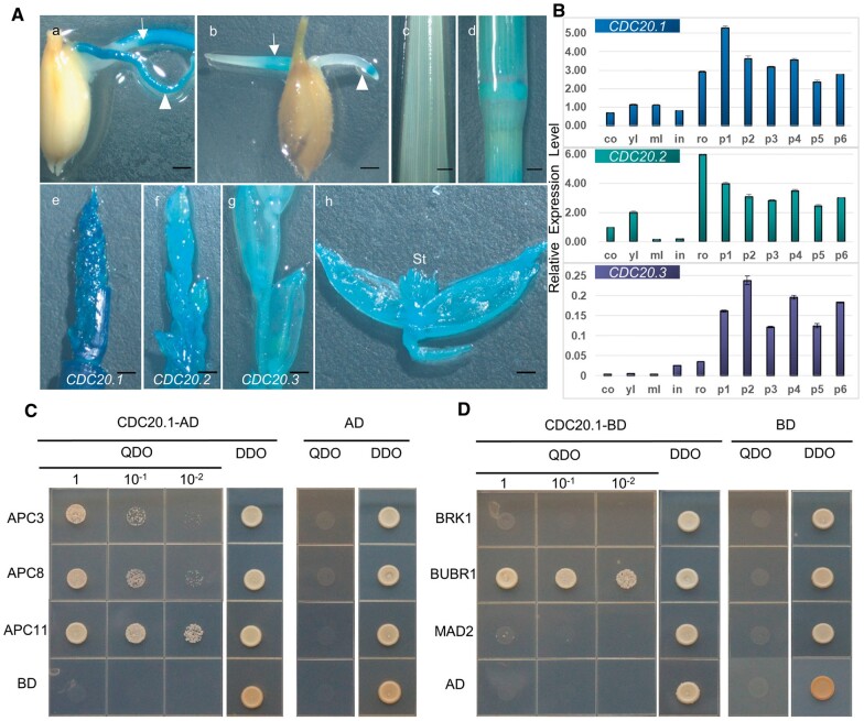
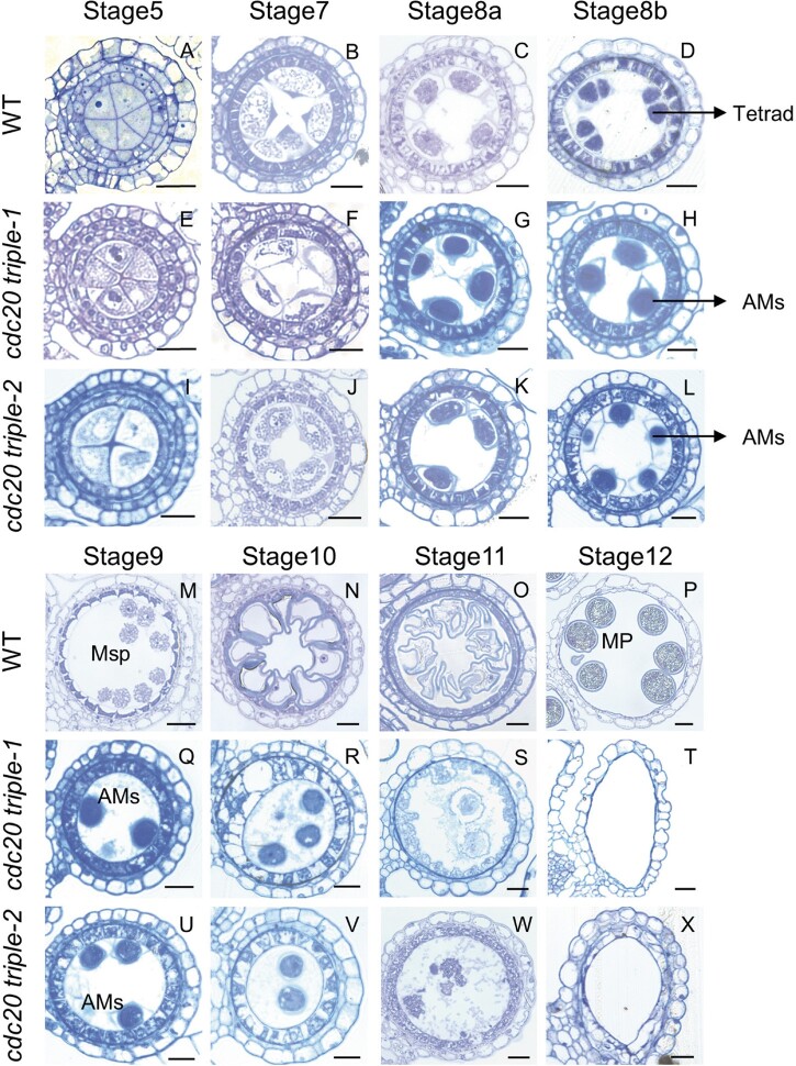
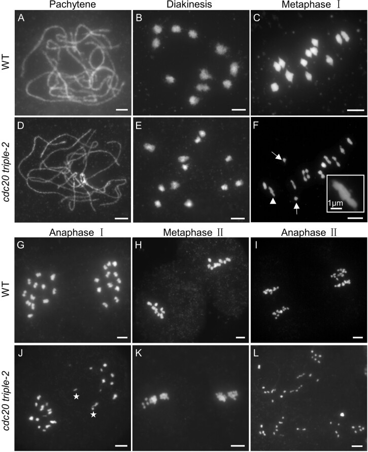
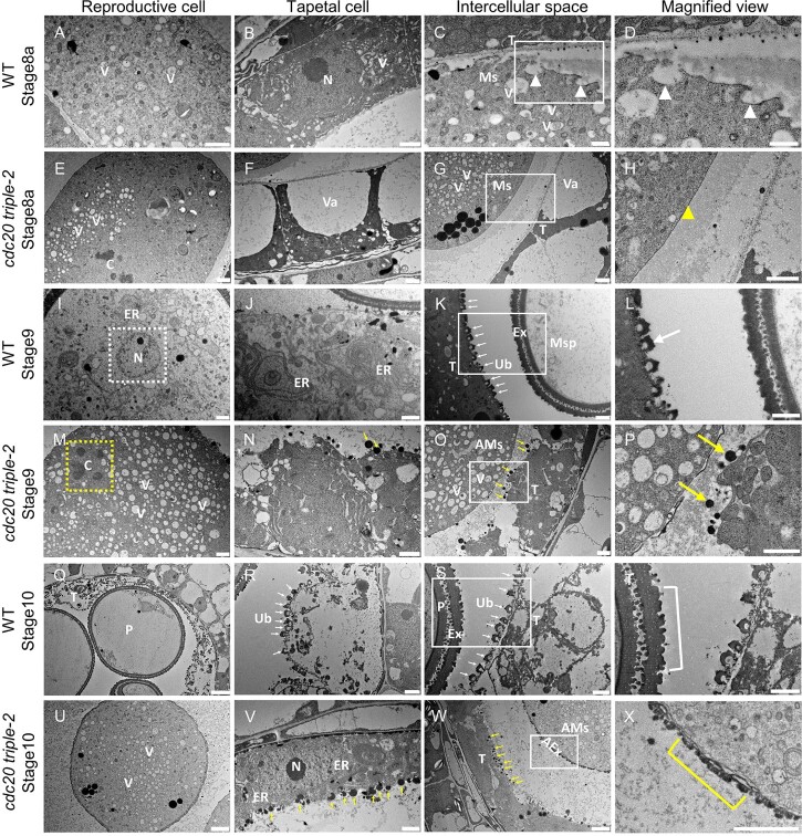
Chromosome segregation must be under strict regulation to maintain chromosome euploidy and stability. Cell Division Cycle 20 (CDC20) is an essential cell cycle regulator that promotes the metaphase-to-anaphase transition and functions in the spindle assembly checkpoint, a surveillance pathway that ensures the fidelity of chromosome segregation. Plant CDC20 genes are present in multiple copies, and whether CDC20s have the same functions in plants as in yeast and animals is unclear, given the potential for divergence or redundancy among the multiple copies. Here, we studied all three CDC20 genes in rice (Oryza sativa) and constructed two triple mutants by clustered regularly interspaced short palindromic repeats (CRISPR)/CRISPR-associated protein 9-mediated genome editing to explore their roles in development. Knocking out all three CDC20 genes led to total sterility but did not affect vegetative development. Loss of the three CDC20 proteins did not alter mitotic division but severely disrupted meiosis as a result of asynchronous and unequal chromosome segregation, chromosome lagging, and premature separation of chromatids. Immunofluorescence of tubulin revealed malformed meiotic spindles in microsporocytes of the triple mutants. Furthermore, cytokinesis of meiosis I was absent or abnormal, and cytokinesis II was completely prevented in all mutant microsporocytes; thus, no tetrads or pollen formed in either cdc20 triple mutant. Finally, the subcellular structures and functions of the tapetum were disturbed by the lack of CDC20 proteins. These findings demonstrate that the three rice CDC20s play redundant roles but are indispensable for faithful meiotic chromosome segregation and cytokinesis, which are required for the production of fertile microspores.


Figures

Figure 1
Figure 1
Rice CDC20 genes are highly expressed in panicles, and CDC20 interacts with APC/C core subunits and MCC members. A, GUS staining of pCDC20:GUS transgenic plants. Blue indicates the expression signal of the GUS reporter gene. CDC20.1 and CDC20.2 are expressed in coleoptiles and roots of germinated seeds (a, b). Arrows indicate the coleoptiles, and arrowheads point to the roots. CDC20.1 expression in a mature leaf (c) and in young node and internode (d). The three CDC20 genes are highly expressed in panicles at different stages (e, CDC20.1, f, CDC20.2, and g, CDC20.3), including the stamens (h, CDC20.2). St, stamen. Bars, 1 mm. B, Relative expression levels of CDC20 genes in different tissues, as examined by RT-qPCR. co, coleoptile; yl, young leaf; ml, mature leaf; in, internode; ro, root; p1–p6, 1- to 6-cm panicles. The experiment was conducted in three technical replicates, and error bars represent SD. C, Yeast two-hybrid assay showing that CDC20.1 interacts with the APC/C core subunits APC3, APC8, and APC11. AD and BD empty vectors were used as negative controls. QDO, SD medium lacking adenine, histidine, leucine, and tryptophan; DDO, SD medium lacking leucine and tryptophan. D, Yeast two-hybrid assay showing that CDC20.1 interacts with the MCC member BUBR1/MAD3.
Figure 2
Figure 2
Homozygous plants of both cdc20 triple mutant lines are totally sterile. A and B, Comparison of the WT and homozygous cdc20 triple mutants (cdc20 triple-1 and cdc20 triple-2) after seed maturation. Bars, 10 cm. C–E, Panicles of the WT (C) and homozygous triple mutants (D and E) at the seed maturation stage. F–H, Dissection of florets reveals abnormal white anthers in homozygous cdc20 triple mutants (G and H) compared to the yellow anthers in the WT (F). St, stamen. Bars, 1 mm. I–K, Stamens were stained with I2-KI solution to examine mature pollen grains. Normal WT anthers were stained dark blue (I), whereas anthers from homozygous plants of the two cdc20 triple mutants did not stain (J and K). Pollen grains were scattered when WT anthers were dissected, whereas the dissected anthers of both cdc20 triple mutants were empty, with no pollen. Bars, 200 μm. L, The seed setting rate of the WT was 95.4% ± 1.8% (n = 10), whereas that of cdc20 triple-1 (n = 30) and cdc20 triple-2 (n = 30) was 0%. Error bars represent SD. M, Average pollen number in a single stamen at the heading stage. There were 592 ± 86 (n = 10) pollen grains per anther in the WT but 0 pollen grains at the same stage in both cdc20 triple-1 (n = 20) and cdc20 triple-2 (n = 27). Error bars represent SD. N, Schematic diagram of the three CDC20 genes and the respective mutation sites in the two cdc20 triple mutants. Black box, coding region; black line, noncoding region; black triangle, target site of CRISPR/Cas9; red triangle, premature translation termination; black letters, WT sequence; red letters, inserted nucleotides or altered amino acid sequence in the mutants; red curve, sequence not shown; black dashed line, no nucleotides in the WT sequence; red dashed line, nucleotide deletion in the mutants.
Figure 3
Figure 3
In situ hybridization reveals the expression patterns of the three rice CDC20 genes in stamens. A–F, In situ hybridization of CDC20.1 mRNA in stamens at Stage 2 (A), Stage 3 (B), Stage 4 (C), Stage 5 (D), Stages 6–7 (E), and Stage 8 (F). The CDC20.1 signal is concentrated in the dividing cells and is distributed in a spotted pattern before meiosis (A–D). The signal becomes focused in meiocytes and the tapetum during meiosis (E and F). H–M, CDC20.2 mRNA expression signals in stamens at the same stages. The signal is concentrated at four corners of the stamen during the early stages (H–J) and is focused in microsporocytes and the tapetum at Stage 5 (K) as well as during meiosis (L and M). O–T, CDC20.3 expression in stamens at the same stages. CDC20.3 is expressed mainly in the germline and in cells adjacent to the germline. Sense probe was used as a negative control (G, N, and U). Bars, 25 μm.
Figure 4
Figure 4
Chromosomal observation demonstrates that mitotic division is not affected in early cdc20 triple mutant anthers. DAPI-stained semithin transverse sections of WT anthers (A–C) and cdc20.3-1 (D–F), cdc20 triple-1 (G–I), and cdc20 triple-2 anthers (J–L). One anther lobe is shown in (A, D, G, and J) to show the normal morphology and structure. White boxes in (B, E, H, and K) show cells undergoing mitotic division at metaphase with all the chromosomes aligned at the cell plate, and insets (b, e, h, and k) show magnified images of the regions in white boxes. Cells at anaphase with sister chromatids segregating toward opposite poles are shown in (C, F, I, and L), and insets (c, f, i, and l) show magnified images of the regions in white boxes. Bars in (A–L) represent 10 μm, and bars in (b, c, e, f, h, i, k, and l) represent 2.5 μm.
Figure 5
Figure 5
Knockout of the three CDC20 genes disrupts meiotic cytokinesis and the subsequent degeneration of microsporocytes. A–D and M–P, Semithin transverse sections of WT anther lobes; (E–H and Q–T) semithin sections of cdc20 triple-1 anthers; (I–L and U–X) semithin sections of cdc20 triple-2 anthers. Anther lobes at Stage 5 (A, E, and I), Stage 7 (B, F, and J), Stage 8a (C, G and K), Stage 8b (D, H, and L), Stage 9 (M, Q, and U), Stage 10 (N, R, and V), Stage 11 (O, S and W), and Stage 12 (P, T, and X) are shown. AMs, abnormal microsporocyte; Msp, microspore; MP, mature pollen. Bars, 25 μm.
Figure 6
Figure 6
The cdc20 triple mutant exhibits severe chromosome missegregation during meiosis. A–C and G–I, Analysis of chromosome behavior in WT microsporocytes; (D–F, J–L) analysis of chromosome behavior in the microsporocytes of a cdc20 triple-2 homozygous mutant at pachytene (A and D), diakinesis (B and E), metaphase I (C and F), anaphase I (G and J), metaphase II (H and K), and anaphase II (I and L) during meiosis. Bars, 5 μm. Arrows in (F) point to precociously segregated homologous chromosomes; arrowhead indicates an abnormal bivalent that is divided into three parts; inset box shows a magnified view of this abnormal bivalent. Asterisks in (J) indicate lagging chromosomes.
Figure 7
Figure 7
The cdc20 triple mutant shows chromosome missegregation and malformed spindle morphology. The chromosomes were stained with DAPI (purple), and microtubules were immunostained with α-tubulin antibody (green) in WT (A and C) and cdc20 triple-2 (B and D) microsporocytes at metaphase I (A and B) and anaphase I (C and D). Bars, 10 μm.
Figure 8
Figure 8
TEM reveals aberrant subcellular structures of the microsporocyte and tapetum in the cdc20 triple mutant. A–H, TEM observation of ultrathin transverse sections of Stage 8a anthers in the WT (A–D) and cdc20 triple-2 (E–H). I–P, Subcellular structures of reproductive and tapetal cells in Stage 9 anthers of the WT (I–L) and cdc20 triple-2 (M–P). Q–X, Subcellular structures of reproductive and tapetal cells in Stage 10 stamens in the WT (Q–T) and cdc20 triple-2 (U–X). Reproductive cells are shown in (A, E, I, M, Q, and U), tapetal cells are shown in (B, F, J, N, R, and V), the intercellular space between the reproductive cell and tapetal cell is shown in (C, G, K, O, S, and W), and magnified views of the images in white boxes are shown in (D, H, L, P, T, and X). White triangles in (C and D) point to fusing vesicles on the plasma membranes of WT microsporocytes. Yellow triangle in (H) indicates the smooth surface of the microsporocyte in cdc20 triple-2. White dashed frame highlights a normal nucleus in the WT (I), and yellow dashed frame highlights the scattered chromosomes in the cdc20 triple mutant (M). White arrows in (K, L, R, and S) point to normal Ubisch bodies on tapetal cells in WT anthers, and yellow arrows in (O, P, V, and W) point to the aberrant round and electron-dense structures out of the surfaces of tapetal cells in cdc20 triple-2. The white bracket in (T) indicates normal bilayer extine, and the yellow bracket in (X) indicates an abnormal one-layer structure outside the plasma membrane of a microsporocyte in the cdc20 triple mutant. AEx, abnormal extine; AMs, abnormal microsporocyte; C, chromosome; ER, endoplasmic reticulum; Ex, extine; Ms, microsporocyte; Msp, microspore; N, nucleus; P, pollen; T, tapetum; Ub, Ubisch body; V, vesicle; Va, vacuole. Bars represent 500 nm in (C and D); 1 μm in (A, B, E–P, R–T, and V); 2 μm in (U, W, and X); 5 μm in (Q).

Similar articles

Cited by

References

    1. Bai W, Wang P, Hong J, Kong W, Xiao Y, Yu X, Zheng H, You S, Lu J, Lei D, et al. (2019) Earlier degraded tapetum1 (EDT1) encodes an ATP-Citrate lyase required for tapetum programmed cell death. Plant Physiol 181:1223–1238 - PMC - PubMed
    1. Basu J, Bousbaa H, Logarinho E, Li Z, Williams BC, Lopes C, Sunkel CE, Goldberg ML (1999) Mutations in the essential spindle checkpoint gene bub1 cause chromosome missegregation and fail to block apoptosis in Drosophila. J Cell Biol 146:13–28 - PMC - PubMed
    1. Capron A, Ökrész L, Genschik P (2003) First glance at the plant APC/C, a highly conserved ubiquitin–protein ligase. Trends Plant Sci 8:83–89 - PubMed
    1. Chernomor O, von Haeseler A, Minh BQ (2016) Terrace aware data structure for phylogenomic inference from supermatrices. Syst Biol 65:997–1008 - PMC - PubMed
    1. Cimini D, Degrassi F (2005) Aneuploidy: a matter of bad connections. Trends Cell Biol 15:442–451 - PubMed

Publication types

LinkOut - more resources