. 2022 Feb 28;186(1):149-162.
doi: 10.1093/toxsci/kfab147.

Neuroinflammatory and Neurometabolomic Consequences From Inhaled Wildfire Smoke-Derived Particulate Matter in the Western United States

Affiliations

Neuroinflammatory and Neurometabolomic Consequences From Inhaled Wildfire Smoke-Derived Particulate Matter in the Western United States

David Scieszka et al. Toxicol Sci. .

Abstract

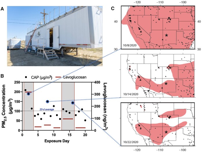
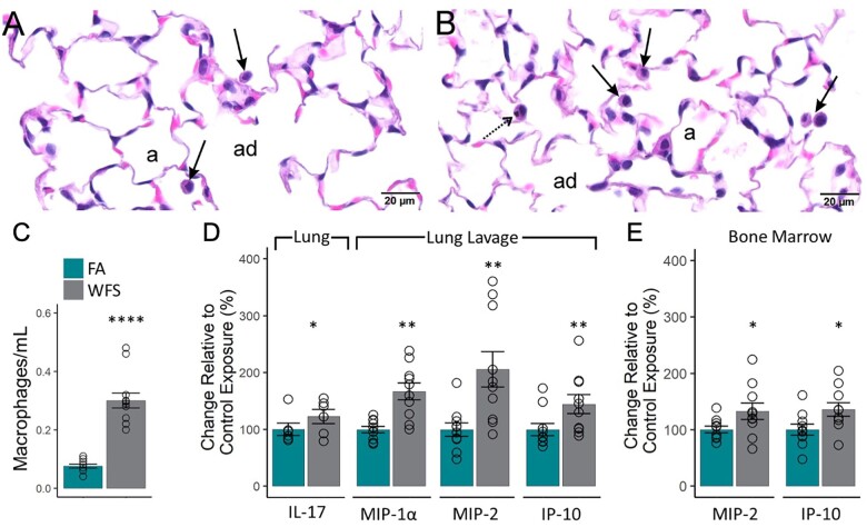
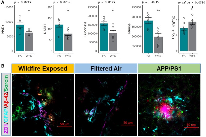
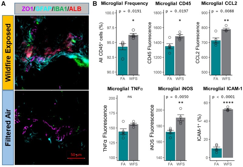
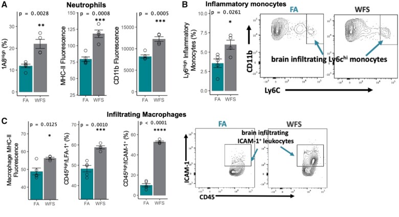
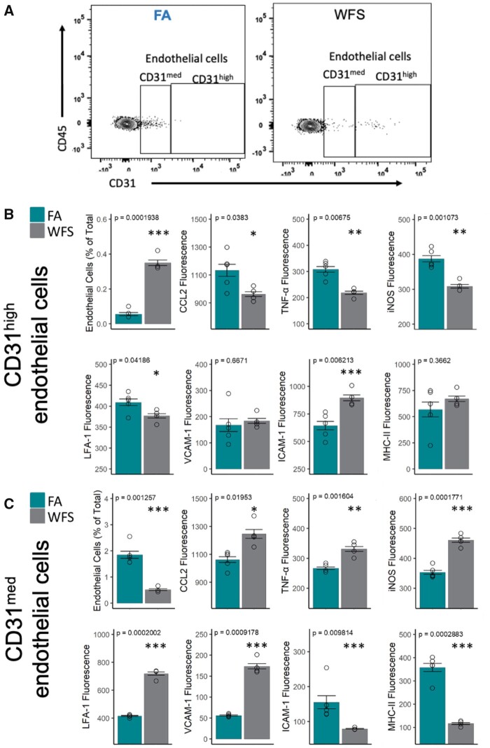
Utilizing a mobile laboratory located >300 km away from wildfire smoke (WFS) sources, this study examined the systemic immune response profile, with a focus on neuroinflammatory and neurometabolomic consequences, resulting from inhalation exposure to naturally occurring wildfires in California, Arizona, and Washington in 2020. After a 20-day (4 h/day) exposure period in a mobile laboratory stationed in New Mexico, WFS-derived particulate matter (WFPM) inhalation resulted in significant neuroinflammation while immune activity in the peripheral (lung, bone marrow) appeared to be resolved in C57BL/6 mice. Importantly, WFPM exposure increased cerebrovascular endothelial cell activation and expression of adhesion molecules (VCAM-1 and ICAM-1) in addition to increased glial activation and peripheral immune cell infiltration into the brain. Flow cytometry analysis revealed proinflammatory phenotypes of microglia and peripheral immune subsets in the brain of WFPM-exposed mice. Interestingly, endothelial cell neuroimmune activity was differentially associated with levels of PECAM-1 expression, suggesting that subsets of cerebrovascular endothelial cells were transitioning to resolution of inflammation following the 20-day exposure. Neurometabolites related to protection against aging, such as NAD+ and taurine, were decreased by WFPM exposure. Additionally, increased pathological amyloid-beta protein accumulation, a hallmark of neurodegeneration, was observed. Neuroinflammation, together with decreased levels of key neurometabolites, reflect a cluster of outcomes with important implications in priming inflammaging and aging-related neurodegenerative phenotypes.

Keywords: VCAM-1; microglia; neuroinflammation; neurovascular unit; particulate matter; smoke.


Figures

Figure 1.
Figure 1.
Exposure to concentrated WFPM particulates in Paguate, New Mexico. A, Mobile laboratory containing rodent exposure chambers and a Harvard-type particle concentrator. B, Daily PM2.5 gravimetric mass concentrations for the 20 days of exposure (black dots), along with levoglucosan measures (red bars) that span 3–4 days, as filters were pooled to provide sufficient material. C, Smoke and wildfire locations for 3 representative days in October 2020 derived from the National Oceanic and Atmospheric Administration Hazard Mapping System Fire and Smoke Product (Ruminski et al., 2006, 2007; Ruminski and Kondragunta, 2006), with dashed lines indicating the link to exposure days in (B). The black star indicates the location of the mobile laboratory in Paguate, New Mexico. Smoke plumes shown in pink were derived manually from geostationary and polar-orbiting satellites and represent a minimum smoke PM2.5 concentration of 5 µg/m3. Fire locations shown by triangles were obtained with automated fire detection algorithms; the nearest fire (on the border of Arizona and New Mexico) was over 300 km away from the mobile laboratory (Ruminski et al., 2006).
Figure 2.
Figure 2.
Significant pulmonary and bone marrow cytokine responses to 20-day exposure to wildfire smoke (WFPM). Light photomicrographs of hematoxylin and eosin-stained lung tissue from the left lung lobe of mice exposed to (A) filtered air (FA) or (B) concentrated fine ambient particles. Slightly more alveolar macrophages (arrows) were present in WFPM-exposed mice than FA control mice. Stippled arrow, particle laden-alveolar macrophage (black cytoplasmic material); solid arrow, alveolar macrophage without cytoplasmic particles; a, alveolus; ad, alveolar duct. (C) Airway macrophages in bronchoalveolar lavage fluid (n = 10) were significantly elevated after WFPM exposure. (D) Whole lung cytokine IL-17 (n = 6), and bronchoalveolar lavage cytokines (n = 10), MIP-1a, MIP-2, and IP-10 were significantly elevated by WFPM. (E) Bone marrow concentrations of MIP-2 and IP-10 were elevated by WFPM. Independent samples; two-tailed t test; mean and SEM shown, *p < .05, **p < .01, ***p < .005, ****p < .001. All measured cytokines are provided in Supplementary Tables 2–5.
Figure 3.
Figure 3.
Markers related to aging and neurodegenerative disease pathogenesis. A, NAD+, NADH, succinate, and taurine were all downregulated (n = 6), and amyloid beta (Aβ, n = 5) levels were upregulated in WFPM-exposed mice (n = 6, cerebellum; n = 5, rest of brain material, log2() correction for normality; independent samples; 2-tailed t test; mean and SEM shown). B, Representative images of neurodegenerative pathogenic markers found at the neocortical neurovascular unit on the sagittal plane starting 1.5 mm from the lateral aspect in WFPM-exposed mice. Brain Aβ-42 (red) increases in WFPM-exposed mice were observed only proximal to the neurovascular unit (endothelial ZO1 and astrocyte GFAP). Additionally, the early pathogenic marker Sorcin increased (green), indicating ER stress/unfolded protein responses proximal to areas of Aβ-42 staining, which was also observed in unexposed APP/PS1 mice of the same age and background as a positive control, though they exhibited higher density Aβ-42 aggregation.
Figure 4.
Figure 4.
Microglial activation following WFPM. A, Imaging of the neurovascular unit on the sagittal plane starting 1.5 mm from the lateral aspect reflected similar pathology as reported with other pollutants previously with more pronounced GFAP (cyan) staining and a greater presence of IBA1+ (green) microglia/macrophages adjacent to large-diameter (>20 µm) cerebrovasculature (ZO1+, purple), along with an increased staining in parenchymal albumin (red) in WFPM-exposed relative to FA control (right hemisphere). B, Percentage of microglia and their surface expression levels of CD45, adhesion molecule ICAM-1 and intracellular levels of proinflammatory factors, iNOS, TNFα, and CCL2 were analyzed, n = 3–5 independent samples, left hemisphere homogenate; 2-tailed t test; mean and SEM shown.
Figure 5.
Figure 5.
Infiltrating immune cells in the brain following WFPM exposure. A, Increased frequency of mature neutrophils (with high expression of 1A8), along with overall increase in MHC-II and CD11b surface expression of all CD45+1A8+ neutrophils in the brain due to WFPM exposure. B, Inflammatory monocytes in the brain were increased in frequency with WFPM exposure. C, CNS infiltrating CD45highCD11b+ macrophage/monocyte population shows increased MHC-II expression and frequency of LFA-1+ and ICAM-1+; peripheral leukocytes were also increased due to WFPM exposure, n = 4–5 independent samples, left hemisphere homogenate; 2-tailed t test; mean and SEM are shown.
Figure 6.
Figure 6.
Cerebrovascular endothelial cells exhibit a heterogeneous inflammatory response to WFPM. A, Representative flow cytometry plots of endothelial cells (with medium and high expression of CD31/PECAM-1 on CD45 cells) in the brain in FA or WFPM exposure groups. These 2 subsets of endothelial cells displayed comparatively different neuroimmune activity profile, as detailed in (B) and (C). B, Frequency of endothelial cells with high expression of CD31 was increased following WFPM exposure; however, these endothelial cells displayed reduced levels of these proinflammatory factors. Increased frequency of ICAM-1 expressing endothelial cells was observed due to WFPM exposure in this endothelial subset despite no changes in MHC-II. C, Frequency of CD31med endothelial cells were reduced following WFPM exposure, however, these cells displayed increased proinflammatory phenotype such as increased levels of CCL2, TNFα, and iNOS, although reduced levels of ICAM-1 and MHC-II were observed, n = 4–5 independent samples, left hemisphere homogenate; 2-tailed t test; mean and SEM are shown.

Similar articles

Cited by

References

    1. Agency USEP. (1997). Reference method for the determination of fine particulate matter as PM2.5 in the atmosphere. EPA 40 CFR Part 50, Washington, DC, Appendix L.
    1. Ahmadi Z., Arababadi M. K., Hassanshahi G. (2013). CXCL10 activities, biological structure, and source along with its significant role played in pathophysiology of type I diabetes mellitus. Inflammation 36, 364–371. - PubMed
    1. Aragon M., Erdely A., Bishop L., Salmen R., Weaver J., Liu J., Hall P., Eye T., Kodali V., Zeidler-Erdely P., et al. (2016a). MMP-9-dependent serum-borne bioactivity caused by multiwalled carbon nanotube exposure induces vascular dysfunction via the CD36 scavenger receptor. Toxicol. Sci. 150, 488–498. - PMC - PubMed
    1. Aragon M. J., Chrobak I., Brower J., Roldan L., Fredenburgh L. E., McDonald J. D., Campen M. J. (2016b). Inflammatory and vasoactive effects of serum following inhalation of varied complex mixtures. Cardiovasc. Toxicol. 16, 163–171. - PMC - PubMed
    1. Aragon M. J., Topper L., Tyler C. R., Sanchez B., Zychowski K., Young T., Herbert G., Hall P., Erdely A., Eye T., et al. (2017). Serum-borne bioactivity caused by pulmonary multiwalled carbon nanotubes induces neuroinflammation via blood-brain barrier impairment. Proc. Natl. Acad. Sci. U.S.A. 114, E1968–E1976. - PMC - PubMed

Publication types