

The morphology of the subacromial and related shoulder bursae. An anatomical and histological study
- PMID: 34865216
- PMCID: PMC9005683
- DOI: 10.1111/joa.13603
The morphology of the subacromial and related shoulder bursae. An anatomical and histological study
Abstract
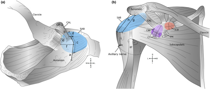
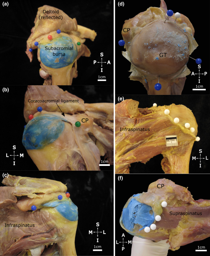
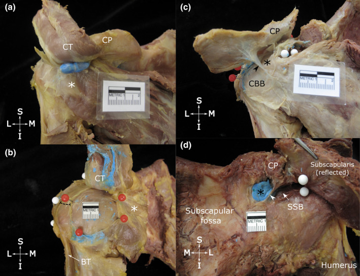
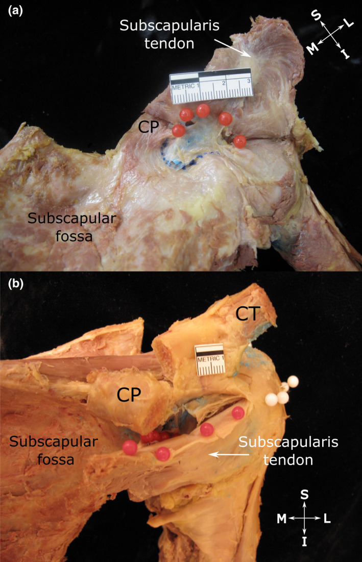
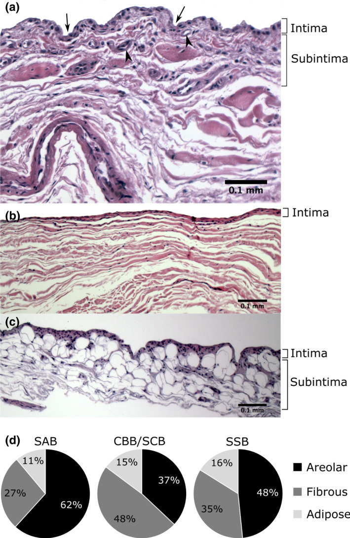
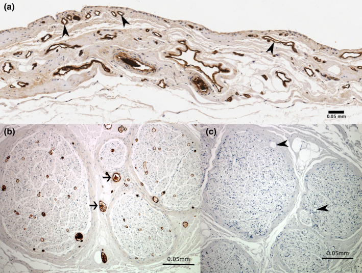
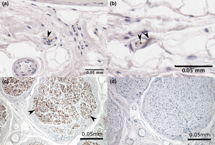
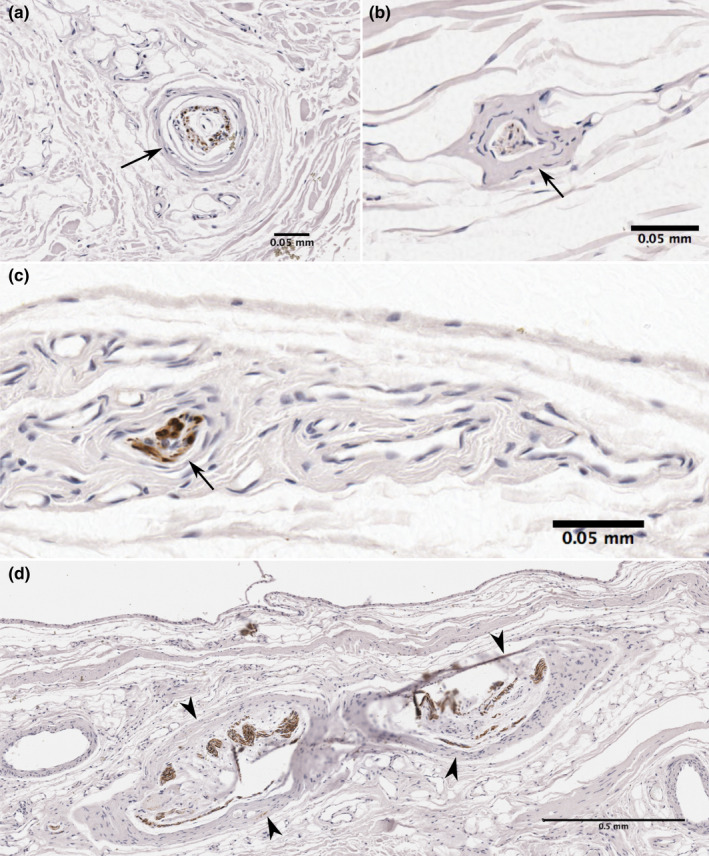
Shoulder bursae are essential for normal movement and are also implicated in the pathogenesis of shoulder pain and dysfunction. The subacromial bursa (SAB), within the subacromial space, is considered a primary source of shoulder pain. Several other bursae related to the subcoracoid space, including the coracobrachial (CBB), subcoracoid (SCB) and subtendinous bursa of subscapularis (SSB), are also clinically relevant. The detailed morphology and histological characteristics of these bursae are not well described. Sixteen embalmed cadaveric shoulders from eight individuals (five females, three males; mean age 78.6 ± 7.9 years) were investigated using macro-dissection and histological techniques to describe the locations, dimensions and attachments of the bursae, their relationship to surrounding structures and neurovascular supply. Bursal sections were stained with haematoxylin and eosin to examine the synovium and with antibodies against von Willebrand factor and neurofilament to identify blood vessels and neural structures respectively. Four separate bursae were related to the subacromial and subcoracoid spaces. The SAB was large, with a confluent subdeltoid portion in all except one specimen, which displayed a distinct subdeltoid bursa. The SAB roof attached to the lateral edge and deep surface of the acromion and coracoacromial ligament, and the subdeltoid fascia; its floor fused with the supraspinatus tendon and greater tubercle. The CBB (15/16 specimens) was deep to the conjoint tendon of coracobrachialis and short head of biceps brachii and the tip of the coracoid process, while the inconstant SCB (5/16 specimens) was deep to the coracoid process. Located deep to the subscapularis tendon, the SSB was a constant entity that commonly displayed a superior extension. Synovial tissue was predominantly areolar (SAB and SSB) or fibrous (CBB and SCB), with a higher proportion of areolar synovium in the bursal roofs compared to their floors. Blood vessels were consistently present in the subintima with a median density of 3% of the tissue surface area, being greatest in the SSB and SAB roofs (4.9% and 3.4% respectively) and least in the SAB floor (1.8%) and CBB roof and floor (both 1.6%). Nerve bundles and free nerve endings were identified in the subintima in approximately one-third of the samples, while encapsulated nerve endings were present in deeper tissue layers. The extensive expanse and attachments of the SAB support adoption of the term subacromial-subdeltoid bursa. Morphologically, the strong attachments of the bursal roofs and floors along with their free edges manifest as fixed and mobile portions, which enable movement in relation to surrounding structures. The presence of neurovascular structures demonstrates that these bursae potentially contribute blood supply to surrounding structures and are involved in mechanoreception. The anatomical details presented in this study clarify the morphology of the shoulder bursae, including histological findings that offer further insight into their potential function.
Keywords: anatomy; blood vessels; bursa; free nerve endings; shoulder; subacromial space; subcoracoid space; synovium.
© 2021 Anatomical Society.
Figures
Similar articles
-
Clinical anatomy of the subacromial and related shoulder bursae: A review of the literature.Clin Anat. 2017 Mar;30(2):213-226. doi: 10.1002/ca.22823. Epub 2017 Feb 9. Clin Anat. 2017. PMID: 28033656 Review.
-
The subscapular and subcoracoid bursae: descriptive and functional anatomy.J Shoulder Elbow Surg. 2004 Jul-Aug;13(4):454-8. doi: 10.1016/j.jse.2004.02.001. J Shoulder Elbow Surg. 2004. PMID: 15220888
-
[Findings of the subcoracoid bursa on MR arthrography and their clinical significance].Nihon Igaku Hoshasen Gakkai Zasshi. 2002 Oct;62(12):690-4. Nihon Igaku Hoshasen Gakkai Zasshi. 2002. PMID: 12462070 Japanese.
-
Pathological formation of subcoracoid bursa effusion on magnetic resonance imaging studies.J Shoulder Elbow Surg. 2025 Jun;34(6):e340-e347. doi: 10.1016/j.jse.2024.09.033. Epub 2024 Nov 26. J Shoulder Elbow Surg. 2025. PMID: 39603382
-
Imaging of bursae around the shoulder joint.Skeletal Radiol. 1996 Aug;25(6):513-7. doi: 10.1007/s002560050127. Skeletal Radiol. 1996. PMID: 8865483 Review.
Cited by
-
Subacromial Bursa: A Neglected Tissue Is Gaining More and More Attention in Clinical and Experimental Research.Cells. 2022 Feb 14;11(4):663. doi: 10.3390/cells11040663. Cells. 2022. PMID: 35203311 Free PMC article. Review.
-
Contribution of pathogenic T helper 1 and 17 cells to bursitis and tenosynovitis in polymyalgia rheumatica.Front Immunol. 2022 Aug 11;13:943574. doi: 10.3389/fimmu.2022.943574. eCollection 2022. Front Immunol. 2022. PMID: 36032100 Free PMC article.
-
The Effectiveness of Ultrasound-Guided Subacromial-Subdeltoid Bursa Combined With Long Head of the Biceps Tendon Sheath Corticosteroid Injection for Hemiplegic Shoulder Pain: A Randomized Controlled Trial.Front Neurol. 2022 Jun 14;13:899037. doi: 10.3389/fneur.2022.899037. eCollection 2022. Front Neurol. 2022. PMID: 35775042 Free PMC article.
References
-
- Apaydin, N. , Uz, A. , Bozkurt, M. & Elhan, A. (2007) The anatomic relationships of the axillary nerve and surgical landmarks for its localization from the anterior aspect of the shoulder. Clinical Anatomy, 20(3), 273–277. - PubMed
-
- Beals, T.C. , Harryman, D.T. & Lazarus, M.D. (1998) Useful boundaries of the subacromial bursa. Arthroscopy, 14(5), 465–470. - PubMed
-
- Birnbaum, K. & Lierse, W. (1992) Anatomy and function of the bursa subacromialis. Acta Anatomica, 145(4), 354–363. - PubMed
-
- Birnbaum, K. , Prescher, A. & Heller, K.D. (1998) Anatomic and functional aspects of the kinetics of the shoulder joint capsule and the subacromial bursa. Surgical and Radiologic Anatomy, 20(1), 41–45. - PubMed
-
- Black, B.M. (1934) The prenatal incidence, structure and development of some human synovial bursae. The Anatomical Record, 60(3), 333–355.
MeSH terms
LinkOut - more resources
Full Text Sources
