. 2022 Jun;129(7):1039-1049.
doi: 10.1111/1471-0528.17040. Epub 2021 Dec 29.

Evaluation of the short-term host response and biomechanics of an absorbable poly-4-hydroxybutyrate scaffold in a sheep model following vaginal implantation

Affiliations

Evaluation of the short-term host response and biomechanics of an absorbable poly-4-hydroxybutyrate scaffold in a sheep model following vaginal implantation

Chantal M Diedrich et al. BJOG. 2022 Jun.

Abstract

Objective: To evaluate the host- and biomechanical response to a fully absorbable poly-4-hydroxybutyrate (P4HB) scaffold in comparison with the response to polypropylene (PP) mesh.

Design: In vivo animal experiment.

Setting: KU Leuven Center for Surgical Technologies.

Population: Fourteen parous female Mule sheep.

Methods: P4HB scaffolds were surgically implanted in the posterior vaginal wall of sheep. The comparative PP mesh data were obtained from an identical study protocol performed previously.

Main outcome measures: Gross necropsy, host response and biomechanical evaluation of explants, and the in vivo P4HB scaffold degradation were evaluated at 60- and 180-days post-implantation. Data are reported as mean ± standard deviation (SD) or standard error of the mean (SEM).

Results: Gross necropsy revealed no implant-related adverse events using P4HB scaffolds. The tensile stiffness of the P4HB explants increased at 180-days (12.498 ± 2.66 N/mm SEM [p =0.019]) as compared to 60-days (4.585 ± 1.57 N/mm) post-implantation, while P4HB degraded gradually. P4HB scaffolds exhibited excellent tissue integration with dense connective tissue and a moderate initial host response. P4HB scaffolds induced a significantly higher M2/M1 ratio (1.70 ± 0.67 SD, score 0-4), as compared to PP mesh(0.99 ± 0.78 SD, score 0-4) at 180-days.

Conclusions: P4HB scaffold facilitated a gradual load transfer to vaginal tissue over time. The fully absorbable P4HB scaffold, in comparison to PP mesh, has a favorable host response with comparable load-bearing capacity. If these results are also observed at longer follow-up in-vivo, a clinical study using P4HB for vaginal POP surgery may be warranted to demonstrate efficacy.

Tweetable abstract: Degradable vaginal P4HB implant might be a solution for treatment of POP.

Keywords: biomechanics; degradable scaffold; host response; pelvic organ prolapse; poly-4-hydroxybutyrate; vaginal surgery.


Conflict of interest statement

CMD, ZG, LH, EV, MZ, EM and JPR declare that they have no competing interests. JD declares that he received a grant from Ethicon (J&J) for an audit of women implanted with Alyte/Ultrapro for abdominal prolapse operations.

Figures

FIGURE 1
FIGURE 1
(A) Schematic and (B) photographic representation of poly‐4‐hydroxybutyrate (P4HB) vaginal implantation in the posterior compartment. Fixation points using non‐resorbable polypropylene (PP) 3/0 sutures and vaginal closure using a running closure with 3/0 polyglactin 910 (Vicryl). (C) After en bloc excision of the vagina: the left panel illustrates the subcutaneous view with the remaining P4HB material; the right panel shows the mucosal side of the vagina with the healed vaginal closure
FIGURE 2
FIGURE 2
(A) Typical behaviour of poly‐4‐hydroxybutyrate (P4HB, red) implants (n = 4) and polypropylene (PP, blue) implants (n = 4) when subjected to a cyclic uniaxial tensile test. (B) Tangent membrane stiffness of P4HB (red) and polypropylene (PP, blue) evaluated at the beginning of the loading curve in the first and the tenth load cycle for tests in dry conditions at room temperature. Error bars represent means ± SDs. An ANOVA test was used to test for differences between groups. Values differing significantly are indicated with asterisks: **p < 0.01
FIGURE 3
FIGURE 3
Vaginal wall contractility after implantation with poly‐4‐hydroxybutyrate (P4HB) at 60 days post‐implantation (n = 7) and at 180 days post‐implantation (n = 7). Error bars represent means ± standard errors of the mean (SEM). A Kruskal–Wallis test was used to test for a difference between the time points. No significant difference was found between 60 and 180 days post‐implantation
FIGURE 4
FIGURE 4
Stiffness (N/mm) of vaginal poly‐4‐hydroxybutyrate (P4HB) explants (n = 7 at 60 days post‐implantation; n = 5 at 180 days post‐implantation) compared with control tissue (tissue harvested from posterior middle vagina) (n = 7 for both time points) at 60 and 180 days post‐implantation. Error bars represent standard errors of the mean (SEM). A Kruskal–Wallis test was used to test for differences between groups and time points. Values differing significantly from the control are indicated with asterisks: *p < 0.05
FIGURE 5
FIGURE 5
In vivo degradation (A, B) and tissue integration (C) of vaginal poly‐4‐hydroxybutyrate (P4HB) explants at 0, 60 and 180 days post‐implantation. (A) Change in molecular weight according to gel permeation chromatography (n = 7 for each time point); data points represent mean values per time point and error bars represent ±standard deviation (SD). (B) Scanning electron microscopy images illustrating microstructural change as a result of degradation over time. (C) Scanning electron microscopy images showing the integration of the P4HB implant in vaginal tissue at 60 and 180 days post‐implantation
FIGURE 6
FIGURE 6
The panels on the left present representative images of haematoxylin and eosin (H&E) staining at low magnification (4×; scale bar, 500 μm), showing the localization of the poly‐4‐hydroxybutyrate (P4HB) scaffolds in the vaginal tissue. Also shown are representative figures of H&E and Masson’s trichrome (MT) staining of P4HB vaginal explants used for scoring (scale bar, 50 μm). Implant structures are indicated with asterisks and foreign body giant cells are indicated with black arrows. MT staining: collagen is stained blue, cell nuclei are stained black and the background is stained red. On the right: host response to vaginal P4HB implants based on H&E and MT staining, in terms of foreign body giant cells (A), polymorphonuclear cells (B), blood vessels (C) and collagen content (D) at 60 and 180 days post‐implantation. At 60 days post‐implantation, n = 7 samples; at 180 days post‐implantation, n = 5 samples. Error bars represent means ± standard deviations (SD). A Kruskal–Wallis test was used to test for differences between time points; no significant differences were found
FIGURE 7
FIGURE 7
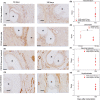
(A, C, E, G) Representative images of immunostained samples at 60 and 180 days post‐implantation. Implant structures are represented by asterisks. (B, D, F, H) Scoring results for endothelial cells of the blood vessels (CD34), leukocytes (CD45), neurons (PGP9.5) and myofibroblast and smooth muscle cells (smooth α‐SMA) of poly‐4‐hydroxybutyrate (P4HB) vaginal explants at 60 and 180 days post‐implantation. At 60 days post‐implantation, n = 7 samples; at 180 days post‐implantation, n = 5 samples. Error bars represent means ± standard deviations (SD). A Kruskal–Wallis statistical test was used to test for differences between time points. Values differing significantly are indicated with asterisks: *p < 0.05
FIGURE 8
FIGURE 8
Representative images of immunostained samples and scoring results for macrophage type 1 (HLA‐DR) and macrophage type 2 (CD163) of poly‐4‐hydroxybutyrate (P4HB) vaginal explants at 60 and 180 days post‐implantation. Implant structures are represented by asterisks in (A) and (C). At 60 days post‐implantation, n = 7 samples; at 180 days post‐implantation, n = 5 samples. Error bars represent means ± standard deviations (SD). A Kruskal–Wallis statistical test was used to test for differences between time points. Values differing significantly between time points are indicated with asterisks: *p < 0.05; **p < 0.01

Similar articles

Cited by

References

    1. Lakeman MM, van der Vaart CH, Laan E, Roovers JPW. The effect of prolapse surgery on vaginal sensibility. J Sex Med. 2011;8(4):1239–45. - PubMed
    1. Weber MA, Lakeman MM, Laan E, Roovers JPW. The effects of vaginal prolapse surgery using synthetic mesh on vaginal wall sensibility, vaginal vasocongestion, and sexual function: a prospective single‐center study. J Sex Med. 2014;11(7):1848–55. - PubMed
    1. Liang R, Abramowitch S, Knight K, Palcsey S, Nolfi A, Feola A, et al. Vaginal degeneration following implantation of synthetic mesh with increased stiffness. BJOG. 2013;120(2):233–43. - PMC - PubMed
    1. Hympanova L, Rynkevic R, Roman S, Mori da Cunha M, Mazza E, Zundel M, et al. Assessment of electrospun and ultra‐lightweight polypropylene meshes in the sheep model for vaginal surgery. Eur Urol Focus. 2020;6(1):190–8. - PubMed
    1. Birch C, Fynes MM. The role of synthetic and biological prostheses in reconstructive pelvic floor surgery. Curr Opin Obstet Gynecol. 2002;14(5):527–35. - PubMed