. 2021 Dec 5;27(1):153.
doi: 10.1186/s10020-021-00413-0.

Microfibrillar-associated protein 5 regulates osteogenic differentiation by modulating the Wnt/β-catenin and AMPK signaling pathways

Affiliations

Microfibrillar-associated protein 5 regulates osteogenic differentiation by modulating the Wnt/β-catenin and AMPK signaling pathways

Haoran Li et al. Mol Med. .

Abstract

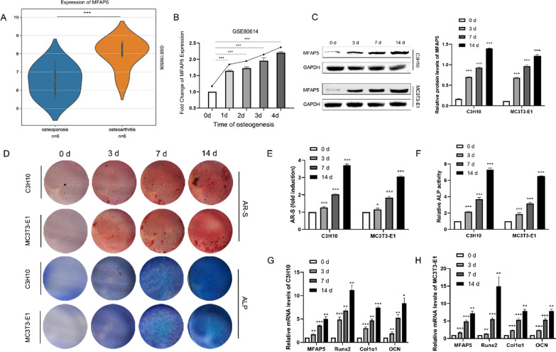
Background: Dysfunctional osteogenesis of bone marrow mesenchymal stem cells (BMSCs) plays an important role in osteoporosis occurrence and development. However, the molecular mechanisms of osteogenic differentiation remain unclear. This study explored whether microfibrillar-associated protein 5 (MFAP5) regulated BMSCs osteogenic differentiation.

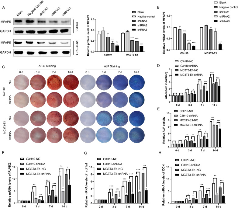
Methods: We used shRNA or cDNA to knock down or overexpress MFAP5 in C3H10 and MC3T3-E1 cells. AR-S- and ALP-staining were performed to quantify cellular osteogenic differentiation. The mRNA levels of the classical osteogenic differentiation biomarkers Runx2, Col1α1, and OCN were quantified by qRT-PCR. Finally, we employed Western blotting to measure the levels of Wnt/β-catenin and AMPK signaling proteins.

Results: At days 0, 3, 7, and 14 after osteogenic induction, AR-S- and ALP-staining was lighter in MFAP5 knockdown compared to control cells, as were the levels of Runx2, Col1α1 and OCN. During osteogenesis, the levels of β-catenin, p-GSK-3β, AMPK, and p-AMPK were upregulated, while that of GSK-3β was downregulated, indicating that Wnt/β-catenin and AMPK signaling were activated. The relevant molecules were expressed at lower levels in the knockdown than control group; the opposite was seen for overexpressing cell lines.

Conclusions: MFAP5 regulates osteogenesis via Wnt/β‑catenin- and AMPK-signaling; MFAP5 may serve as a therapeutic target in patients with osteoporosis.

Keywords: AMPK; Bone marrow mesenchymal stem cells (BMSCs); MFAP5; Osteogenesis; Osteoporosis; Wnt/β-catenin.


Conflict of interest statement

The authors declare that they have no competing interests.

Figures

Fig. 1
Fig. 1
MFAP5 expression was associated with osteogenesis. A MFAP5 expression was lower in the BMSCs of patients with osteoporosis compared to those with osteoarthritis. B Osteogenic induction of human mesenchymal stromal cells (hMSCs) at 0, 1, 2, 3, and 4 days showed that the level of mRNA encoding MFAP5 was positively associated with osteogenesis. C The expression patterns of MFAP5 in C3H10 and MC3T3-E1 cells during osteogenic differentiation, as revealed by Western blotting and the quantitative results of Western blotting. D Mineralized nodules and ALP expression revealed by AR-S and ALP staining during the osteogenesis of C3H10 and MC3T3-E1 cells. E The relative ALP expression levels of the two cell lines. F Quantitation of AR-S staining of C3H10 and MC3T3-E1 cells. G and H The levels of mRNAs encoding MFAP5, Runx2, Col1α1, and OCN of both cell types during osteogenesis. Values are expressed as means ± standard deviations (*P < 0.05, **P < 0.01, ***P < 0.001)
Fig. 2
Fig. 2
MFAP5 knockdown inhibited osteogenesis. A The knockdown efficiency of the three shRNAs; protein levels were quantified by Western blotting. B The mRNA level of MFAP5 in C3H10 and MC3T3-E1 cells, as revealed by qRT-PCR. C AR-S and ALP staining showed that MFAP5 knockdown inhibited the osteogenic capacity of C3H10 and MC3T3-E1 cells. D and E Quantitative analyses of ALP expression and AR-S staining. FH The mRNA levels of classical biomarkers of osteogenic differentiation (Runx2, Col1α1, and OCN) in the shRNA and NC groups. Values are expressed as means ± standard deviations (*P < 0.05, **P < 0.01, ***P < 0.001)
Fig. 3
Fig. 3
MFAP5 knockdown inhibited osteogenesis by affecting the Wnt/β-catenin and AMPK signaling pathways. A These pathways were activated in C3H10 and MC3T3-E1 cells during osteogenic induction; the levels of MFAP5, β-catenin, p-GSK-3β, AMPK, and p-AMPK increased, while that of GSK-3β decreased, as osteogenic induction proceeded. Wnt/β-catenin and AMPK signaling were inhibited in shRNA-transformed C3H10 and MC3T3-E1 cells throughout osteogenic differentiation. BD Relative protein levels of β-catenin, p-GSK-3β/ GSK-3β and p-AMPK/AMPK
Fig. 4
Fig. 4
MFAP5 overexpression promoted osteogenesis, which was mediated by the Wnt/β-catenin and AMPK signaling pathways. AB C3H10 cells overexpressed MFAP5 in protein and mRNA. C The C3H10-cDNA group had more calcified nodules and higher ALP expression than the other groups during osteogenic induction. DE The ALP activity level and AR-S staining revealed that the osteogenic capacity of the two groups differed. F The MFAP5, β-catenin, p-GSK-3β, GSK-3β, AMPK, and p-AMPK expression levels indicated that the Wnt/β-catenin and AMPK signaling pathways were more highly activated in the C3H10-cDNA group. GI Relative protein levels of β-catenin, p-GSK-3β/ GSK-3β and p-AMPK/AMPK. JL The levels of the classical osteogenic biomarkers Runx2, Col1α1, and OCN in the C3H10-NC and C3H10-cDNA groups. Values are expressed as means ± standard deviations (*P < 0.05, **P < 0.01, ***P < 0.001)

Similar articles

Cited by

References

    1. Albig AR, Becenti DJ, Roy TG, Schiemann WP. Microfibril-associate glycoprotein-2 (MAGP-2) promotes angiogenic cell sprouting by blocking notch signaling in endothelial cells. Microvasc Res. 2008 doi: 10.1016/j.mvr.2008.01.001. - DOI - PMC - PubMed
    1. Ann E-J, et al. Inhibition of Notch1 signaling by Runx2 during osteoblast differentiation. J Bone Miner Res. 2011;26:317–330. doi: 10.1002/jbmr.227. - DOI - PubMed
    1. Ao Q, et al. Fibrin Glue/Fibronectin/Heparin-based delivery system of BMP2 induces osteogenesis in MC3T3-E1 cells and bone formation in rat calvarial critical-sized defects. ACS Appl Mater Interfaces. 2020;12:13400–13410. doi: 10.1021/acsami.0c01371. - DOI - PubMed
    1. Augusciak-Duma A, et al. Mutations in the COL1A1 and COL1A2 genes associated with osteogenesis imperfecta (OI) types I or III. Acta Biochim Pol. 2018;65:79–86. doi: 10.18388/abp.2017_1612. - DOI - PubMed
    1. Black DM, Rosen CJ. Clinical practice. Postmenopausal osteoporosis. N Engl J Med. 2016;374:254–262. doi: 10.1056/NEJMcp1513724. - DOI - PubMed

Publication types