. 2021 Dec 5;12(1):599.
doi: 10.1186/s13287-021-02665-3.

Optogenetically controlled human functional motor endplate for testing botulinum neurotoxins

Affiliations

Optogenetically controlled human functional motor endplate for testing botulinum neurotoxins

Juliette Duchesne de Lamotte et al. Stem Cell Res Ther. .

Abstract

Background: The lack of physiologically relevant and predictive cell-based assays is one of the major obstacles for testing and developing botulinum neurotoxins (BoNTs) therapeutics. Human-induced pluripotent stem cells (hiPSCs)-derivatives now offer the opportunity to improve the relevance of cellular models and thus the translational value of preclinical data.

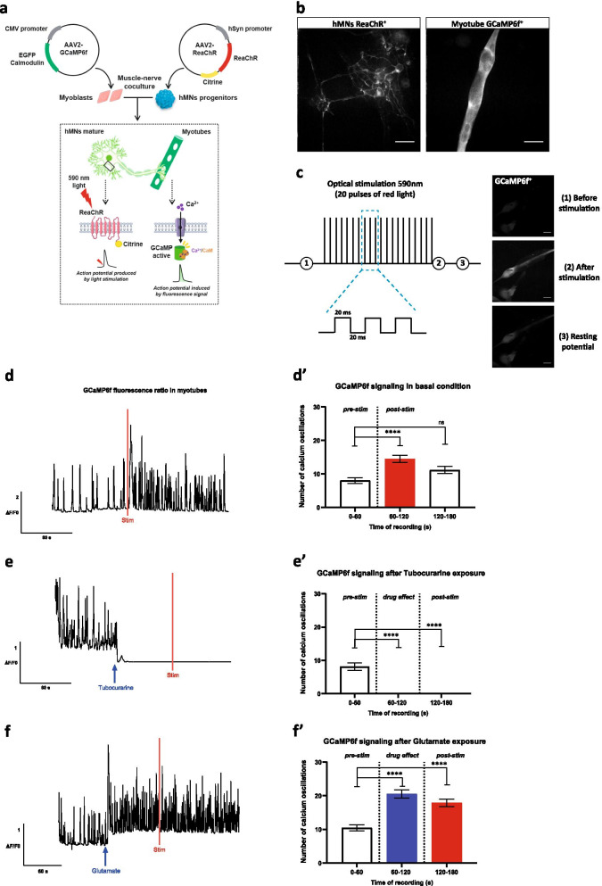
Methods: We investigated the potential of hiPSC-derived motor neurons (hMNs) optical stimulation combined with calcium imaging in cocultured muscle cells activity to investigate BoNT-sensitivity of an in vitro model of human muscle-nerve system.

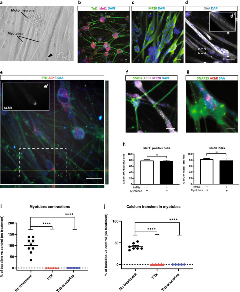
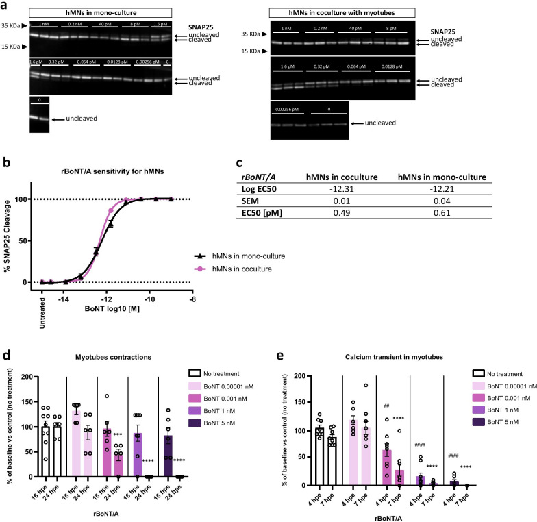
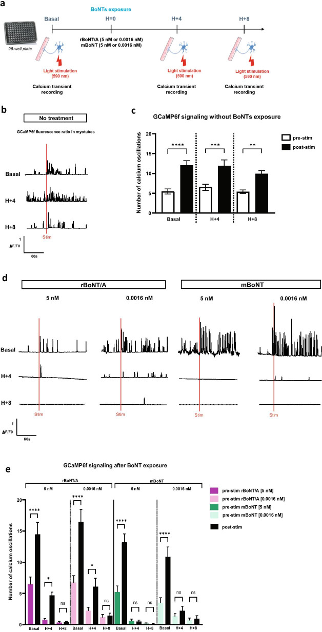
Results: Functional muscle-nerve coculture system was developed using hMNs and human immortalized skeletal muscle cells. Our results demonstrated that hMNs can innervate myotubes and induce contractions and calcium transient in muscle cells, generating an in vitro human motor endplate showing dose-dependent sensitivity to BoNTs intoxication. The implementation of optogenetics combined with live calcium imaging allows to monitor the impact of BoNTs intoxication on synaptic transmission in human motor endplate model.

Conclusions: Altogether, our findings demonstrate the promise of optogenetically hiPSC-derived controlled muscle-nerve system for pharmaceutical BoNTs testing and development.

Keywords: Botulinum neurotoxins; Calcium indicators; Functional; Human-induced pluripotent stem cells; Motor endplate; Optogenetics.


Conflict of interest statement

The authors declare that they have no competing interests. CN and JDL are IPSEN employees.

Figures

Fig. 1
Fig. 1
Generation of an in vitro functional muscle-nerve coculture system at DIV 15. a Representative images of the muscle-nerve coculture where hMNs neurites made contacts with myotubes (black triangle in the phase contrast image). b hMNs express Islet and Tuj1. c, d/d’ Myotubes express MF20 and SAA. e/e’g The coculture system expresses SYN (right and lower panels represent cross sections of myotubes in the orthogonal view), SMI32, SNAP25 and presents AChR clusters. ag Scale bars: 100 µm. h Quantification of Islet1+ hMNs and quantification of fusion index in myotubes. Data are represented as mean ± SD (N = 3 independent experiments, each performed in triplicate n = 3). Mann–Whitney test (ns., not significant). i Disruption of myotubes contractions after treatment with 5 µM TTX and 150 µM tubocurarine compared to control condition with no treatment. Each drug was added at DIV 15, the recording of myotubes contractions was performed before treatment (baseline) and 30 min after treatment. j Disruption of Ca2+ oscillations in myotubes after treatment with 5 µM TTX and 150 µM tubocurarine compared to control condition with no treatment. For Ca2+, measurement cells were stained with 2 µM Cal520 dye the day of the recording. Each drug was added at DIV 15, the recording of Ca2+ oscillations was performed before treatment (baseline) and 30 min after treatment. i, j Data are represented as mean ± SEM (N = 3 independent experiments, each performed in triplicate n = 3). ANOVA with Sidak’s post hoc tests (****p < 0.0001)
Fig. 2
Fig. 2
Effect of botulinum neurotoxin on muscle-nerve coculture function. a Detection by Western blot of cleaved-SNAP25 from hMNs mono-culture (left) or coculture (right) treated with serial doses of rBoNT/A, compared to toxin-free control dose (untreated) using an antibody recognizing cleaved and uncleaved form of SNAP25 protein. b EC50 curve for hMNs in mono-culture or in coculture. DIV 15 hMNs were exposed to rBoNT/A for 24 h before cell lysates were harvested, followed by Western blot to quantify SNAP25 cleavage. c EC50 for hMNs in coculture is 0.49 pM (10–12.31) and EC50 for hMNs in mono-culture is 0.61 pM (10–12.21). Data are represented as mean ± SEM (N = 3 independent experiments, each performed in triplicate n = 3). d Effect of rBoNT/A on myotubes contractions 16 hpe (hour post-exposure) and 24 hpe. Coculture was treated at DIV 15 with different doses of rBoNT/A (5 nM, 1 nM, 0.001 nM, 0.00001 nM), and recordings of myotubes contractions were performed before treatment (baseline), 16 h and 24 h after treatment. Data are represented as mean ± SEM (N = 3 independent experiments, each performed in triplicate n = 3). ANOVA with Sidak’s post hoc tests (***p < 0.001; ****p < 0.0001). Significant statistics only 24 hpe, each dose was compared to untreated condition. e Effect of rBoNT/A on myotubes Ca2+ oscillations 4 hpe and 7 hpe. Cells were stained with 2 µM Cal520 dye the day of the recording. Coculture was treated at DIV 15 with different doses of rBoNT/A (5 nM, 1 nM, 0.001 nM, 0.00001 nM), and recordings of Ca2+ oscillations were performed before treatment (baseline), 4 h and 7 h after treatment. d-e Data are represented as mean ± SEM (N = 3 independent experiments, each performed in triplicate n = 3). ANOVA with Sidak’s post hoc tests (##p < 0.01; ####,****p < 0.0001). Significant statistics 4 hpe (represented by #) and 7 hpe (represented by *), each dose was compared to untreated condition
Fig. 3
Fig. 3
Effect of the optogenetic stimulation of hMNs on the myotubes Ca2+ dynamic. a ReaChR was transduced into hMNs progenitors to enable ion channel activation by light allowing for control of neuronal activity by red light stimulation (590 nm). Optogenetic activation was confirmed by reading the Ca2+ response generated in myoblasts transduced with GCaMP6f by quantifying fluorescent intensity. b AAV2-hSyn-ReaChR-citrine expression in hMNs for optogenetic control and AAV2-CMV-GCaMP6f-WPRE-SV40pA expression in myotubes. Scale bars: 50 µm. c Representative schema of the optical stimulation protocol at 590 nm: 20 red light pulses, each pulse was 20 ms long. Optogenetics activation was confirmed by GCaMP6f in myotubes by quantifying fluorescent intensity. Scale bars: 50 µm. d-d’ Representative traces of normalized GCaMP6f fluorescence before and after red light stimulation (red bar) and quantification of Ca2+ oscillations. Data are represented as mean ± SEM (N = 3 independent experiments, 3 myofibers/well, 3 wells/condition). ANOVA with Sidak’s post hoc tests (****p < 0.0001; ns, not significant), with the recording times 60–120 and 120–180 compared to the time 0–60. e-e’ Effect of the addition of 150 µM tubocurarine before and after red light stimulation (red bar) and quantification of Ca2+ oscillations. Data are represented as mean ± SEM (N = 3 independent experiments, 3 myofibers/well, 3 wells/condition). ANOVA with Sidak’s post hoc tests (****p < 0.0001), with the recording times 60–120 and 120–180 compared to the time 0–60. ff’ Effect of the addition of 10–50 µM range of glutamate before and after red light stimulation (red bar) and quantification of Ca2+ oscillations. Data are represented as mean ± SEM (N = 3 independent experiments, 3 myofibers/well, 3 wells/condition). ANOVA with Sidak’s post hoc tests (****p < 0.0001), with the recording times 60–120 and 120–180 compared to the time 0–60
Fig. 4
Fig. 4
BoNTs effects on optogenetic controlled muscle-nerve system. a Representative schema of the optogenetic procedure before (baseline/before treatment) and 4 h and 8 h after exposure of BoNTs (H + 4 and H + 8). The addition of both BoNTs was notified by H = 0. Cells were exposed to two doses (5 nM or 0.0016 nM) of rBoNT/A or mBoNT. b, c Representative traces of normalized GCaMP6f fluorescence in untreated condition before (pre-stim) and after (post-stim) red light stimulation (red bar) over the time and quantification of Ca2+ oscillations. Data are represented as mean ± SEM (N = 2 independent experiments, 3 myofibers/well, 3 wells/condition). ANOVA with Sidak’s post hoc tests (****p < 0.0001; ***p < 0.001; **p < 0.01), each pre-stim recordings were compared to post-stim recordings. d, e Representative traces of normalized GCaMP6f fluorescence at 5 nM rBoNT/A, 0.0016 nM rBoNT/A, 5 nM mBoNT and 0.0016 nM mBoNT treatments before and after red light stimulation (red bar) over the time and quantification of Ca2+ oscillations. Data are represented as mean ± SEM (N = 2 independent experiments, 3 myofibers/well, 3 wells/condition). ANOVA with Sidak’s post hoc tests (****p < 0.0001; *p < 0.05; ns, not significant), each pre-stim recordings were compared to post-stim recordings

Similar articles

Cited by

References

    1. Schiavo G, Matteoli M, Montecucco C. Neurotoxins affecting neuroexocytosis. Physiol Rev. 2000;80:717–766. doi: 10.1152/physrev.2000.80.2.717. - DOI - PubMed
    1. Popoff MR, Poulain B. Bacterial toxins and the nervous system: neurotoxins and multipotential toxins interacting with neuronal cells. Toxins. 2010;2:683–737. doi: 10.3390/toxins2040683. - DOI - PMC - PubMed
    1. Pirazzini M, Rossetto O, Eleopra R, Montecucco C. Botulinum neurotoxins: biology, pharmacology, and toxicology. Pharmacol Rev. 2017;69:200–235. doi: 10.1124/pr.116.012658. - DOI - PMC - PubMed
    1. Rossetto O. Botulinum toxins: molecular structures and synaptic physiology. In: Jabbari B, editor. Botulinum toxin treatment in clinical medicine. Cham: Springer International Publishing; 2018. pp. 1–12.
    1. Orsini M, Leite MAA, Chung TM, Bocca W, De Souza JA, De Souza OG, Moreira RP, Bastos VH, Teixeira S, Oliveira AB, et al. Botulinum neurotoxin type A in neurology: update. Neurol Int. 2015 doi: 10.4081/ni.2015.5886. - DOI - PMC - PubMed

Publication types

Substances