Two Cases of Hemorrhagic Ampullary Lesions Successfully Treated by Endoscopic Papillectomy
- PMID: 34866100
- PMCID: PMC9259823
- DOI: 10.2169/internalmedicine.8294-21
Two Cases of Hemorrhagic Ampullary Lesions Successfully Treated by Endoscopic Papillectomy
Abstract
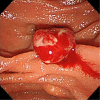
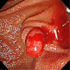
We herein report two cases of hemorrhagic ampullary lesions in which endoscopic papillotomy was performed to control bleeding and resulted in successful treatment. Both patients were pathologically diagnosed with an underlying pathology characterized by inflammatory cell infiltration and capillary proliferation. They also had disposing factors for bleeding, such as antithrombotic therapy and idiopathic thrombocytopenic purpura. Endoscopic treatment was selected because the risk of surgical resection was high due to the patients' hemorrhagic condition. Both patients were successfully treated without any serious adverse events and had an uneventful postoperative course with no relapse of bleeding.
Keywords: ampulla of Vater; ampullary lesion; bleeding; endoscopic papillectomy; hemorrhagic.
Conflict of interest statement
Figures


Similar articles
-
Ampullary neuroendocrine tumor diagnosed by endoscopic papillectomy in previously confirmed ampullary adenoma.World J Gastroenterol. 2016 Apr 7;22(13):3687-92. doi: 10.3748/wjg.v22.i13.3687. World J Gastroenterol. 2016. PMID: 27053861 Free PMC article.
-
Clinical outcomes of endoscopic papillectomy of ampullary adenoma: A multi-center study.World J Gastroenterol. 2022 May 7;28(17):1845-1859. doi: 10.3748/wjg.v28.i17.1845. World J Gastroenterol. 2022. PMID: 35633905 Free PMC article.
-
Therapeutic outcomes of endoscopic papillectomy for ampullary neoplasms: retrospective analysis of a multicenter study.BMC Gastroenterol. 2017 May 30;17(1):69. doi: 10.1186/s12876-017-0626-5. BMC Gastroenterol. 2017. PMID: 28558658 Free PMC article.
-
Three Cases of Ampullary Neuroendocrine Tumor Treated by Endoscopic Papillectomy: A Case Report and Literature Review.Intern Med. 2020 Oct 1;59(19):2369-2374. doi: 10.2169/internalmedicine.4568-20. Epub 2020 Jun 30. Intern Med. 2020. PMID: 32611953 Free PMC article. Review.
-
Successful treatment for ampullary submucosal bleeding-induced pancreatitis: a rare sequla of hereditary hemorrhagic telangiectasia.Hepatogastroenterology. 2005 Jan-Feb;52(61):270-3. Hepatogastroenterology. 2005. PMID: 15783047 Review.
References
-
- Yamamoto K, Itoi T, Sofuni A, et al. . Expanding the indication of endoscopic papillectomy for T1a ampullary carcinoma. Dig Endosc 31: 188-196, 2019. - PubMed
-
- Niido T, Itoi T, Harada Y, et al. . Carcinoid of major duodenal papilla. Gastrointest Endosc 61: 106-107, 2005. - PubMed
-
- Suzuki K, Kantou U, Murakami Y. Two cases with ampullary cancer who underwent endoscopic excision. Prog Dig Endosc 23: 236-239, 1983.

