. 2022 Feb;18(6):e2105640.
doi: 10.1002/smll.202105640. Epub 2021 Dec 5.

Extracellular Vimentin as a Target Against SARS-CoV-2 Host Cell Invasion

Affiliations

Extracellular Vimentin as a Target Against SARS-CoV-2 Host Cell Invasion

Łukasz Suprewicz et al. Small. 2022 Feb.

Abstract

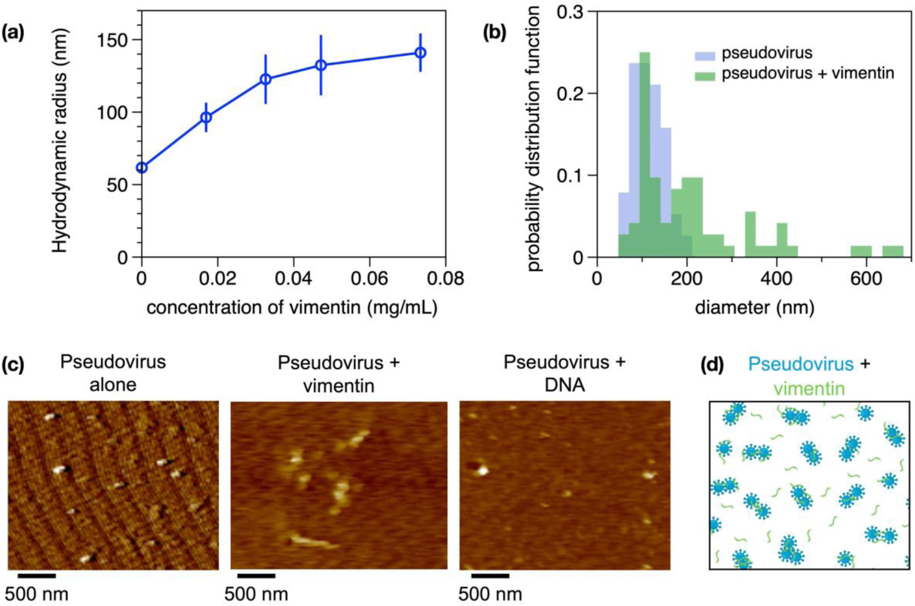
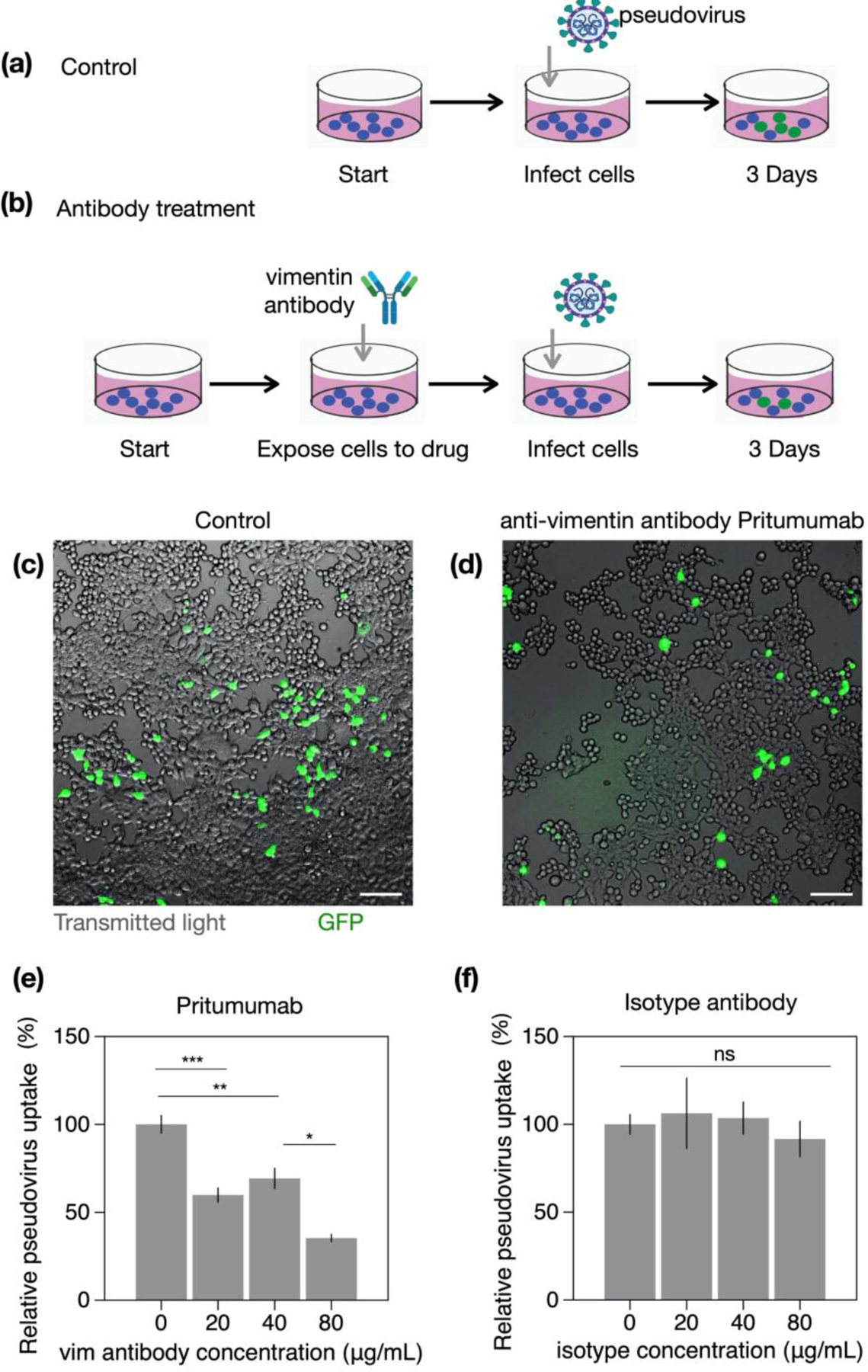
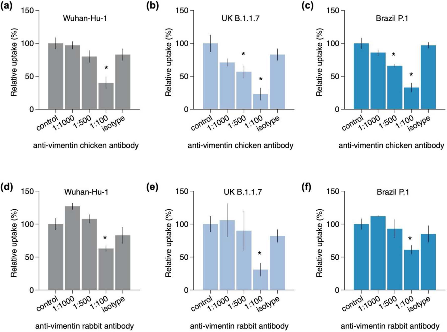
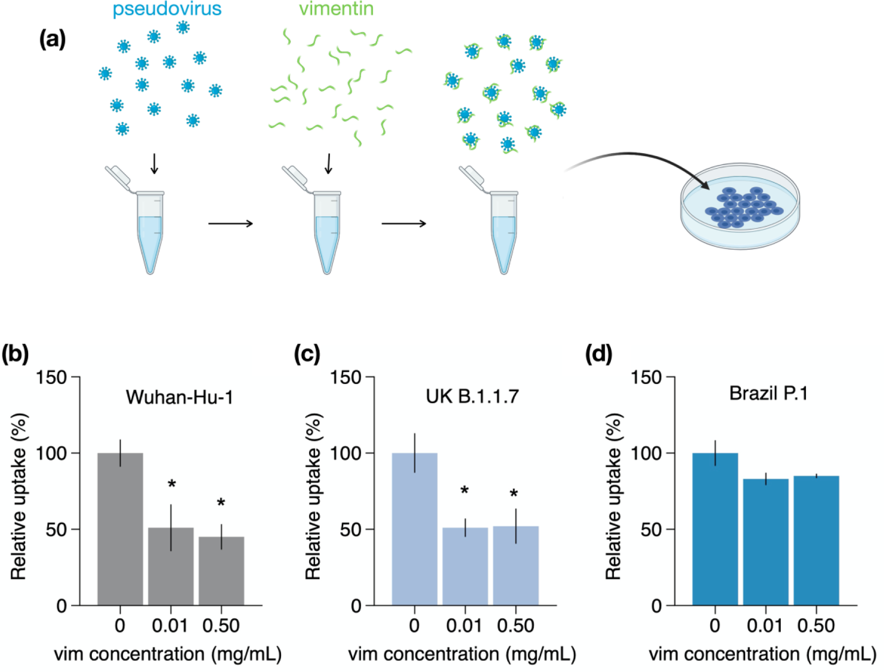
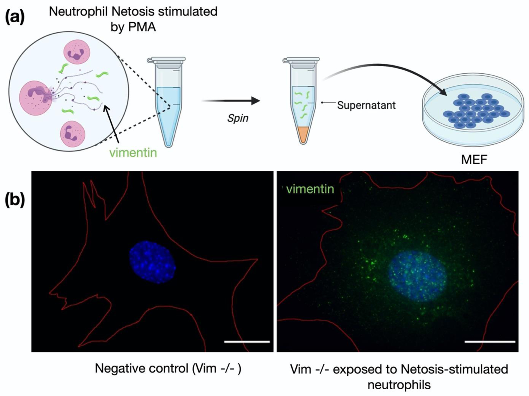
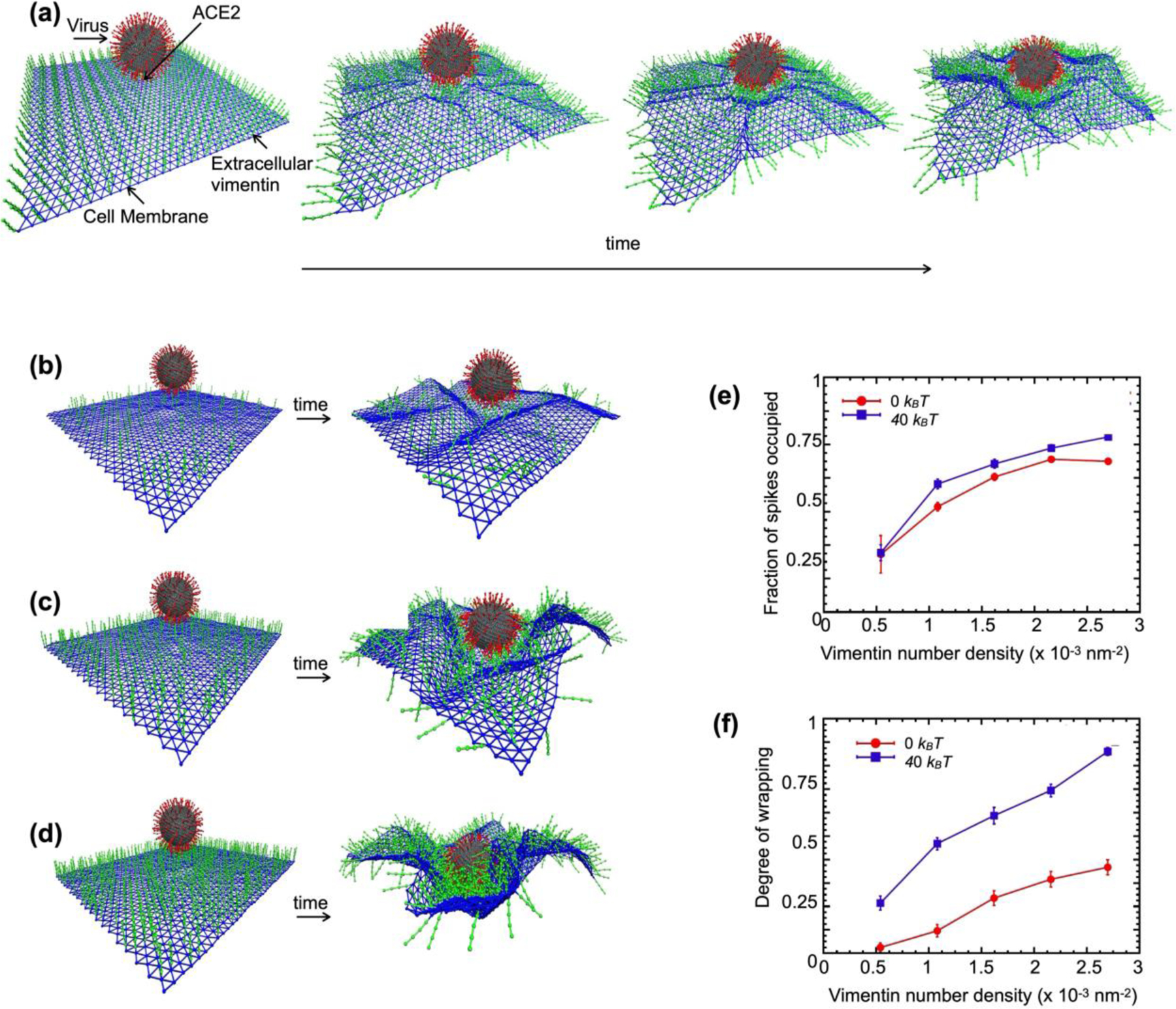
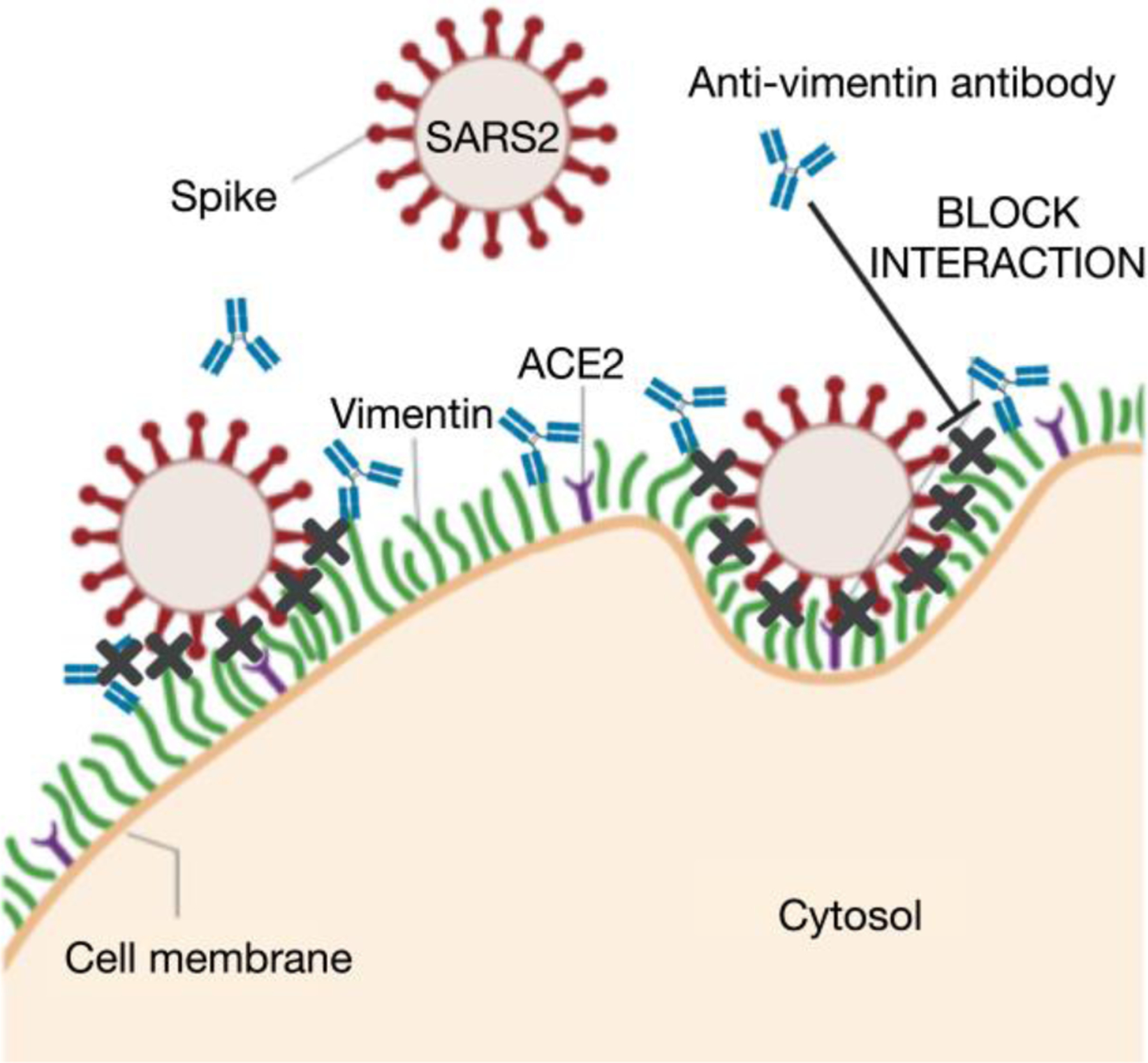
Infection of human cells by pathogens, including SARS-CoV-2, typically proceeds by cell surface binding to a crucial receptor. The primary receptor for SARS-CoV-2 is the angiotensin-converting enzyme 2 (ACE2), yet new studies reveal the importance of additional extracellular co-receptors that mediate binding and host cell invasion by SARS-CoV-2. Vimentin is an intermediate filament protein that is increasingly recognized as being present on the extracellular surface of a subset of cell types, where it can bind to and facilitate pathogens' cellular uptake. Biophysical and cell infection studies are done to determine whether vimentin might bind SARS-CoV-2 and facilitate its uptake. Dynamic light scattering shows that vimentin binds to pseudovirus coated with the SARS-CoV-2 spike protein, and antibodies against vimentin block in vitro SARS-CoV-2 pseudovirus infection of ACE2-expressing cells. The results are consistent with a model in which extracellular vimentin acts as a co-receptor for SARS-CoV-2 spike protein with a binding affinity less than that of the spike protein with ACE2. Extracellular vimentin may thus serve as a critical component of the SARS-CoV-2 spike protein-ACE2 complex in mediating SARS-CoV-2 cell entry, and vimentin-targeting agents may yield new therapeutic strategies for preventing and slowing SARS-CoV-2 infection.

Keywords: SARS-CoV2; cell membranes; endocytosis; extracellular vimentin; pseudoviruses; spike proteins.


Conflict of interest statement

Competing Interests:

The authors declare no competing interests.

Figures

Figure 1.
Figure 1.. Presence of extracellular vimentin in human lung, airway fluids, and fat tissue.
(a) Positive staining for extracellular vimentin (green) in human lung, fat tissue, and sputum obtained from cystic fibrosis (CF) patients. Vimentin appears on the apical side of type I and type II pneumocytes. DNA stained with DAPI. (b) There are numerous internal and exogenous pathways by which vimentin may be found in lung epithelia and other tissues, in either intracellular or cell surface forms (shown as green filaments). Vimentin is expressed directly by mesenchymal cells, cells having undergone EMT, cancer cells, senescent fibroblasts, and interestingly by cells bound and infected by the SARS-CoV virus (see Table 1). Exogenous sources of vimentin are largely related to immune response and tissue injury in the form of vimentin exported by neutrophils, T-lymphocytes, monocytes/macrophages, and exosomes. Schematics generated with Biorender.com.
Figure 2.
Figure 2.. Binding of vimentin to SARS-CoV-2 pseudovirus.
Purified human recombinant vimentin was added to suspensions of SARS-CoV-2 spike protein-containing pseudovirus, and their size was measured by dynamic light scattering (a) and atomic force microscopy (b and c). The size of the pseudovirus as measured by DLS increased from 60 nm to 150 nm after addition of 0.07 mg/ml vimentin (panel a). Panel c: The pseudoviruses were imaged using atomic force microscopy before and after addition of either vimentin (0.07 mg/ml) or DNA (0.08 mg/mL). The probability distribution functions (b) show that the average size of pseudovirus imaged by AFM confirms the change in size detected by DLS. (d) Schematic representation indicating how binding of vimentin to SARS-CoV-2 spike protein might couple particles together and increase their effective radii. Error bars denote standard deviation.
Figure 3.
Figure 3.. Intracellular and extracellular vimentin expression in mouse embryonic fibroblasts (MEF) and HEK-293T-hsACE2.
(a-b) MEF stained for vimentin either (a) intracellularly with triton permeabilization before exposure to vimentin antibodies or (b) extracellularly, where cells were exposed to anti-vimentin antibodies before fixation, then permeabilized and stained for actin. Images show vimentin (green), actin (red), and DNA (blue). (c-d) Positive staining for intracellular and extracellular vimentin was detected in human kidney epithelial cells HEK 293T-hs ACE2.
Figure 4.
Figure 4.. Distribution of intracellular and extracellular vimentin in mouse embryonic fibroblasts (MEF).
Super resolution confocal images showing MEF cells exposed first to a primary anti-vimentin antibody (human, Pritumumab) to bind to vimentin on the extracellular surface of the cells, then fixed, permeabilized and exposed to a second primary anti-vimentin antibody (chicken, NOVUS) to bind to intracellular vimentin. This immunofluorescence technique reveals the two distinct organization patterns of intracellular cytoskeletal vimentin and extracellular vimentin. Scale bar; 10 μm.
Figure 5.
Figure 5.. Anti-vimentin antibodies block uptake of Wuhan-Hu-1 SARS-CoV-2 pseudovirus in HEK 293T-hsACE2.
(a) HEK 293T-hsACE were exposed to pseudovirus particles bearing the SARS-CoV-2 spike protein and a GFP reporter. Cells were imaged three days after exposure to detect the number of cells expressing GFP, indicating the number of cells transfected by pseudoviruses. (b) In the antibody treatment case, cells were pre-exposed to anti-vimentin antibodies before infection by the pseudovirus. (c-d) Fluoresence images showing cells expressing GFP after pseudovirus exposure with and without Pritumumab treatment. Scale bar; 100 μm. (e) Pritumumab inhibits cellular infection by up to 60%. (f) Use of an isotype antibody does not inhibit infection, suggesting a specific interaction between the SARS-CoV-2 spike protein and extracellular vimentin. Error bars denote standard deviation. Denotations: *, P ≤ 0.05; **, P < 0.01; ***, P < 0.001; NS, P > 0.05.
Figure 6.
Figure 6.. Effect of vimentin antibodies on cellular uptake of pseudoviruses bearing spike protein from three different strains of SARS-CoV-2.
The effect of (a-c) chicken polyclonal anti-vimentin antibodies (C-terminal, multiple epitopes) and (d-f) rabbit monoclonal (against C-terminal 425–466) and their respective isotypes were tested in the uptake of pseudo-virus baring spike proteins from three different strains: Wuhan-Hu-1, UK B.1.1.7, and Brazil P.1. Isotypes were diluted 1:100. Experiments were conducted in HEK 293T-hsACE and measured via plate readers (Methods). Error bars denote standard deviation. Denotations: *, P ≤ 0.05.
Figure 7.
Figure 7.. Effect of soluble vimentin on SARS-CoV-2 pseudovirus uptake in HEK293T hsACE2.
The effect of vimentin itself on SARS-CoV-2 pseudovirus uptake was assessed by pre-exposing pseudoviruses with recombinant human vimentin (NOVUS Biological) for 20 minutes before their addition to HEK 293T-hsACE2 cells. (a) Schematic of vimentin neutralization experiment. Vimentin blocked approximately 50% of uptake in (b) the native Wuhan-Hu-1 strain and (c) the UK B.1.1.7 variant, but did not block uptake in (d) the Brazil P.1. variant. Error bars denote standard deviation. Denotations: *, P ≤ 0.05.
Figure 8.
Figure 8.. Acquisition of cell surface vimentin from extracellular environment.
(a) To determine whether vimentin released from neutrophil NETosis could be acquired on the surface of other cell types, we stimulated neutrophil NETosis with phorbol myristate acetate (PMA), centrifuged, and collected the supernatant. The neutrophil-stimulated supernatant was then presented to vimentin-null (vim−/−) MEF, which were subsequently fixed and stained to detect acquired vimentin. (b) Immunofluorescence images of vimentin-null mEF staining positive for extracellular vimentin after exposure to supernatant of NETosis-activated neutrophils, indicating the acquisition of extracellular vimentin by cells that do not express vimentin. Cell boundary is marked in red. Scale bar, 20 µm. Schematics generated with Biorender.com.
Figure 9.
Figure 9.. Vimentin’s involvement in spike-ACE2 interactions.
(a) Cell surface vimentin acts as a co-receptor that enhances binding to the SARS-CoV-2 virus in either direct fusion or endocytic pathways to enhance wrapping and endocytosis of the virus. A molecular dynamics simulation of the SARS2-CoV-2 virus attached to a cell membrane shows that binding of extracellular vimentin with the virus spike protein facilitates wrapping of cell membrane around the virus. Snapshots are shown at t = 0, 170, 330, and 500 ms. (b-d) Representative snapshots of simulations with cell surface vimentin densities of (b) 0.5 × 10⁻³ (b), (c) 1.6 x x 10−3, and (d) 2.7 × 10⁻³ nm-2. First (t=0ms) and final frame (t=500ms) are shown. Both the fraction of spikes bound to surface vimentin (e) and the degree of membrane wrapping (f) increases as the number density of surface vimentin increases. Finite membrane bending rigidity (40 kBT, blue squares) enhances wrapping compared to the case without bending rigidity (0 kBT, red circles).
Figure 10.
Figure 10.. Extracellular vimentin as a potential target for inhibiting SARS-CoV-2 entry.
A diagram of proposed mechanism of action. Here, cell surface vimentin (green) acts as a co-receptor that binds to SARS-CoV-2 spike protein. Blocking this interaction via the anti-vimentin antibodies reduces cell surface binding of the virus and cellular infection. Schematics generated with Biorender.com.

Update of

Similar articles

Cited by

References

    1. Wrapp D; Wang N; Corbett KS; Goldsmith JA; Hsieh CL; Abiona O; Graham BS; McLellan JS, Cryo-EM structure of the 2019-nCoV spike in the prefusion conformation. Science 2020, 367 (6483), 1260–1263. - PMC - PubMed
    1. Walls AC; Park Y-J; Tortorici MA; Wall A; McGuire AT; Veesler D, Structure, function, and antigenicity of the SARS-CoV-2 spike glycoprotein. Cell 2020. - PMC - PubMed
    1. Synowiec A; Szczepański A; Barreto-Duran E; Lie LK; Pyrc K, Severe Acute Respiratory Syndrome Coronavirus 2 (SARS-CoV-2): a Systemic Infection. Clinical Microbiology Reviews 2021, 34 (2), e00133–20. - PMC - PubMed
    1. Hikmet F; Méar L; Edvinsson Å; Micke P; Uhlén M; Lindskog C, The protein expression profile of ACE2 in human tissues. Mol Syst Biol 2020, 16 (7), e9610. - PMC - PubMed
    1. Aguiar JA; Tremblay BJ-M; Mansfield MJ; Woody O; Lobb B; Banerjee A; Chandiramohan A; Tiessen N; Cao Q; Dvorkin-Gheva A; Revill S; Miller MS; Carlsten C; Organ L; Joseph C; John A; Hanson P; Austin RC; McManus BM; Jenkins G; Mossman K; Ask K; Doxey AC; Hirota JA, Gene expression and in situ protein profiling of candidate SARS-CoV-2 receptors in human airway epithelial cells and lung tissue. European Respiratory Journal 2020, 56 (3), 2001123. - PMC - PubMed

Publication types

LinkOut - more resources