. 2021 Dec;28(12):7105-7116.
doi: 10.1016/j.sjbs.2021.08.003. Epub 2021 Aug 8.

Acaudina molpadioides mediates lipid uptake by suppressing PCSK9 transcription and increasing LDL receptor in human liver cells

Affiliations

Acaudina molpadioides mediates lipid uptake by suppressing PCSK9 transcription and increasing LDL receptor in human liver cells

Allicia Jack et al. Saudi J Biol Sci. 2021 Dec.

Abstract

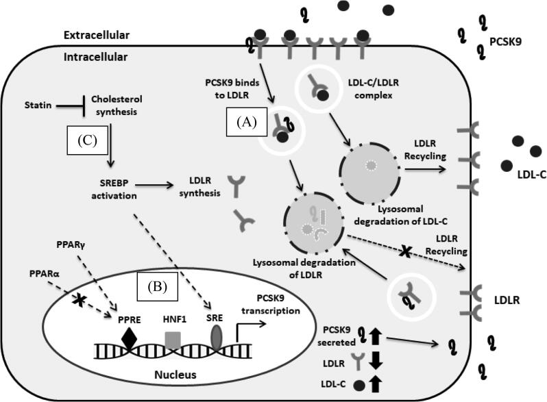
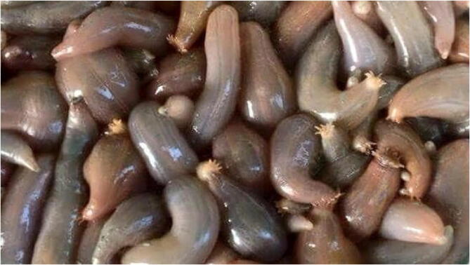
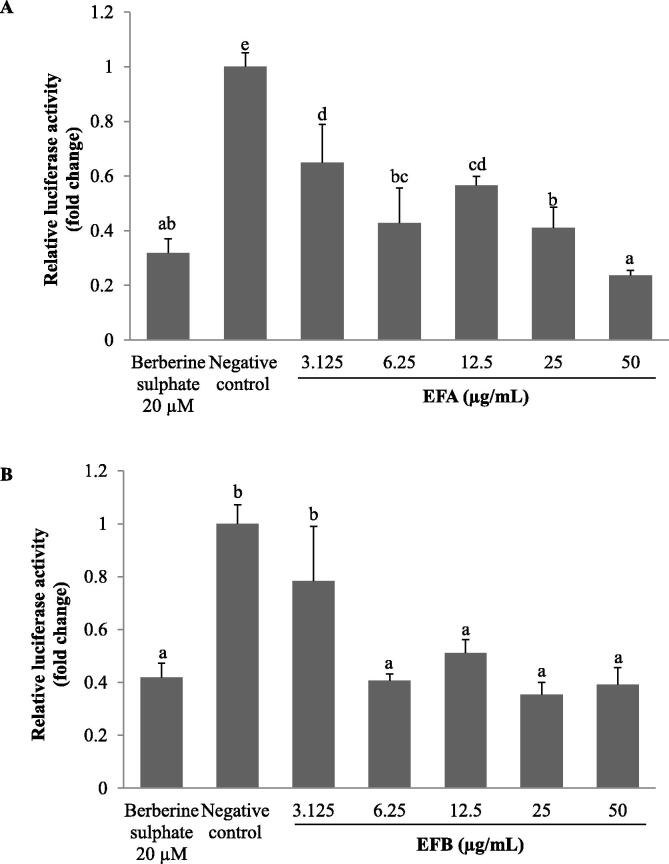
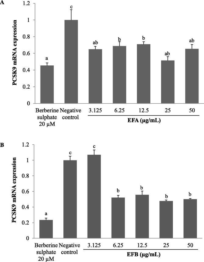
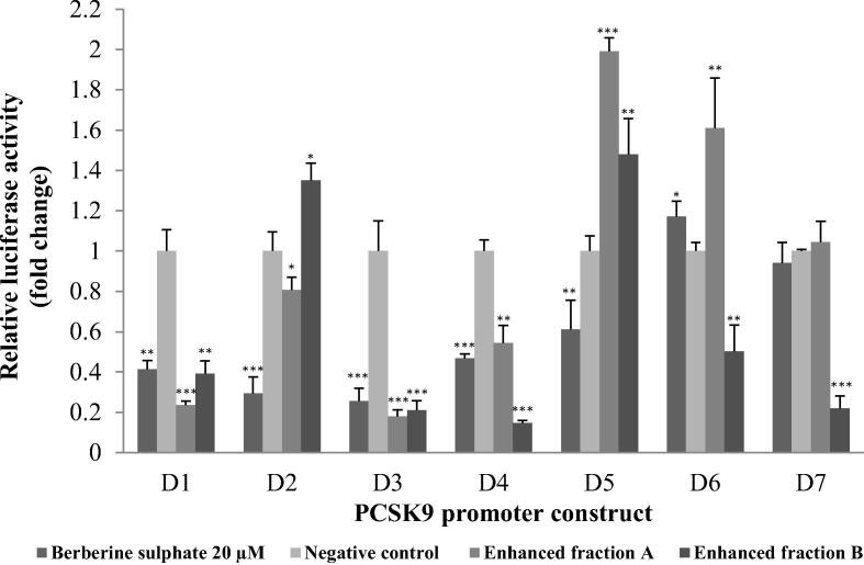
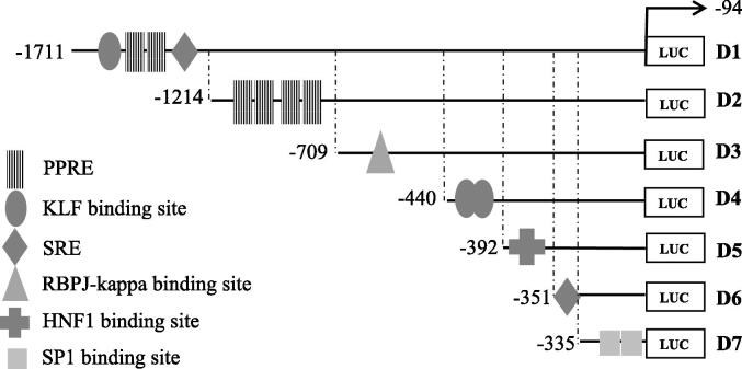
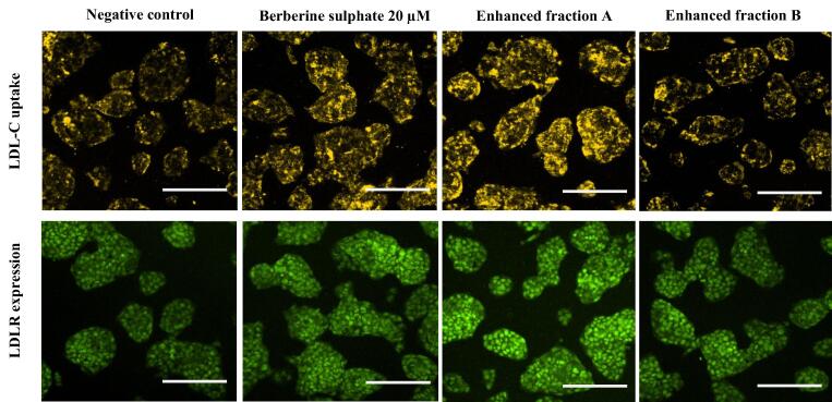
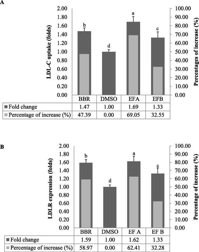
Acaudina molpadioides has been long used as traditional medicinal resources and reported to demonstrate various important bioactivities such as anticoagulation, antithrombosis, anti-hyperglycemia and anticancer. However, its lipid lowering activity is yet to be fully explored. Proprotein convertase subtilisin/kexin type 9 (PCSK9) is an enzyme that enhances the lysosomal degradation of hepatic low density lipoprotein receptor (LDLR) resulting in excessive accumulation of the plasma levels of LDL-cholesterols (LDL-C) which subsequently accelerate atherosclerosis. In the present study, A. molpadioides fractions were subjected to promoter-reporter luciferase assay to determine its role as PCSK9 inhibitors. It was found both fractions (EFA and EFB) reduced the transcriptional activity of PCSK9 promoter. Among the seven 5'end deletion constructs of PCSK9 promoter, fragments D1 (-1,711/-94), D3 (-709/-94) and D4 (-440/-94), were suppressed in the presence of both fractions whereas D2 (-1,214/-94), and, D6 (-351/-94) as well as D7 (-335/-94) were inhibited only by EFA and EFB, respectively. Further transcription factor binding sites prediction using MatInspector software discovered various potential cis-regulatory elements namely, PPAR, KLFs, RBPJ-kappa and SREBP that may potentially be involved in ameliorating the transcriptional activity of PCSK9. Immunofluorescence staining was used to evaluate the effects of both fractions on LDL-C and LDLR. Results showed that levels of LDL-C uptake in EFA-treated cells were 69.1% followed by EFB at 32.6%, as compared to untreated control after 24 h treatment. The LDLR protein distribution was induced by 62.41% and 32.2%, which corresponded to an increase in LDL-C uptake in both EFA and EFB treatment, respectively. Hence, the inhibition of PCSK9 by bioactive compounds in EFA and EFB could be another promising therapeutic agent in reducing the cholesterol levels and atherosclerosis by targeting PCSK9.

Keywords: Acaudina molpadioides; Atherosclerosis; Low density lipoprotein; PCSK9; Promoter-reporter based assay.


Conflict of interest statement

The authors declare that they have no known competing financial interests or personal relationships that could have appeared to influence the work reported in this paper.

Figures

Fig. 1
Fig. 1
The correlation of PCSK9 and statin in regulating the metabolism of LDL-C in the PCSK9 and LDLR are synthesized in the liver. The secreted PCSK9 protein binds to the extracellular domain of LDLR at the cell surface. The PCSK9/LDLR-LDL-C complex enters via the endosomal pathway and is directed to the lysosomal compartment for degradation of both PCSK9 and LDLR, decreasing the number of LDLRs available for clearance of LDL-C particles (A). The gene expression of PCSK9 is subjected to the regulation by the binding of SREBP2, HNF1 and PPAR to their corresponding binding sites on PCSK9 promoter (B). Activation of SREBP2, under conditions of intracellular cholesterol depletion due to inhibitory activity of statin, increases the expression of both PCSK9 and LDLR (C).
Fig. 2
Fig. 2
Acaudina molpadioides is an invertebrate marine organism, commonly known as sea potato and belongs to the Holothuroidea family. It is widely consumed by locals in South Eastern Asian countries as traditional healthy food. The figure shows sea potato that was collected from the intertidal zone of Pulau Langkawi, Kedah, Malaysia.
Fig. 3
Fig. 3
The effects of A. molpadioides enhanced fractions, EFA (A) and EFB (B) on human proprotein convertase subtilisin/kexin type 9 (PCSK9) promoter activity. HepG2 cells were transfected with a recombinant pGL3 reporter plasmid comprising the human PCSK9 promoter sequence linked to firefly luciferase gene. The transfected cells were then treated with various concentrations of EFA and EFB for 24 h, and subsequently subjected to luciferase assay. The transcriptional activity of PCSK9 promoter was decreased in dose dependent manner in both EFA and EFB (except at 3.125 μg/mL)-treated HepG2 cells with 20 µM berberine sulphate as positive control and 1% (v/v) dimethyl sulfoxide (DMSO) as negative control. Each value represents the mean ± SD of three independent experiments (each in triplicate reaction). Multiple comparisons were done using one-way analysis of variance (ANOVA) where means with different letters were separated with Duncan’s multiple range test group at p < 0.05.
Fig. 4
Fig. 4
The effect of A. molpadioides fractions, EFA (A) and EFB (B) on proprotein convertase subtilisin/kexin type 9 (PCSK9) mRNA expression. HepG2 cells were cultured until 80% confluence prior to treatment with EFA and EFB, 20 µM berberine sulphate as positive control and 1% (v/v) dimethyl sulfoxide (DMSO) as negative control, for 24 h. Total cellular RNA was isolated and subjected to real time-PCR analysis. Both EFA and EFB produced the lowest PCSK9 mRNA level at 25 µg/mL. Multiple comparisons were done using one-way analysis of variance (ANOVA) where means with different letters were separated with Duncan’s multiple range test group at p < 0.05.
Fig. 5
Fig. 5
Proprotein convertase subtilisin/kexin type 9 (PCSK9) promoter constructs analysis. HepG2 cells that were transfected with each fragment (D1-D7) was treated for 24 h with 25 µg/mL of A. molpadiodes EFA and EFB, with 20 µM berberine sulphate as positive control and 1% (v/v) dimethyl sulfoxide (DMSO) as negative control. Both EFA and EFB significantly decreased the transcriptional activity of promoter fragments D1(-1711/-94 bp), D3(-709/-94 bp) and D4(-440/-94 bp) which indicated that cis-acting elements presence in between these fragment regions were responsible in mediating the inhibitory effect of A. molpadioides on PCSK9 transcriptional activity. Values are means ± standard deviation and unpaired t-test was perform with * p < 0.05; **p < 0.001 and ***p < 0.001.
Fig. 6
Fig. 6
Schematic of the distribution of the predicted potential transcription binding sites on the PCSK9 promoter. The transcriptional start site (TSS) is indicated by the arrow. MatInspector software was used to predict transcription factor binding sites on the PCSK9 promoter. PPRE, peroxisome proliferator response element ; KLF, Krüppel like factor; SRE, sterol regulatory element; RBPJ-kappa, recombination signal binding protein for immunoglobin kappa J; HNF1, hepatocyte nuclear factor 1; SP1, specificity protein 1.
Fig. 7
Fig. 7
LDL-C uptake and LDLR protein expression on HepG2 cells treated with 25 µg/mL A. molpadioides EFA and EFB, 20 µM berberine sulphate as positive control and 1% (v/v) dimethyl sulfoxide (DMSO) as negative control. HepG2 cells were treated with the mediators for 24 h and the LDL-C uptake was observed. At the end of the treatment duration, the culture medium was replaced with 100 µL/well LDL-DyLight™ 550 working solution and incubated for additional 3 to 4 h. Stained cells were observed with high content screening (HCS) with filters capable of measuring excitation and emission wavelengths 540 and 570 nm, and acquired with MetaXpress® 5.1. EFA treatment produced the highest intensity in cells stained with LDL-DyLight™ 550 (yellow fluorescence) indicating an increase in LDL-C uptake as compared to EFB treatment, in line with the increase in LDLR expression (green fluorescence). For LDLR expression, HepG2 cells were incubated for one hour with 100 µL/well of diluted Rabbit Anti-LDL Receptor Primary Antibody and were subsequently incubated in the dark for one hour with 100 µL/well of diluted DyLight™ 488-Conjugated Secondary Antibody. Stained cells were observed with high content screening (HCS) fluorescein detection (excitation/emission = 485/535 nm) and acquired with MetaXpress® 5.1. Fluorescence intensity was analysed and measured with ImageJ. Scale bar: 50 µm.
Fig. 8
Fig. 8
The effect of A. molpadioides enhanced fractions A and B (EFA and EFB) on LDL-C uptake (A) and LDLR translocation (B). ImageJ analysis was carried out by measuring the fold change value of the fluorescence intensity of treated cells over the fluorescence intensity of the untreated cells or negative control, 1% (v/v) dimethyl sulfoxide (DMSO). EFA treatment induced the highest level of LDL-C uptake followed by the positive control, 20 µM berberine sulphate (BBR). Collectively, EFA appeared to show better potential in upregulating LDL-C uptake concomitantly with an increase in LDLR as compared to EFB. Multiple groups were analysed with one-way analysis of variance (ANOVA) where means with different letters were separated with Duncan’s multiple range test group at p < 0.05.

Similar articles

Cited by

References

    1. Abifadel M., Varret M., Rabès J.-P., Allard D., Ouguerram K., Devillers M., Cruaud C., Benjannet S., Wickham L., Erlich D., Derré A., Villéger L., Farnier M., Beucler I., Bruckert E., Chambaz J., Chanu B., Lecerf J.-M., Luc G., Moulin P., Weissenbach J., Prat A., Krempf M., Junien C., Seidah N.G., Boileau C. Mutations in PCSK9 cause autosomal dominant hypercholesterolemia. Nat. Genet. 2003;34(2):154–156. doi: 10.1038/ng1161. - DOI - PubMed
    1. Adams M., Reginato M.J., Shao D., Lazar M.A., Chatterjee V.K. Transcriptional activation by peroxisome proliferator-activated receptor γ is inhibited by phosphorylation at a consensus mitogen-activated protein kinase site. J. Biol. Chem. 1997;272(8):5128–5132. doi: 10.1074/jbc.272.8.5128. - DOI - PubMed
    1. Adorni M.P., Zimetti F., Lupo M.G., Ruscica M., Ferri N. Naturally occurring PCSK9 inhibitors. Nutrients. 2020;12:1–30. - PMC - PubMed
    1. Allard D., Amsellem S., Abifadel M., Trillard M., Devillers M., Luc G., Krempf M., Reznik Y., Girardet J.-P., Fredenrich A., Junien C., Varret M., Boileau C., Benlian P., Rabès J.-P. Novel mutations of the PCSK9 gene cause variable phenotype of autosomal dominant hypercholesterolemia. Hum. Mutat. 2005;26(5):497. doi: 10.1002/(ISSN)1098-100410.1002/humu.v26:510.1002/humu.9383. - DOI - PubMed
    1. Azemi N.A., Ismail N., Abu-Baka L., Tengku Muhammad T.S. Sterol composition and anti-atherosclerosis effects of Xestospongia muta extracts by increasing transcriptional activity of SR-B1 promoter. Int. J. Pharm. Sci. Res. 2019;10:2892–2897. doi: 10.13040/IJPSR.0975-8232.10(6).2892-97. - DOI