

Tight Junctions of the Neurovascular Unit
- PMID: 34867185
- PMCID: PMC8640090
- DOI: 10.3389/fnmol.2021.752781
Tight Junctions of the Neurovascular Unit
Abstract
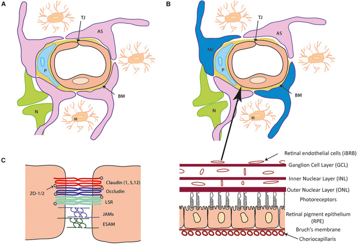
The homeostatic balance of the brain and retina is maintained by the presence of the blood-brain and inner blood-retinal barrier (BBB/iBRB, respectively) which are highly specialized barriers. Endothelial cells forming the lining of these blood vessels are interconnected by the presence of tight junctions which form the BBB and iBRB. These tight junctions, formed of numerous interacting proteins, enable the entry of molecules into neural tissues while restricting the entry of harmful material such as anaphylatoxins, bacteria and viruses. If the tight junction complex becomes dysregulated due to changes in expression levels of one or more of the components, this can have detrimental effects leading to brain and retinal pathology.
Keywords: blood brain barrier; endothelial cells; inner blood-retinal barrier; neurovasculature; tight junction.
Copyright © 2021 Hudson and Campbell.
Conflict of interest statement
The authors declare that the research was conducted in the absence of any commercial or financial relationships that could be construed as a potential conflict of interest.
Figures
References
-
- Antonetti D. A., Barber A. J., Hollinger L. A., Wolpert E. B., Gardner T. W. (1999). Vascular endothelial growth factor induces rapid phosphorylation of tight junction proteins occludin and zonula occluden 1. A potential mechanism for vascular permeability in diabetic retinopathy and tumors. J. Biol. Chem. 274 23463–23467. 10.1074/jbc.274.33.23463 - DOI - PubMed
-
- Antonetti D. A., Barber A. J., Khin S., Lieth E., Tarball J. M., Gardner T. W. (1998). Vascular permeability in experimental diabetes is associated with reduced endothelial occludin content: vascular endothelial growth factor decreases occludin in retinal endothelial cells. Penn State Retina Research Group. Diabetes 47 1953–1959. 10.2337/diabetes.47.12.1953 - DOI - PubMed
-
- Armulik A., Genové G., Mäe M., Nisancioglu M. H., Wallgard E., Niaudet C., et al. (2010). Pericytes regulate the blood brain barrier. Nature 468 557–561. - PubMed
Publication types
LinkOut - more resources
Full Text Sources