. 2021 Nov 11:14:763777.
doi: 10.3389/fnmol.2021.763777. eCollection 2021.

Disease Duration Influences Gene Expression in Neuromelanin-Positive Cells From Parkinson's Disease Patients

Affiliations

Disease Duration Influences Gene Expression in Neuromelanin-Positive Cells From Parkinson's Disease Patients

Katarína Tiklová et al. Front Mol Neurosci. .

Abstract

Analyses of gene expression in cells affected by neurodegenerative disease can provide important insights into disease mechanisms and relevant stress response pathways. Major symptoms in Parkinson's disease (PD) are caused by the degeneration of midbrain dopamine (mDA) neurons within the substantia nigra. Here we isolated neuromelanin-positive dopamine neurons by laser capture microdissection from post-mortem human substantia nigra samples recovered at both early and advanced stages of PD. Neuromelanin-positive cells were also isolated from individuals with incidental Lewy body disease (ILBD) and from aged-matched controls. Isolated mDA neurons were subjected to genome-wide gene expression analysis by mRNA sequencing. The analysis identified hundreds of dysregulated genes in PD. Results showed that mostly non-overlapping genes were differentially expressed in ILBD, subjects who were early after diagnosis (less than five years) and those autopsied at more advanced stages of disease (over five years since diagnosis). The identity of differentially expressed genes suggested that more resilient, stably surviving DA neurons were enriched in samples from advanced stages of disease, either as a consequence of positive selection of a less vulnerable long-term surviving mDA neuron subtype or due to up-regulation of neuroprotective gene products.

Keywords: Parkinson’s disease; RNA sequencing; cell death; disease duration; gene expression; neurodegenerative disease; neuroprotection.


Conflict of interest statement

The authors declare that the research was conducted in the absence of any commercial or financial relationships that could be construed as a potential conflict of interest.

Figures

FIGURE 1
FIGURE 1
LCM and RNA-seq analysis of mDA neurons from postmortem cases of Parkinson’s disease, incidental Lewy body disease and age-matched controls. (A) Schematic overview of experimental design. Midbrain sections were used to isolate neuromelanin positive DA neurons by LCM and processed for RNA-seq. (B) Micrographs of ventral midbrain neuromelanin positive cells before and after LCM. (C) Parkinson’s disease, incidental Lewy body disease and control samples analyzed in the study.
FIGURE 2
FIGURE 2
RNA-seq analysis of neuromelanin positive cells in comparison to cortex. (A) Two dimensional PCA (PC1, PC2) of NM+ neurons (252 samples) and cortical neurons (18 samples). Samples are color-coded by cell type. (B) Volcano plot showing fold changes of individual genes in the DESeq2 analysis. Red: higher expression in NM+ samples (right side of the plot) or cortex samples (left side of the plot), (padj < 0.05, RPKM > 1, fold change > 1.5), Black: no significant difference. (C–E) Expression level per cell type for indicated genes in log2(RPKM+1) in y-axes. Genes represent markers for pan-neuronal cells (C), dopamine neuron (D), and dopamine transcription factors (E).
FIGURE 3
FIGURE 3
Segregation of Parkinson’s disease samples influenced by disease progression. (A) Two dimensional PCA (PC2, PC5) of Control, Parkinson’s disease and incidental Lewy body disease samples. PC5 separates Parkinson’s disease from Control and incidental Lewy body disease samples. Samples are color-coded by disease. (B,C) Two dimensional PCA (PC4, PC5) of Control and Parkinson’s disease samples. Samples are color-coded by disease (B) and disease duration (C). PC5 separates Parkinson’s disease from Control samples. (D) Two dimensional PCA (PC2, PC3) of PDearly and PDlate samples. PC2 separates PDlate from PDearly samples. Samples are color-coded by disease duration.
FIGURE 4
FIGURE 4
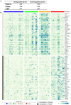
Gene expression changes in PDearly, PDlate and incidental Lewy body disease. (A) Number of up- and down-regulated genes in PDearly, PDlate and incidental Lewy body disease (padj < 0.05, RPKM > 1, fold change > 1.5). (B) Heatmap visualizing expression of top-upregulated genes in PDearly and PDlate. Sample types are indicated by color coding. Genes upregulated early are in the top part of the heatmap (light gray) while genes upregulated late are in the bottom part (dark gray).
FIGURE 5
FIGURE 5
Dopamine related gene expression changes in PDearly, PDlate and incidental Lewy body disease. (A–C) Arrows indicate significant (padj < 0.05, RPKM in controls > 1) up- or down-regulation of typical DA markers (A), PD-associated genes (B) and SNC/VTA genes (C) in PDearly, PDlate and incidental Lewy body disease samples. Hyphen indicates nonsignificant change.
FIGURE 6
FIGURE 6
Up- and down-regulated biological processes in PDearly, PDlate and incidental Lewy body disease. Top significant biological process categories from differentially expressed genes (padj < 0.05, RPKM in controls > 1, fold change > 1.5) as analyzed by ToppGene suite. Heatmap indicates significance of each individual GO-term as revealed by Bonferroni test.

References

    1. Adler C. H., Connor D. J., Hentz J. G., Sabbagh M. N., Caviness J. N., Shill H. A., et al. (2010). Incidental lewy body disease: clinical comparison to a control cohort. Mov. Disord. 25 642–646. 10.1002/mds.22971 - DOI - PMC - PubMed
    1. Armstrong M. J., Okun M. S. (2020). Diagnosis and treatment of Parkinson disease. JAMA 323 548–560. 10.1001/jama.2019.22360 - DOI - PubMed
    1. Beach T. G., Adler C. H., Sue L. I., Peirce J. B., Bachalakuri J., Dalsing-Hernandez J. E., et al. (2008). Reduced striatal tyrosine hydroxylase in incidental Lewy body disease. Acta Neuropathol. 115 445–451. 10.1007/s00401-007-0313-7 - DOI - PMC - PubMed
    1. Beach T. G., Adler C. H., Sue L. I., Serrano G., Shill H. A., Walker D. G., et al. (2015). Arizona study of aging and neurodegenerative disorders and brain and body donation program. Neuropathology 35 354–389. 10.1111/neup.12189 - DOI - PMC - PubMed
    1. Bernardini J. P., Brouwer J. M., Tan I. K., Sandow J. J., Huang S., Stafford C. A., et al. (2019). Parkin inhibits BAK and BAX apoptotic function by distinct mechanisms during mitophagy. EMBO J. 38:e99916. 10.15252/embj.201899916 - DOI - PMC - PubMed