. 2021 Nov 18:13:715811.
doi: 10.3389/fnsyn.2021.715811. eCollection 2021.

Synaptic Density and Neuronal Metabolic Function Measured by Positron Emission Tomography in the Unilateral 6-OHDA Rat Model of Parkinson's Disease

Affiliations

Synaptic Density and Neuronal Metabolic Function Measured by Positron Emission Tomography in the Unilateral 6-OHDA Rat Model of Parkinson's Disease

Nakul Ravi Raval et al. Front Synaptic Neurosci. .

Abstract

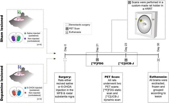
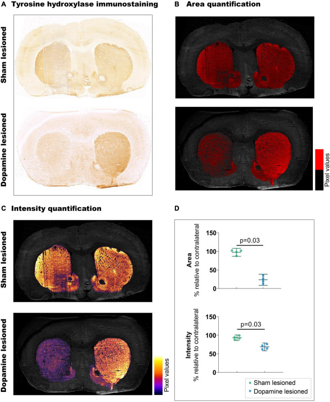
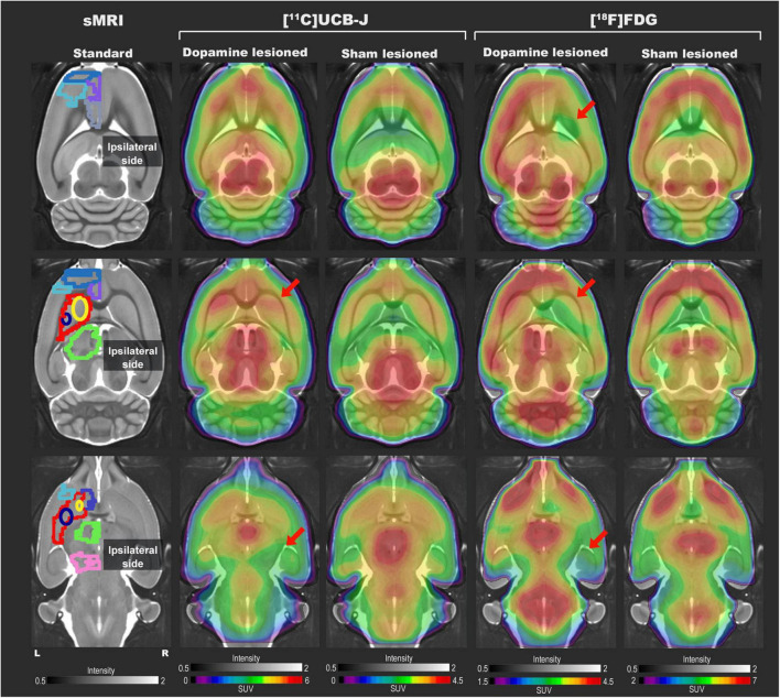
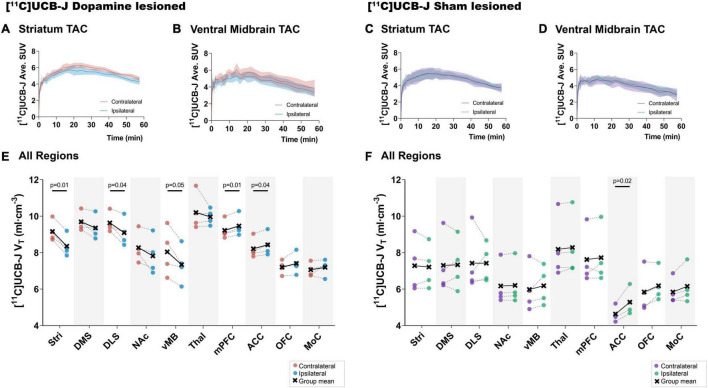
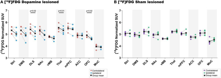
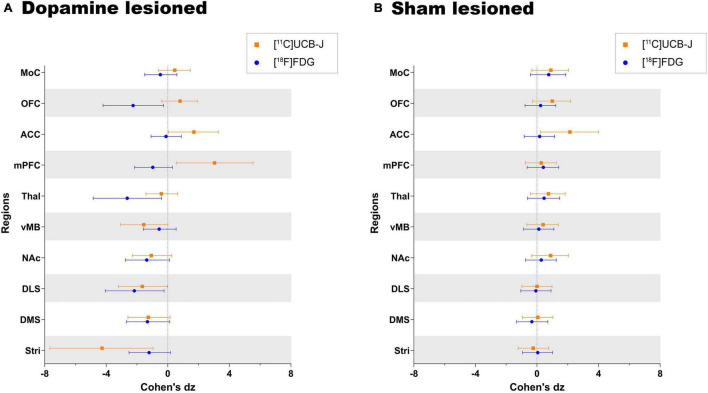
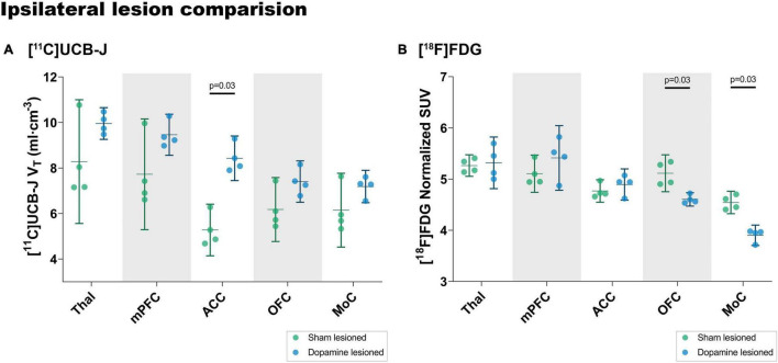
Parkinson's disease (PD) is caused by progressive neurodegeneration and characterised by motor dysfunction. Neurodegeneration of dopaminergic neurons also causes aberrations within the cortico-striato-thalamo-cortical (CSTC) circuit, which has been hypothesised to lead to non-motor symptoms such as depression. Individuals with PD have both lower synaptic density and changes in neuronal metabolic function in the basal ganglia, as measured using [11C]UCB-J and [18F]FDG positron emission tomography (PET), respectively. However, the two radioligands have not been directly compared in the same PD subject or in neurodegeneration animal models. Here, we investigate [11C]UCB-J binding and [18F]FDG uptake in the CSTC circuit following a unilateral dopaminergic lesion in rats and compare it to sham lesioned rats. Rats received either a unilateral injection of 6-hydroxydopamine (6-OHDA) or saline in the medial forebrain bundle and rostral substantia nigra (n = 4/group). After 3 weeks, all rats underwent two PET scans using [18F]FDG, followed by [11C]UCB-J on a separate day. [18F]FDG uptake and [11C]UCB-J binding were both lower in the ipsilateral striatal regions compared to the contralateral regions. Using [11C]UCB-J, we could detect an 8.7% decrease in the ipsilateral ventral midbrain, compared to a 2.9% decrease in ventral midbrain using [18F]FDG. Differential changes between hemispheres for [11C]UCB-J and [18F]FDG outcomes were also evident in the CSTC circuit's cortical regions, especially in the orbitofrontal cortex and medial prefrontal cortex where higher synaptic density yet lower neuronal metabolic function was observed, following lesioning. In conclusion, [11C]UCB-J and [18F]FDG PET can detect divergent changes following a dopaminergic lesion in rats, especially in cortical regions that are not directly affected by the neurotoxin. These results suggest that combined [11C]UCB-J and [18F]FDG scans could yield a better picture of the heterogeneous cerebral changes in neurodegenerative disorders.

Keywords: 6-OHDA = 6-hydroxydopamine; CSTC = cortico-striato-thalamo-cortical; FDG – PET; Parkinson’s disease (PD); SV2 proteins; SV2A; UCB-J; dopamine neurodegeneration.


Conflict of interest statement

MP: Compass Pathways Plc (research collaboration), GK: H. Lundbeck A/S (research collaboration), Compass Pathways Plc (research collaboration), Elysis (research collaboration), Novo Nordisk, Novozymes, Chr. Hansen (stockholder), Sage Therapeutics and Sanos (Advisor). GK is currently the president of the European College of Neuropsychopharmacology. The remaining authors declare that the research was conducted in the absence of any commercial or financial relationships that could be construed as a potential conflict of interest.

Figures

FIGURE 1
FIGURE 1
Study design. Eight rats received two intracranial injections of either 6-OHDA or saline in the medial forebrain bundle (MFB) and rostral to substantia nigra and hence divided into two groups sham lesioned (saline) or dopamine lesioned (6-OHDA). Approximately 21 days after the injections, all rats underwent an [18F]FDG PET scan followed by [11C]UCB-J PET scan 2 days after in a Siemen’s high-resolution research tomography (HRRT). All animals were euthanised 30 days after the intracranial injection.
FIGURE 2
FIGURE 2
Confirmation of 6-OHDA-induced dopaminergic lesions. (A) Representative example of tyrosine hydroxylate immunostaining: upper section from sham lesioned rats, lower from dopamine lesioned rats. (B) Quantification of the stained area, threshold emphasised in red. (C) Quantification of staining intensity, intensity scale insert. (D) Quantification of intensity and area relative to contralateral striatum (n = 4/group). Error bar denotes the mean and the 95% confidence interval. P values demonstrated from the Mann-Whitney tests.
FIGURE 3
FIGURE 3
Representative [11C]UCB-J and [18F]FDG PET SUV horizontal brain slices from a dopamine and a sham lesioned rat. Standard structural MRI (for illustrative purposes) slices show the selected volumes of interest in one hemisphere; mPFC (medium blue), OFC (purple), motor cortex (light blue), ACC (grey), striatum (red), dorsomedial striatum (yellow), dorsolateral striatum (navy blue), thalamus (green), NAc (dark blue), and ventral midbrain (pink). For [11C]UCB-J, the SUV image represents the sum of 15–60 min; for [18F]FDG, it is the sum of all 45 min. The red arrow shows decreased tracer uptake in dopamine lesioned animals.
FIGURE 4
FIGURE 4
Average [11C]UCB-J binding. Average time-activity curves from all the animals in the striatum (A,C) and ventral midbrain (B,D). Comparison of ipsilateral and contralateral [11C]UCB-J VT values in the selected regions of interest within the dopamine lesioned (E) and sham lesioned (F) rats. Notable differences are marked with their p values. Stri = striatum, DMS = dorsomedial striatum, DLS = dorsolateral striatum, NAc = nucleus accumbens, vMB = ventral midbrain, Thal = thalamus, mPFC = medial prefrontal cortex, ACC = anterior cingulate cortex, OFC = orbitofrontal cortex, MoC = motor cortex.
FIGURE 5
FIGURE 5
[18F]FDG uptake. Direct comparison between ipsilateral and contralateral hemispheres of normalised [18F]FDG uptake in all regions of interest within the dopamine lesioned (A) and sham lesioned (B) rats. Notable differences were marked with their uncorrected p values. Stri = striatum, DMS = dorsomedial striatum, DLS = dorsolateral striatum, NAc = nucleus accumbens, vMB = ventral midbrain, Thal = thalamus, mPFC = medial prefrontal cortex, ACC = anterior cingulate cortex, OFC = orbitofrontal cortex, MoC = motor cortex.
FIGURE 6
FIGURE 6
Direct comparison of effect size (Cohen’s dz values) as measured by [11C]UCB-J and [18F]FDG PET in (A) dopamine lesioned and (B) sham lesioned animals. All regions within the dopamine and sham lesioned animals in the study are compared. Error bar denotes the mean and the 95% confidence interval. Stri = striatum, DMS = dorsomedial striatum, DLS = dorsolateral striatum, NAc = nucleus accumbens, vMB = ventral midbrain, Thal = thalamus, mPFC = medial prefrontal cortex, ACC = anterior cingulate cortex, OFC = orbitofrontal cortex, and MoC = motor cortex.
FIGURE 7
FIGURE 7
Analysis of [11C]UCB-J VT values (A) and [18F]FDG (B) uptake in the ipsilateral side of dopamine lesioned and sham lesioned animals. Error bar denotes the mean and the 95% confidence interval. Thal = thalamus, mPFC = medial prefrontal cortex, ACC = anterior cingulate cortex, OFC = orbitofrontal cortex, MoC = motor cortex.

References

    1. Bajjalieh S. M., Frantz G. D., Weimann J. M., McConnell S. K., Scheller R. H. (1994). Differential expression of synaptic vesicle protein 2 (SV2) isoforms. J. Neurosci. 14 5223–5235. 10.1523/jneurosci.14-09-05223.1994 - DOI - PMC - PubMed
    1. Bertoglio D., Verhaeghe J., Miranda A., Kertesz I., Cybulska K., Korat Š, et al. (2020). Validation and noninvasive kinetic modeling of [11C]UCB-J PET imaging in mice. J. Cereb. Blood Flow Metab. 40 1351–1362. 10.1177/0271678X19864081 - DOI - PMC - PubMed
    1. Binda K. H., Lillethorup T. P., Real C. C., Bærentzen S. L., Nielsen M. N., Orlowski D., et al. (2021). Exercise protects synaptic density in a rat model of Parkinson’s disease. Exp. Neurol. 342:113741. 10.1016/j.expneurol.2021.113741 - DOI - PubMed
    1. Blandini F., Armentero M. T., Martignoni E. (2008). The 6-hydroxydopamine model: news from the past. Parkinsonism Relat. Disord. 14 S124–S129. 10.1016/j.parkreldis.2008.04.015 - DOI - PubMed
    1. Bliss T. V. P., Collingridge G. L., Kaang B. K., Zhuo M. (2016). Synaptic plasticity in the anterior cingulate cortex in acute and chronic pain. Nat. Rev. Neurosci. 17 485–496. 10.1038/nrn.2016.68 - DOI - PubMed

LinkOut - more resources