. 2021 Nov 19:12:748367.
doi: 10.3389/fphys.2021.748367. eCollection 2021.

Analysis of Cardiac Vibration Signals Acquired From a Novel Implant Placed on the Gastric Fundus

Affiliations

Analysis of Cardiac Vibration Signals Acquired From a Novel Implant Placed on the Gastric Fundus

Henry Areiza-Laverde et al. Front Physiol. .

Abstract

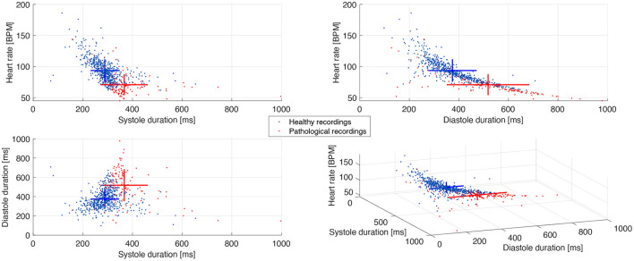
The analysis of cardiac vibration signals has been shown as an interesting tool for the follow-up of chronic pathologies involving the cardiovascular system, such as heart failure (HF). However, methods to obtain high-quality, real-world and longitudinal data, that do not require the involvement of the patient to correctly and regularly acquire these signals, remain to be developed. Implantable systems may be a solution to this observability challenge. In this paper, we evaluate the feasibility of acquiring useful electrocardiographic (ECG) and accelerometry (ACC) data from an innovative implant located in the gastric fundus. In a first phase, we compare data acquired from the gastric fundus with gold standard data acquired from surface sensors on 2 pigs. A second phase investigates the feasibility of deriving useful hemodynamic markers from these gastric signals using data from 4 healthy pigs and 3 pigs with induced HF with longitudinal recordings. The following data processing chain was applied to the recordings: (1) ECG and ACC data denoising, (2) noise-robust real-time QRS detection from ECG signals and cardiac cycle segmentation, (3) Correlation analysis of the cardiac cycles and computation of coherent mean from aligned ECG and ACC, (4) cardiac vibration components segmentation (S1 and S2) from the coherent mean ACC data, and (5) estimation of signal context and a signal-to-noise ratio (SNR) on both signals. Results show a high correlation between the markers acquired from the gastric and thoracic sites, as well as pre-clinical evidence on the feasibility of chronic cardiovascular monitoring from an implantable cardiac device located at the gastric fundus, the main challenge remains on the optimization of the signal-to-noise ratio, in particular for the handling of some sources of noise that are specific to the gastric acquisition site.

Keywords: biomedical signal processing; cardiac vibration signals; heart failure; implantable devices; seismocardiogram (SCG).


Conflict of interest statement

CD is employed by SentinHealth SA. The remaining authors declare that the research was conducted in the absence of any commercial or financial relationships that could be construed as a potential conflict of interest.

Figures

Figure 1
Figure 1
Implant prototypes used for acquiring the ECG and ACC data. (A) Schematic representation of the implant prototype V0. (B) Physical design of the gastric implant prototype V0. (C) Physical design of the external module of prototype V0. (D) Schematic representation of the implant prototype V1. (E) Physical design of the implant prototype V1. (F) Schematic representation of the implant prototype V2. (G) Physical design of the gastric module of the implant prototype V2.
Figure 2
Figure 2
Global diagram of the processing chain applied to the acquired data. Dashed-line arrows represent the ECG signal pipeline and solid-line arrows represent the ACC axes pipeline.
Figure 3
Figure 3
Data processing chain applied to the electrophysiological and mechanical cardiac data. (A) ECG signal after applying baseline removal, filtering, normalization, and QRS detection processes. (B) ACC axes after applying the baseline removal and filtering processes, the N-axis represents the norm. (C) Segmented and aligned ECG cardiac cycles in the left, and the corresponding coherent mean cardiac cycle in the right. (D) Coherent mean cardiac cycle on each ACC axis with their respective envelopes, including candidate detections for S1 and S2. The vertical dotted lines represent t1, t2, t3, and t4 in red color for Abs and black for Sqr.
Figure 4
Figure 4
Example of the final detection instants estimated for S1 and S2 on a representative recording. Vertical dotted lines represent the start and the end of S1 in red and S2 in black.
Figure 5
Figure 5
Representation of the signal of interest and noise segments of the cardiac cycle in the ECG and ACC signals to compute the SNR.
Figure 6
Figure 6
Example of implant and gold standard signals comparison. Dash-dotted lines correspond to the implant signals and solid lines correspond to the gold standard reference signals. (A) Coherent mean cycles of ECG signals taken from one representative recording. Note that the differences in signal morphology and amplitude between the surface and gastric devices are mainly explained by the significantly different dipoles that are observed. (B) Coherent mean cycles of ACC and PCG signals during both respiratory phases taken from the recording 2.
Figure 7
Figure 7
Example of implant (ACC) and gold standard (PCG) signals evolution over time. Dash-dotted lines correspond to the implant signals and solid lines correspond to the gold standard reference signals. The white background corresponds to the CPP stage and the gray background corresponds to the apnea stage. (A) Time-profiles of the duration of S1 and S2 measured on the recording 1. (B) Time-profiles of the peak-to-peak values of S1 and S2 measured on the recording 1.
Figure 8
Figure 8
Signal acceptance distribution over time. Green dots represent the recordings finally preserved, black dots represent all discarded recordings.
Figure 9
Figure 9
Scatter plot of the cardiac cycle duration between healthy and pathological pigs. The crosses represent the mean and standard deviation of each population along the three axes.
Figure 10
Figure 10
Statistical distribution and scatter plot of the norm of the peak-to-peak values between all ACC axes and the duration of S1 and S2 between healthy and pathological pigs. The crosses in the scatter plot represent the mean and standard deviation of each population.

References

    1. Ashouri H., Hersek S., Inan O. T. (2017). Universal pre-ejection period estimation using seismocardiography: quantifying the effects of sensor placement and regression algorithms. IEEE Sensors J. 18, 1665–1674. 10.1109/JSEN.2017.2787628 - DOI - PMC - PubMed
    1. Boehmer J. P., Hariharan R., Devecchi F. G., Smith A. L., Molon G., Capucci A., et al. . (2017). A multisensor algorithm predicts heart failure events in patients with implanted devices: results from the multisense study. JACC Heart Fail. 5, 216–225. 10.1016/j.jchf.2016.12.011 - DOI - PubMed
    1. Bordachar P., Garrigue S., Ritter P., Ploux S., Labrousse L., Casset C., et al. . (2011). Contributions of a hemodynamic sensor embedded in an atrial lead in a porcine model. J. Cardiovasc. Electrophysiol. 22, 579–583. 10.1111/j.1540-8167.2010.01930.x - DOI - PubMed
    1. Bordachar P., Labrousse L., Ploux S., Thambo J.-B., Lafitte S., Reant P., et al. . (2008). Validation of a new noninvasive device for the monitoring of peak endocardial acceleration in pigs: implications for optimization of pacing site and configuration. J. Cardiovasc. Electrophysiol. 19, 725–729. 10.1111/j.1540-8167.2008.01105.x - DOI - PubMed
    1. Calvo M., Bonnet J.-L., Le Rolle V., Lemonnier M., Yasuda S., Oosterlinck W., et al. . (2018). Evaluation of three-dimensional accelerometers for the study of left ventricular contractility, in 2018 Computing in Cardiology Conference (CinC), Vol. 45, (Maastricht: IEEE; ), 1–4.