. 2021 Nov 17:12:720320.
doi: 10.3389/fneur.2021.720320. eCollection 2021.

Study on the Application of Super-Resolution Ultrasound for Cerebral Vessel Imaging in Rhesus Monkeys

Affiliations

Study on the Application of Super-Resolution Ultrasound for Cerebral Vessel Imaging in Rhesus Monkeys

Li Yan et al. Front Neurol. .

Abstract

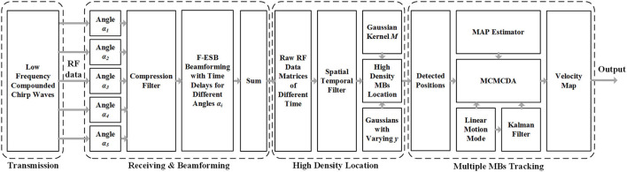
Background: Ultrasound is ideal for displaying intracranial great vessels but not intracranial microvessels and terminal vessels. Even with contrast agents, the imaging effect is still unsatisfactory. In recent years, significant theoretical advances have been achieved in super-resolution imaging. The latest commonly used ultrafast plane-wave ultrasound Doppler imaging of the brain and microbubble-based super-resolution ultrasound imaging have been applied to the imaging of cerebral microvessels and blood flow in small animals such as mice but have not been applied to in vivo imaging of the cerebral microvessels in monkeys and larger animals. In China, preliminary research results have been obtained using super-resolution imaging in certain fields but rarely in fundamental and clinical experiments on large animals. In recent years, we have conducted a joint study with the Xi'an Jiaotong University to explore the application and performance of this new technique in the diagnosis of cerebrovascular diseases in large animals. Objective: To explore the characteristics and advantages of microbubble-based super-resolution ultrasound imaging of intracranial vessels in rhesus monkeys compared with conventional transcranial ultrasound. Methods: First, the effectiveness and feasibility of the super-resolution imaging technique were verified by modular simulation experiments. Then, the imaging parameters were adjusted based on in vitro experiments. Finally, two rhesus monkeys were used for in vivo experiments of intracranial microvessel imaging. Results: Compared with conventional plane-wave imaging, super-resolution imaging could measure the inner diameters of cerebral microvessels at a resolution of 1 mm or even 0.7 mm and extract blood flow information. In addition, it has a better signal-to-noise ratio (5.625 dB higher) and higher resolution (~30-fold higher). The results of the experiments with rhesus monkeys showed that microbubble-based super-resolution ultrasound imaging can achieve an optimal resolution at the micron level and an imaging depth >35 mm. Conclusion: Super-resolution imaging can realize the monitoring imaging of high-resolution and fast calculation of microbubbles in the process of tissue damage, providing an important experimental basis for the clinical application of non-invasive transcranial ultrasound.

Keywords: cerebral vessel; microbubbles; monkey; super-resolution imaging; ultrasound.


Conflict of interest statement

The authors declare that the research was conducted in the absence of any commercial or financial relationships that could be construed as a potential conflict of interest.

Figures

Figure 1
Figure 1
Schematic view of transcranial ultrasound vascular imaging (TUVl) based on low frequency compounded chirp waves and the Markov chain Monte Carlo data association (MCMCDA) tracking algorithm. The compounded chirp waves enhance the sensitivity of the microbubble (MB) detection process while the Fast eigenspace-based beamforming (F-ESB) improves the fundamental imaging quality with low computational complexity. Based on the results of the spatial-temporal filtering, high-density MBs can be located. The map of the final vessels with blood flow velocity is achieved by tracking the detected MBs based on the MCMCDA algorithm.
Figure 2
Figure 2
Phantom imaging experimental framework.
Figure 3
Figure 3
Experimental platform for the in vivo imaging of the brain microvessels in rhesus monkeys.
Figure 4
Figure 4
Simulations with very low bubble density (up row, an initial concentration of 1 × 10² MBs per ml) and high bubble density (bottom row, an initial concentration of 2 × 109 MB per ml), respectively.
Figure 5
Figure 5
Results and comparison of the super-resolution imaging results of the cerebrovascular phantom (14). (A) Super-resolution imaging results of the phantom. (B) The compound image of the super-resolution imaging results and 5-angle compound chirp waves (CCW-5) results.
Figure 6
Figure 6
Comparison of images of the transparietal region in rhesus monkeys obtained by different imaging methods. (A) Low-frequency phased-array imaging. (B) High-frequency linear-array imaging. (C) Conventional contrast-enhanced ultrasound. (D) Super-resolution imaging results.
Figure 7
Figure 7
Comparison of the transtemporal window images of the rhesus monkeys with different imaging methods. (A) Low-frequency phased-array imaging. (B) High-frequency linear-array imaging. (C) Conventional contrast-enhanced ultrasound. (D) Super-resolution imaging results. RMCA, right middle cerebral artery.
Figure 8
Figure 8
Super-resolution transparietal imaging results of the cerebral arterial circle in rhesus monkeys. (A) Super-resolution imaging results. (B) The compound image superimposing super-resolution imaging results and STF imaging results.
Figure 9
Figure 9
Super-resolution transtemporal imaging results of the cerebral arterial circle in rhesus monkeys. (A) Super-resolution imaging results. (B) The compound image superimposing super-resolution imaging results and STF imaging results.

Similar articles

Cited by

References

    1. Harput S, Christensen-Jeffries K, Brown J, Li Y, Williams KJ, Davies AH, et al. . Two-stage motion correction for super-resolution ultrasound imaging in human lower limb. IEEE Trans Ultrason Ferroelectr Freq Control. (2018) 65:803–14. 10.1109/TUFFC.2018.2824846 - DOI - PubMed
    1. Couture O, Montaldo BB, Fink G, Tanter aM. Microbubble ultrasound super-localization imaging (MUSLI). In: 2011 IEEE International Ultrasonics Symposium. (2011). p. 1285–7. 10.1109/ULTSYM.2011.6293576 - DOI - PubMed
    1. Viessmann OM, Eckersley RJ, Christensen-Jeffries K, Tang MX, Dunsby C. Acoustic super-resolution with ultrasound and microbubbles. Phys Med Biol. (2013) 58:6447–58. 10.1088/0031-9155/58/18/6447 - DOI - PubMed
    1. Osmanski BF, Pezet S, Ricobaraza A, Lenkei Z, Tanter M. Functional ultrasound imaging of intrinsic connectivity in the living rat brain with high spatiotemporal resolution. Nat Commun. (2014) 5:5023. 10.1038/ncomms6023 - DOI - PMC - PubMed
    1. Errico C, Pierre J, Pezet S, Desailly Y, Lenkei Z, Couture O, et al. . Ultrafast ultrasound localization microscopy for deep super-resolution vascular imaging. Nature. (2015) 527:499–502. 10.1038/nature16066 - DOI - PubMed