. 2021 Nov 12:12:769597.
doi: 10.3389/fmicb.2021.769597. eCollection 2021.

Raman Imaging of Pathogenic Candida auris: Visualization of Structural Characteristics and Machine-Learning Identification

Affiliations

Raman Imaging of Pathogenic Candida auris: Visualization of Structural Characteristics and Machine-Learning Identification

Giuseppe Pezzotti et al. Front Microbiol. .

Abstract

Invasive fungal infections caused by yeasts of the genus Candida carry high morbidity and cause systemic infections with high mortality rate in both immunocompetent and immunosuppressed patients. Resistance rates against antifungal drugs vary among Candida species, the most concerning specie being Candida auris, which exhibits resistance to all major classes of available antifungal drugs. The presently available identification methods for Candida species face a severe trade-off between testing speed and accuracy. Here, we propose and validate a machine-learning approach adapted to Raman spectroscopy as a rapid, precise, and labor-efficient method of clinical microbiology for C. auris identification and drug efficacy assessments. This paper demonstrates that the combination of Raman spectroscopy and machine learning analyses can provide an insightful and flexible mycology diagnostic tool, easily applicable on-site in the clinical environment.

Keywords: Candida auris; Raman imaging; Raman spectroscopy; ergosterol; glucans; machine-learning.


Conflict of interest statement

The authors declare that the research was conducted in the absence of any commercial or financial relationships that could be construed as a potential conflict of interest.
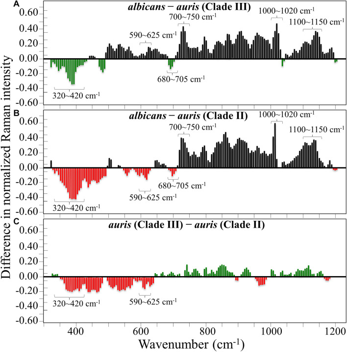
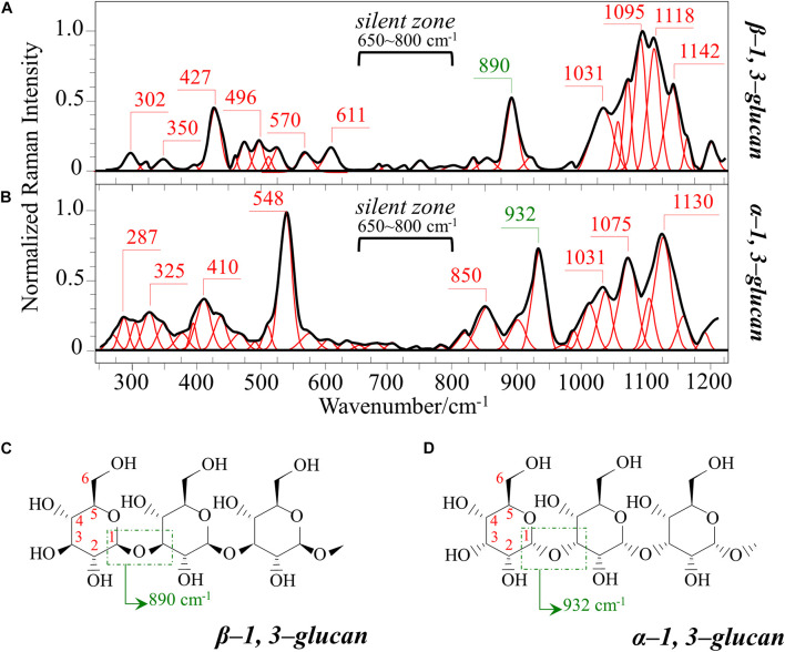
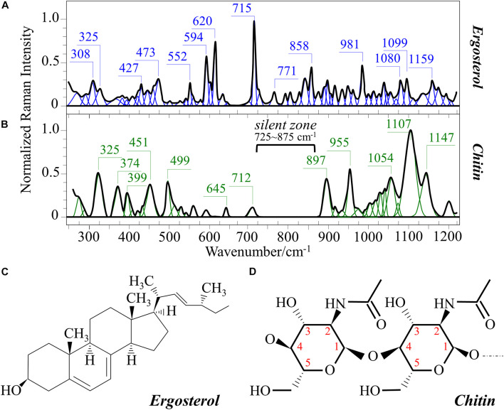
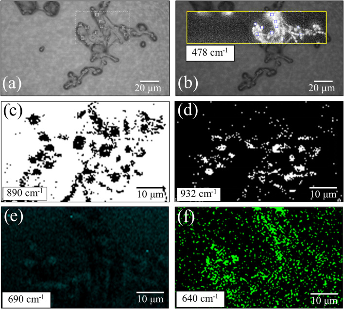
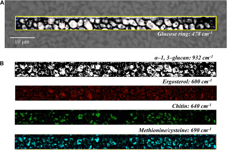
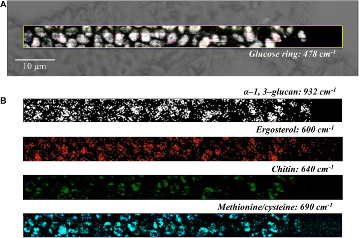
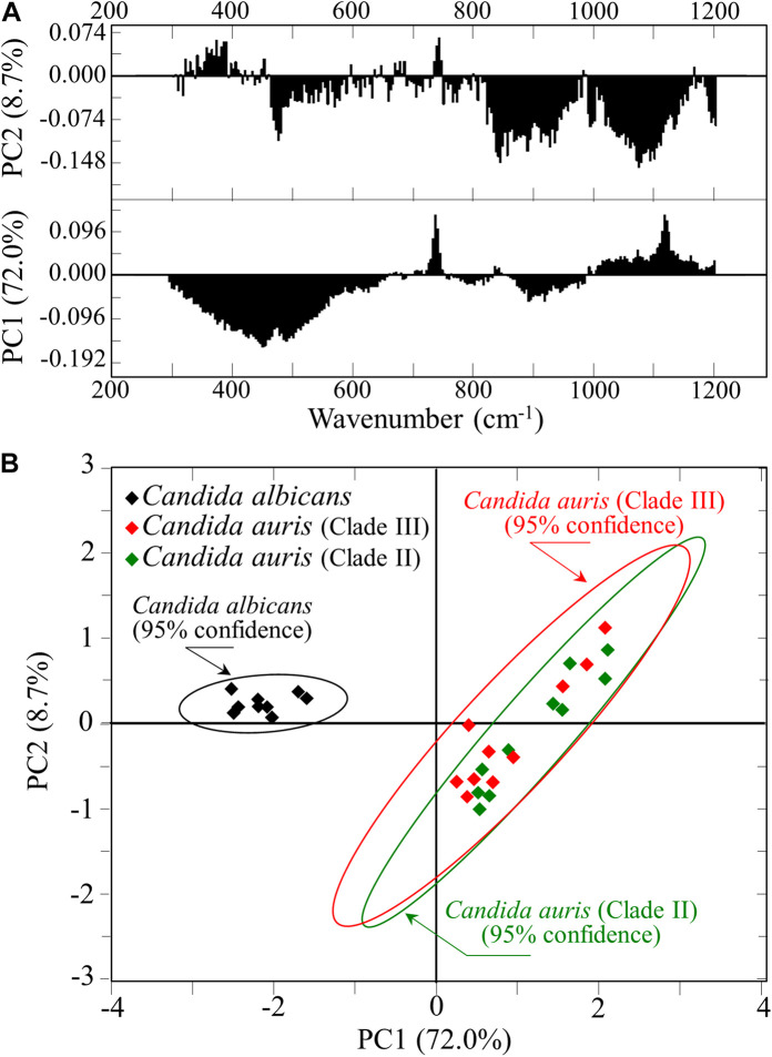
Figures

FIGURE 1
FIGURE 1
Average Raman spectra of (A) C. albicans (ATCC® 90028), (B) C. auris (Clade III), and (C) C. auris (Clade II) in the wavenumber interval 300–1200 cm–1; spectra are normalized to the glucose ring band at ∼478 cm–1 and deconvoluted into Voigtian sub-bands by means of the machine-learning algorithm described in section “Chemometric Analysis.” The wavenumbers and the assignments of the main bands discussed in the text are given with labels in inset.
FIGURE 2
FIGURE 2
Results of subtraction of the average normalized spectra in Figure 1: (A) subtraction of the C. auris (Clade III) spectrum from the C. albicans spectrum; (B) subtraction of the C. auris (Clade II) spectrum from the C. albicans spectrum; (C) subtraction of the C. auris (Clade II) spectrum from the C. auris (Clade III) spectrum.
FIGURE 3
FIGURE 3
Reference Raman spectra of: (A) β-1, 3-glucans and (B) α-1, 3-glucans (frequencies in cm–1 of the main bands given by labels in inset); (C,D) the respective glucan structures (cf. labels in inset). The spectra are deconvoluted in series of Voigtian bands.
FIGURE 4
FIGURE 4
Reference Raman spectra of: (A) ergosterol and (B) chitin (frequencies in cm– 1 of the main bands given in inset); (C,D) give the respective molecular structures (cf. labels in inset). The spectra are deconvoluted in series of Voigtian bands.
FIGURE 5
FIGURE 5
(a) Optical micrograph of cultured C. albicans cells; spatially resolved Raman maps in: (b) glucose rings in membrane polysaccharides at 478 cm–1 (in the yellow square region), (c) β-1, 3-glucans (in the white broken-line square region) at 890 cm–1 (black spots on white background), (d) α-1, 3-glucans (in the broken-line square region) at 932 cm–1 (white spots on black background), (e) S-containing amino acids (in the white broken-line square region) at 690 cm–1, and (f) chitin at 640 cm–1 (in the white broken-line square region). The 10 smaller square areas located in a,b represent the regions used for PCA analyses.
FIGURE 6
FIGURE 6
(A) Optical micrograph and Raman map of glucose rings in membrane polysaccharides at 478 cm–1 (in the yellow square region) of C. auris (Clade III) culture; in (B) from top to bottom (cf. labels), α-1, 3-glucans at 932 cm–1 (white spots on black background), ergosterol at 600 cm–1, chitin at 640 cm–1, and S-containing amino acids at 690 cm–1. The maps in (B) correspond to the same square region in (A). The 10 smaller square areas located in (A) represent the regions used for PCA analyses.
FIGURE 7
FIGURE 7
(A) Optical micrograph and Raman map of glucose rings in membrane polysaccharides at 478 cm–1 (in the yellow square region) of C. auris (Clade II) culture; in (B) from top to bottom (cf. labels), α-1, 3-glucans at 932 cm–1 (white spots on black background), ergosterol at 600 cm–1, chitin at 640 cm–1, and S-containing amino acids at 690 cm–1. The maps in (B) correspond to the same square region in (A). The 10 smaller square areas located in (A) represent the regions used for PCA analyses.
FIGURE 8
FIGURE 8
(A) First and second principal components (PC1 and PC2, respectively) of a PCA analysis of Raman spectra at the 10 selected locations from maps of C. albicans and two C. auris clades (Figures 5–7); (B) plot of the loading vectors PC1 and PC2 for the spectral region 300–1200 cm–1.

Similar articles

Cited by

References

    1. Bal A. M., McGill M. (2018). Rapid species identification of Candida directly from blood culture broths by Sepsityper-MALDI-TOF mass spectrometry: impact on antifungal therapy. J. R. Coll. Phys. Edinb. 48 114–119. 10.4997/JRCPE.2018.203 - DOI - PubMed
    1. Bednarova L., Palacky J., Bauerova V., Hruskova-Heidingsfeldova O., Pichova I., Mojzes P. (2012). Raman microspectroscopy of yeast vacuoles. Spectrosc. Int. J. 27 503–507. 10.1002/tcr.201200008 - DOI - PubMed
    1. Biswas S., Van Dijck P., Datta A. (2007). Environmental sensing and signal transduction pathways regulating morphopathogenic determinants. Microbiol. Mol. Biol. Rev. 71 348–376. 10.1128/MMBR.00009-06 - DOI - PMC - PubMed
    1. Borman A. M., Fraser M., Johnson E. M. (2021). CHROMagarTM Candida Plus: a novel chromogenic agar that permits the rapid identification of Candida auris. Med. Mycol. 59 253–258. 10.1093/mmy/myaa049 - DOI - PubMed
    1. Borman A. M., Szekely A., Johnson E. M. (2016). Comparative pathogenicity of United Kingdom isolates of the emerging pathogen Candida auris and other key pathogenic Candida species. Clin. Sci. Epidem. 1 e189–e116. - PMC - PubMed