Review
. 2021 Nov 11:12:777343.
doi: 10.3389/fmicb.2021.777343. eCollection 2021.

Malaria in Pregnancy: From Placental Infection to Its Abnormal Development and Damage

Affiliations
Review

Malaria in Pregnancy: From Placental Infection to Its Abnormal Development and Damage

Caroline Lin Lin Chua et al. Front Microbiol. .

Abstract

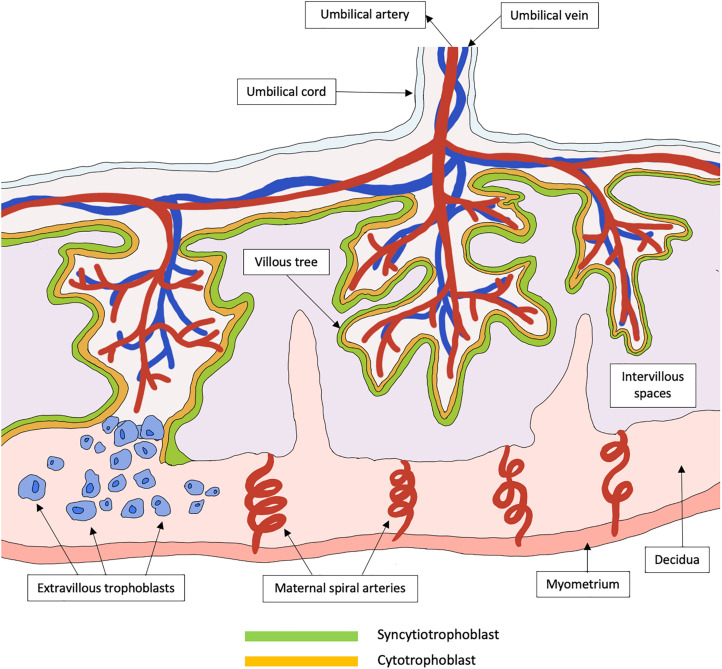
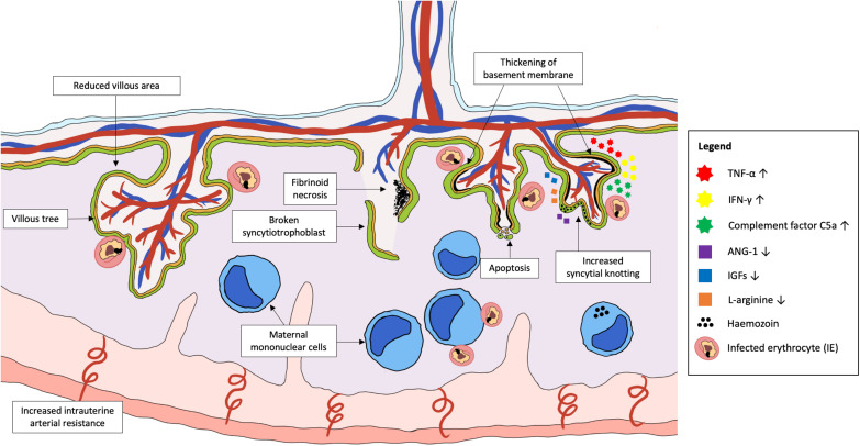
Malaria remains a global health burden with Plasmodium falciparum accounting for the highest mortality and morbidity. Malaria in pregnancy can lead to the development of placental malaria, where P. falciparum-infected erythrocytes adhere to placental receptors, triggering placental inflammation and subsequent damage, causing harm to both mother and her infant. Histopathological studies of P. falciparum-infected placentas revealed various placental abnormalities such as excessive perivillous fibrinoid deposits, breakdown of syncytiotrophoblast integrity, trophoblast basal lamina thickening, increased syncytial knotting, and accumulation of mononuclear immune cells within intervillous spaces. These events in turn, are likely to impair placental development and function, ultimately causing placental insufficiency, intrauterine growth restriction, preterm delivery and low birth weight. Hence, a better understanding of the mechanisms behind placental alterations and damage during placental malaria is needed for the design of effective interventions. In this review, using evidence from human studies and murine models, an integrated view on the potential mechanisms underlying placental pathologies in malaria in pregnancy is provided. The molecular, immunological and metabolic changes in infected placentas that reflect their responses to the parasitic infection and injury are discussed. Finally, potential models that can be used by researchers to improve our understanding on the pathogenesis of malaria in pregnancy and placental pathologies are presented.

Keywords: Plasmodium falciparum; fetal growth restriction; low birth weight; malaria; placental insufficiency; pregnancy; preterm birth; syncytiotrophoblast.


Conflict of interest statement

The authors declare that the research was conducted in the absence of any commercial or financial relationships that could be construed as a potential conflict of interest.

Figures

FIGURE 1
FIGURE 1
Schematic representation of a healthy functional unit of the human placenta. The placenta is a specialized organ that is primarily involved in the exchange of metabolites between the mother and her fetus. In the early trimester, the trophoblast differentiates to form primary syncytium that further divides into the cytotrophoblast and syncytiotrophoblast. The syncytiotrophoblast will then expand to form a continuous layer of tree-like villous that are in contact with maternal blood in the intervillous spaces. These villous trees greatly increase the surface areas available for the exchange of metabolites. Cytotrophoblasts can further differentiate into extravillous trophoblasts that invade the decidua, which is the maternal uterine tissue, to promote immune tolerance between the mother and her fetus. The extravillous trophoblasts also migrate up the maternal spiral arteries and promote spiral artery remodeling, to form large vessels of low resistance that is required to sustain a healthy pregnancy.
FIGURE 2
FIGURE 2
Plasmodium parasite infection in the placenta that cause abnormal placental development and damage. Placental malaria is an infection in the placenta by Plasmodium spp., commonly P. falciparum. Emerging evidence suggests that pregnant women are at the highest risk of PM in the first trimester, which coincides with the period of placental growth and development. Abnormalities in the placenta during PM include permanent damage to placental structures such as broken syncytiotrophoblast, thickening of basement membrane, increased syncytial knotting, increased areas of fibrinoid necrosis and dysregulated apoptosis of trophoblasts. This can result in placental insufficiency that contributes to the increased risk of fetal growth restriction, low birth weight and pre-eclampsia. The pathology is mainly associated with the activation of a local inflammatory response, characterized by the accumulation of activated maternal mononuclear cells within the intervillous spaces. The subsequent pathways leading to placental pathologies include the increased production of pro-inflammatory cytokines, chemokines, and complement proteins, as well as reduced levels of important angiogenic and placental growth factors. Together, these factors may disrupt the development of placenta if infection takes place early in pregnancy, leading to shallow spiral artery development and increased intrauterine arterial resistance.

References

    1. Abrahams V. M., Visintin I., Aldo P. B., Guller S., Romero R., Mor G. (2005). A role for TLRs in the regulation of immune cell migration by first trimester trophoblast cells. J. Immunol. 175 8096–8104. 10.4049/jimmunol.175.12.8096 - DOI - PubMed
    1. Agostinis C., Bulla R., Tripodo C., Gismondi A., Stabile H., Bossi F., et al. (2010). An alternative role of C1q in cell migration and tissue remodeling: contribution to trophoblast invasion and placental development. J. Immunol. 185 4420–4429. 10.4049/jimmunol.0903215 - DOI - PubMed
    1. Aluvihare V. R., Kallikourdis M., Betz A. G. (2004). Regulatory T cells mediate maternal tolerance to the fetus. Nat. Immunol. 5 266–271. 10.1038/ni1037 - DOI - PubMed
    1. Ashkar A. A., Di Santo J. P., Croy B. A. (2000). Interferon gamma contributes to initiation of uterine vascular modification, decidual integrity, and uterine natural killer cell maturation during normal murine pregnancy. J. Exp. Med. 192 259–270. 10.1084/jem.192.2.259 - DOI - PMC - PubMed
    1. Avery J. W., Smith G. M., Owino S. O., Sarr D., Nagy T., Mwalimu S., et al. (2012). Maternal malaria induces a procoagulant and antifibrinolytic state that is embryotoxic but responsive to anticoagulant therapy. PLoS One 7:e31090. 10.1371/journal.pone.0031090 - DOI - PMC - PubMed

LinkOut - more resources