. 2021 Nov 16:12:790742.
doi: 10.3389/fmicb.2021.790742. eCollection 2021.

Structure, Function and Regulation of a Second Pyruvate Kinase Isozyme in Pseudomonas aeruginosa

Affiliations

Structure, Function and Regulation of a Second Pyruvate Kinase Isozyme in Pseudomonas aeruginosa

Yassmin Abdelhamid et al. Front Microbiol. .

Abstract

Pseudomonas aeruginosa (PA) depends on the Entner-Doudoroff pathway (EDP) for glycolysis. The main enzymatic regulator in the lower half of the EDP is pyruvate kinase. PA contains genes that encode two isoforms of pyruvate kinase, denoted PykAPA and PykFPA. In other well-characterized organisms containing two pyruvate kinase isoforms (such as Escherichia coli) each isozyme is differentially regulated. The structure, function and regulation of PykAPA has been previously characterized in detail, so in this work, we set out to assess the biochemical and structural properties of the PykFPA isozyme. We show that pykF PA expression is induced in the presence of the diureide, allantoin. In spite of their relatively low amino acid sequence identity, PykAPA and PykFPA display broadly comparable kinetic parameters, and are allosterically regulated by a very similar set of metabolites. However, the x-ray crystal structure of PykFPA revealed significant differences compared with PykAPA. Notably, although the main allosteric regulator binding-site of PykFPA was empty, the "ring loop" covering the site adopted a partially closed conformation. Site-directed mutation of the proline residues flanking the ring loop yielded apparent "locked on" and "locked off" allosteric activation phenotypes, depending on the residue mutated. Analysis of PykFPA inter-protomer interactions supports a model in which the conformational transition(s) accompanying allosteric activation involve re-orientation of the A and B domains of the enzyme and subsequent closure of the active site.

Keywords: Entner-Doudoroff pathway; Pseudomonas aeruginosa; bacterial metabolism; glycolysis; pykF; pyruvate kinase; x-ray crystallography.


Conflict of interest statement

The authors declare that the research was conducted in the absence of any commercial or financial relationships that could be construed as a potential conflict of interest.

Figures

FIGURE 1
FIGURE 1
Genetic context and expression of PykF. (A) ORFs associated with the PA1489-PA1502 cluster and their predicted function. (B) Structure of allantoin. (C) Predicted metabolic reactions catalyzed by each encoded enzyme in the gene cluster. Note that glyoxylate and urea are the breakdown products of allantoin degradation. (D) PykF is not expressed in wild-type P. aeruginosa in the presence of glucose but is expressed in the presence of allantoin. The figure shows western blots of cell extracts of the indicated P. aeruginosa derivatives (wild-type PAO1, pykA mutant, or pykF mutant, as indicated) obtained after overnight growth in M9 medium containing glucose, glucose plus allantoin, or allantoin alone. The blot in the upper panel was probed with anti-PykA antibodies and the blot in the lower panel was probed with anti-PykF antibodies. The identity of the cross-reacting bands with higher molecular mass than PykA or PykF in each panel is not known, but their presence in the pykA and pykF mutant extracts indicates that they are unrelated to these pyruvate kinases. (E) Allantoin-induced PykF expression is abolished in a pykF mutant. The figure shows a western blot of cell extracts from a pykA or a pykF mutant (as indicated) grown in the presence or absence of glucose and/or allantoin (as indicated). To confirm minimal cross-reactivity of the antibodies, purified PykA or PykF (as indicated) were loaded on the right-hand side of each blot.
FIGURE 2
FIGURE 2
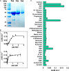
Kinetic characterization of PykF. (A) SDS-PAGE gel showing purified, untagged PykF (51.5 kDa). The gel was prepared with 12% (w/v) acrylamide and stained with Coomassie Brilliant Blue G250 [The minor bands at around 30 and 40 kDa molecular mass correspond to degradation products of PykF]. (B) PykF kinetics with respect to PEP and ADP. The PEP titration was performed using 2 mM ADP (i.e., a saturating concentration) whereas the ADP titration was performed using 5 mM PEP (also a saturating concentration). The R2 values for the curve fit to the PEP and ADP titrations were 0.96 in both cases. The 95% confidence interval estimates for the kinetic parameters are shown in Supplementary Table 4. (C) The effect of different metabolic regulators on PykF activity at low [PEP] (0.3 mM) and 2 mM ADP. Putative regulators were added at 1 mM final concentration, except for R5P, X5P, and RL5P which were used at 0.15, 0.5, and 0.5, respectively. The aim of this experiment was to identify potential activators. Data in panels (B,C) represent the mean and standard deviation of three independent experiments.
FIGURE 3
FIGURE 3
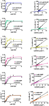
The metabolic regulation of PykF. (Left panel) Michaelis-Menten plots showing that the indicated regulators primarily convert PykF from sigmoidal to hyperbolic kinetics. Data represent the mean and standard deviation of three independent experiments. (Right panel) Lineweaver-Burk plots showing that the indicated regulators primarily act to decrease S0.5 of PykF compared with the control. “Control” indicates the reaction kinetics in the absence of added regulators. The changes in PykF kinetic constants elicited by each regulator are shown in Table 1. The 95% confidence interval estimates for the kinetic parameters are provided in Supplementary Table 4.
FIGURE 4
FIGURE 4
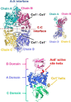
X-ray crystal structure of PykF. (Top diagrams) Front and side views of the PykF homotetramer. Chain A and B were already present in the asymmetric unit of PykF, whereas chains C and D were generated using symmetry coordinates. The A-A and the C-C interfaces are shown. The Cα1′ helices across the A-A interface are shown in dashed ovals. (Bottom diagram) Domain organization of a PykF subunit. A-, B- and C-domains of PykF are colored in blue, pink, and green, respectively. The active site helix and Cα1′ are colored in red and orange, respectively.
FIGURE 5
FIGURE 5
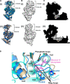
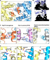
The active and allosteric sites of PykF. (A) (i) Cartoon representation of the crystal structure of a presumed inactive conformation of pyruvate kinase (PykFPA; 7OO1, blue) overlain onto the crystal structure of a presumed active configuration of the enzyme (PykAPA; 6QXL, cyan). (ii) Cartoon representation of the crystal structure of PykFPA (inactive configuration, 7OO1, blue) overlain onto the crystal structure of PykFEC (inactive configuration, 1PKY, yellow). (B) Surface representation of the crystal structure of PykAPA [(i) 6QXL] and PykFPA [(ii) 7OO1, blue]. (C) (i) Cross-section through the active site of PykAPA (active configuration of the enzyme, 6QXL) The substrate analog, malonate, is shown in yellow. (ii) Cross-section through the active site of PykFPA (inactive configuration of the enzyme, 7OO1). The superimposed binding mode of the malonate from 6QXL is shown in yellow. (D) Partial closure of the allosteric site of PykFPA. Superposition of the allosteric site in PykFPA (7OO1, blue), PykFEC (1PKY, yellow), and PykAPA (6QXL, cyan) showing disposition of the ring loop of PykFPA toward the allosteric site, probably determined by the configuration of Pro455 and Pro459. The G6P bound to PykAPA is shown as pink sticks.
FIGURE 6
FIGURE 6
The A-A interface of PykF. (A) Secondary structures present at the A-A interface in PykF. The Cα1′ helices are shown in coral. (B) The interlocking of α-helices Aα6 and Aα7 at the A-A interface (upper and lower panels show a cross-section through the interface at different tilt angles). (C) Close-up view of the interactions at the Cα1′-Cα1′ interspace in bacterial species that have a Cα1′-like structure. (D) Close-up view of the A-A interface in PykF showing salt bridge formation across the interface. Of note, the A-A interface of E. coli PykF does not contain a Cα1′-like structure or salt bridges.
FIGURE 7
FIGURE 7
Secondary structures of the C-C interface of PykFPA (blue), PykFEC (wheat) and PykAPA (cyan). The diagram shows that the C-C interface in PykFPA adopts almost the same configuration as the C-C interface in PykFEC, likely due to the absence of bound regulator in the allosteric site. The G6P bound to PykAPA is shown as pink sticks.
FIGURE 8
FIGURE 8
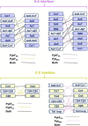
The inter-protomer interfaces in PykF. The top diagram shows a comparison between the A-A interface in PykFPA, and (left) PykFEC and (right) PykMtb. The bottom panel shows a comparison between the C-C interface in PykFPA and (left) PykFEC and (right) PykMtb. Interactions unique to PykFPA (black dashed lines), PykFEC (yellow dashed lines), and PykMtb (pink dashed lines) are shown. Interactions present in PykFPA and any of the two structures are shown as solid black lines.

Similar articles

Cited by

References

    1. Abdelhamid Y., Brear P., Greenhalgh J., Chee X., Rahman T., Welch M. (2019). Evolutionary plasticity in the allosteric regulator binding site of pyruvate kinase isoform PykA from Pseudomonas aeruginosa. J. Biol. Chem. 294 15505–15516. 10.1074/jbc.RA119.009156 - DOI - PMC - PubMed
    1. Adams P. D., Afonine P. V., Bunkóczi G., Chen V. B., Davis I. W., Echols N., et al. (2010). PHENIX: a comprehensive Python-based system for macromolecular structure solution. Acta Crystallogr. Sect. D Biol. Crystallogr. 66 213–221. 10.1107/S0907444909052925 - DOI - PMC - PubMed
    1. Al Mahmud H., Baishya J., Wakeman C. A. (2021). interspecies metabolic complementation in cystic fibrosis pathogens via purine exchange. Pathogens 10:146. 10.3390/pathogens10020146 - DOI - PMC - PubMed
    1. Al-Zaid Siddiquee K., Arauzo-Bravo M. J., Shimizu K. (2004). Metabolic flux analysis of pykF gene knockout Escherichia coli based on 13C-labeling experiments together with measurements of enzyme activities and intracellular metabolite concentrations. Appl. Microbiol. Biotechnol. 63 407–417. 10.1007/s00253-003-1357-9 - DOI - PubMed
    1. Baek Y. H., Nowak T. (1982). Kinetic evidence for a dual cation role for muscle pyruvate kinase. Arch. Biochem. Biophys. 217 491–497. 10.1016/0003-9861(82)90529-X - DOI - PubMed

LinkOut - more resources