Review
. 2021 Nov 15:12:656797.
doi: 10.3389/fimmu.2021.656797. eCollection 2021.

Epstein-Barr Virus and the Origin of Myalgic Encephalomyelitis or Chronic Fatigue Syndrome

Affiliations
Review

Epstein-Barr Virus and the Origin of Myalgic Encephalomyelitis or Chronic Fatigue Syndrome

Manuel Ruiz-Pablos et al. Front Immunol. .

Abstract

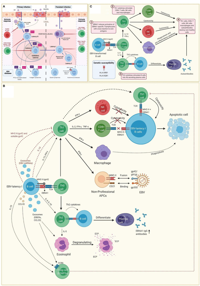
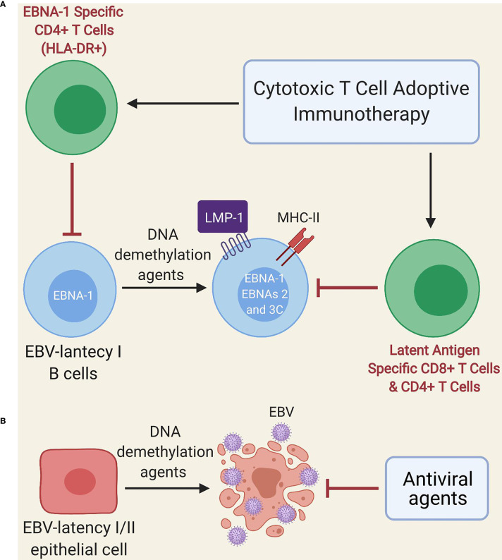
Myalgic encephalomyelitis or chronic fatigue syndrome (ME/CFS) affects approximately 1% of the general population. It is a chronic, disabling, multi-system disease for which there is no effective treatment. This is probably related to the limited knowledge about its origin. Here, we summarized the current knowledge about the pathogenesis of ME/CFS and revisit the immunopathobiology of Epstein-Barr virus (EBV) infection. Given the similarities between EBV-associated autoimmune diseases and cancer in terms of poor T cell surveillance of cells with EBV latency, expanded EBV-infected cells in peripheral blood and increased antibodies against EBV, we hypothesize that there could be a common etiology generated by cells with EBV latency that escape immune surveillance. Albeit inconclusive, multiple studies in patients with ME/CFS have suggested an altered cellular immunity and augmented Th2 response that could result from mechanisms of evasion to some pathogens such as EBV, which has been identified as a risk factor in a subset of ME/CFS patients. Namely, cells with latency may evade the immune system in individuals with genetic predisposition to develop ME/CFS and in consequence, there could be poor CD4 T cell immunity to mitogens and other specific antigens, as it has been described in some individuals. Ultimately, we hypothesize that within ME/CFS there is a subgroup of patients with DRB1 and DQB1 alleles that could confer greater susceptibility to EBV, where immune evasion mechanisms generated by cells with latency induce immunodeficiency. Accordingly, we propose new endeavors to investigate if anti-EBV therapies could be effective in selected ME/CFS patients.

Keywords: CD4+ CTL; EBV EBNA-1; HLA-II alleles; autoimmunity; cancer; chronic fatigue syndrome; immunotherapy; myalgic encephalomyelitis.


Conflict of interest statement

The authors declare that the research was conducted in the absence of any commercial or financial relationships that could be construed as a potential conflict of interest.

Figures

Figure 1
Figure 1
Immunopathobiology of Epstein-Barr virus (EBV) infection. (A) EBV is transmitted to the host through saliva from a carrier individual, and infects pharyngeal epithelial cells followed by naïve tonsillar B cells through interactions between gp350 and gp42 glycoproteins of the viral envelope with CD21 and MHC class II molecules (MHC-II), respectively. Lytic infection produces new viral particles that infect more epithelial cells. Subsequently, these EBV-infected B cells enter into a latency phase in the periphery, where they express a specific set of viral genes, including LMP1, LMP2A, EBNAs and EBER (latency III). These latent III B cells progress through the germinal center reaction into latency II and emerge as memory B cells with I/0 latencies that establish a lifelong latent infection. The immune response of the healthy host is sufficient to maintain control of the EBV infection. NK cells in tonsil produce high levels of IFN-γ that withhold the transformation of B cells by EBV during earlier stages of the infection. Both, type III and type II latent B cells are controlled by NK and T cells specific to latent proteins. By contrast, memory B cells with type I latency are only controlled by activated EBNA-1 specific CD4 T cells. EBV-infected plasma cells can periodically enter in lytic phase, but are controlled by CD4 and CD8 T cells with specificity for EBV lytic proteins. The programs of the viral latent cycle are expressed in various EBV-associated diseases. Latency I is found in Burkitt lymphoma, latency II in Hodgkin lymphoma and latency III in post-transplantation lymphoproliferative disease (PTLD) and AIDS-associated diffuse large B-cell lymphoma (DLBCL). EBV-latency I B cells escaping the surveillance of EBNA-1-specific CD4 T cells could lead to autoimmunity by presenting EBNA-1 in MHC-II/gp42, which may cause cross-reaction with own antigens. (B) EBNA1 is presented to CD4 T cells on MHC class II molecules in EBV-infected B cells. Both, MHC-II bound gp42 and soluble gp42 facilitate immune evasion by preventing activation and recognition of T cell receptors (TCR) in CD4 T cells. In addition, some EBV miRNAs could directly reduce CD4 T cell cytotoxicity through the intercellular exosomal pathway or inhibit MHC class II-mediated antigen processing and presentation in the host cell. By contrast, activation of Th1 CD4 T cells, would favor co-stimulation of CD8 T cells, NK cells and macrophages. Increased levels of IFN-γ in response to EBV infection can induce expression of MHC class II molecules in other cells such as epithelial cells, endothelial cells, pancreatic beta cells, fibroblasts, keratinocytes and glial cells, allowing them to act as non-professional antigen-presenting cells that can become infected by EBV through gp42/MHC-II interaction. If these cells also express low levels of CD21 (thymocytes, a subset of peripheral T lymphocytes, follicular dendritic cells, astrocytes and some epithelial cells), they may further facilitate EBV entry by interacting gp350 with CD21. IFN-γ released by NK cells may withhold the transformation of B cells by EBV during the early stages of infection, but it fails to inhibit the proliferation of fully transformed EBV-infected B cells (latency). CD8 T cells do not recognize EBNA1 in EBV-transformed cells, since it is presented in MHC class II molecules. Only EBNA1-specific CD4 T cells have cytotoxic activity against EBNA1-expressing B cells, causing them to enter apoptosis and become phagocyted by macrophages. However, EBV transformed B cells release IL-10, TGF-β, CCL20 and exosomes (containing EBERs and CCL20), which attract T regulatory (Treg) cells to the site of infection inhibiting antigen-stimulated CD4 effector T cells. IL-10 released by EBV transformed B cells during EBNA1 presentation, favors a Th2 (over Th1) immune response that also inhibits CD8 T cells and NK cells. These EBNA1 specific Th2 cells further induce antibody secretion by plasma cells. EBERs released on exosomes can activate other cells such as eosinophils that degranulate and release the cationic eosinophilic protein (ECP). (C) EBNA1 is one of the main candidates in the generation of autoantibodies and EBNA1 specific self-reactive cytotoxic Th1 cells in genetically predisposed individuals. EBNA-1 is subjected to citrullination and presented in MHC class II molecules after macroautophagy in EBV-infected B cells. As gp42 binds peripherally to the β1 domain of the β chain of HLA-DR -DQ, it may confer greater susceptibility or resistance to this interaction, depending on the host DRB1* and DQB1* allele. Post-translational modifications, such as citrullination, may form neoantigens that can generate autoimmunity when recognized by CD4 T cells. If polarized into a Th2 phenotype, CD4 T cells will stimulate B cell differentiation into plasma cells that could secrete autoantibodies. Antigen-specific CD4 T cells with a Th1 cytokine pattern may have cytotoxic activity, apart from co-stimulating CD8 T cells, NK cells and macrophages. Together with autoantibodies produced by plasma cells, all these cell types would participate in the destruction of healthy cells and the development of autoimmunity.
Figure 2
Figure 2
Schematic model of treatment with DNA demethylation agents followed by adoptive immunotherapy of EBV-specific T cells. (A) Administration of low-dose DNA demethylation agents (e.g. decitabine) restores the expression of MHC class II molecules and induces expression of LMP1, EBNA-2 and EBNA-3C in EBV-latency I B cells, improving the recognition of these cells by EBV-specific T cells (either autogenous or after adoptive immunotherapy). EBNA-1-specific CD4 T cells can only recognize latent I cells exhibiting EBNA-1 in MHC class II molecules since EBNA-1 is poorly immunogenic. (B) DNA demethylation agents (e.g. decitabine) induce lytic infection and apoptosis in EBV-transformed epithelial cells. Antiviral agents prevent viral replication.

Similar articles

Cited by

References

    1. Cortes Rivera M, Mastronardi C, Silva-Aldana C, Arcos-Burgos M, Lidbury B. Myalgic Encephalomyelitis/Chronic Fatigue Syndrome: A Comprehensive Review. Diagnostics (2019) 9(3):91. - PMC - PubMed
    1. Lorusso L, Mikhaylova SV, Capelli E, Ferrari D, Ngonga GK, Ricevuti G. Immunological Aspects of Chronic Fatigue Syndrome. Autoimmun Rev Elsevier (2009) 8:287–91. doi: 10.1016/j.autrev.2008.08.003 - DOI - PubMed
    1. Kerr JR. Epstein-Barr Virus Induced Gene-2 Upregulation Identifies a Particular Subtype of Chronic Fatigue Syndrome/Myalgic Encephalomyelitis. Front Pediatrics Front Media SA (2019) 7:59. doi: 10.3389/fped.2019.00059 - DOI - PMC - PubMed
    1. Bakken IJ, Tveito K, Gunnes N, Ghaderi S, Stoltenberg C, Trogstad L, et al. Two Age Peaks in the Incidence of Chronic Fatigue Syndrome/Myalgic Encephalomyelitis: A Population-Based Registry Study From Norway 2008-2012. BMC Med (2014) 12(1):167. doi: 10.1186/s12916-014-0167-5 - DOI - PMC - PubMed
    1. Collin SM, Bakken IJ, Nazareth I, Crawley E, White PD. Trends in the Incidence of Chronic Fatigue Syndrome and Fibromyalgia in the UK, 2001–2013: A Clinical Practice Research Datalink Study. J R Soc Med (2017) 110(6):231–44. doi: 10.1177/0141076817702530 - DOI - PMC - PubMed

Publication types

MeSH terms