. 2021 Nov 18:12:777860.
doi: 10.3389/fimmu.2021.777860. eCollection 2021.

Adoptive Transfer of Immune Cells Into RAG2IL-2Rγ-Deficient Mice During Litomosoides sigmodontis Infection: A Novel Approach to Investigate Filarial-Specific Immune Responses

Affiliations

Adoptive Transfer of Immune Cells Into RAG2IL-2Rγ-Deficient Mice During Litomosoides sigmodontis Infection: A Novel Approach to Investigate Filarial-Specific Immune Responses

Anna Wiszniewsky et al. Front Immunol. .

Abstract

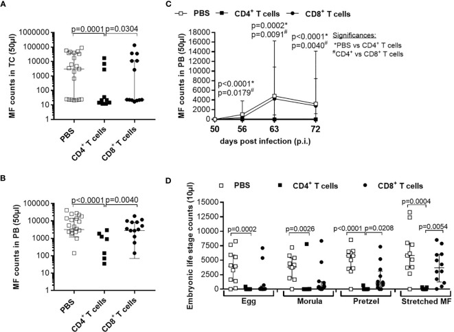
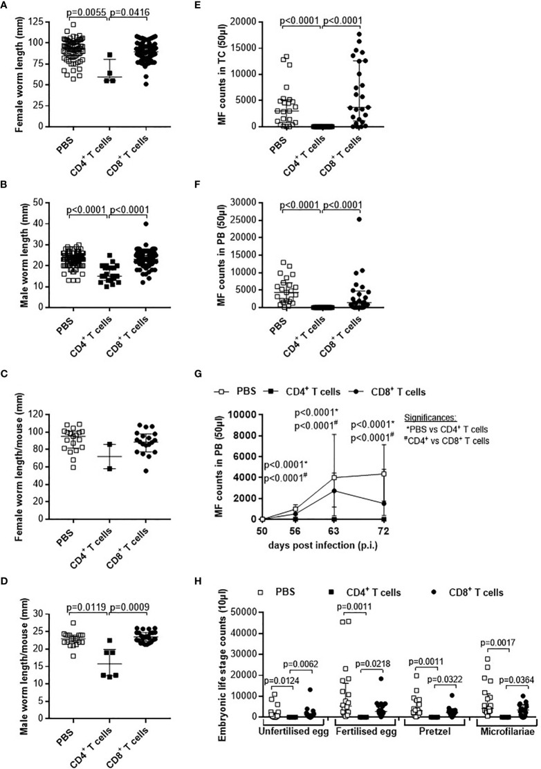
Despite long-term mass drug administration programmes, approximately 220 million people are still infected with filariae in endemic regions. Several research studies have characterized host immune responses but a major obstacle for research on human filariae has been the inability to obtain adult worms which in turn has hindered analysis on infection kinetics and immune signalling. Although the Litomosoides sigmodontis filarial mouse model is well-established, the complex immunological mechanisms associated with filarial control and disease progression remain unclear and translation to human infections is difficult, especially since human filarial infections in rodents are limited. To overcome these obstacles, we performed adoptive immune cell transfer experiments into RAG2IL-2Rγ-deficient C57BL/6 mice. These mice lack T, B and natural killer cells and are susceptible to infection with the human filaria Loa loa. In this study, we revealed a long-term release of L. sigmodontis offspring (microfilariae) in RAG2IL-2Rγ-deficient C57BL/6 mice, which contrasts to C57BL/6 mice which normally eliminate the parasites before patency. We further showed that CD4+ T cells isolated from acute L. sigmodontis-infected C57BL/6 donor mice or mice that already cleared the infection were able to eliminate the parasite and prevent inflammation at the site of infection. In addition, the clearance of the parasites was associated with Th17 polarization of the CD4+ T cells. Consequently, adoptive transfer of immune cell subsets into RAG2IL-2Rγ-deficient C57BL/6 mice will provide an optimal platform to decipher characteristics of distinct immune cells that are crucial for the immunity against rodent and human filarial infections and moreover, might be useful for preclinical research, especially about the efficacy of macrofilaricidal drugs.

Keywords: CD4+ and CD8+ T cells; Filariae; Litomosoides sigmodontis; Th17 polarization; adoptive transfer; anti-filarial immunity.


Conflict of interest statement

The authors declare that the research was conducted in the absence of any commercial or financial relationships that could be construed as a potential conflict of interest.

Figures

Figure 1
Figure 1
Adoptive transfer of naïve T cells do not alter parasite burden and thoracic cavity pathology in L. sigmodontis-infected RAG2IL-2Rγ-deficient C57BL/6 mice. CD4+ or CD8+ T cells from naïve C57BL/6 donor mice were injected intravenously (i.v.) into the tail vein of RAG2IL-2Rγ-deficient C57BL/6 mice one day prior to L. sigmodontis infection. On day 72 p.i. mice were analysed to assess (A) total, (B) female, (C) male and (D) encapsulated worm counts as well as (E) nodule counts in the thoracic cavity. In addition, H&E-stained lung and diaphragm sections were microscopically analysed to assess the (F) inflammation score. Graphs show dot blots with median and interquartile ranges from individual mice from (A–E) four independent experiments including (no adoptive transfer (PBS; n=25) and adoptive transfer of CD4+ (n=21) and CD8+ T cells (n=17) and (F) two independent experiments including PBS (n=9) and adoptive transfer of CD4+ (n=12) and CD8+ T cells (n=9) into RAG2IL-2Rγ-deficient C57BL/6 mice. Significant differences between the groups were determined by Kruskal-Wallis-test followed by a Dunn’s multiple comparison test.
Figure 2
Figure 2
Adoptive transfer of naïve CD4+ T cells impair worm growth. On day 72 p.i., RAG2IL-2Rγ-deficient C57BL/6 mice that were adoptively transferred with CD4+ or CD8+ T cell from naïve C57BL/6 donor mice were analysed for (A) worm length to assess individual (B) female and (C) male worm length and (D) female and (E) male worm length per mouse. (B–E) Graphs show dot blots with median and interquartile ranges and are pooled from four independent experiments including no adoptive transfer (PBS; n=25) and adoptive transfer of CD4+ (n=21) and CD8+ T cell (n=17) into RAG2IL-2Rγ-deficient C57BL/6 mice. Significant differences between the groups were determined by Kruskal-Wallis-test followed by a Dunn’s multiple comparison test.
Figure 3
Figure 3
Adoptive transfer of naïve CD4+ T cells impair embryogenic stage development accompanied with reduce microfilariae release. Microfilaria (MF) counts from RAG2IL-2Rγ-deficient C57BL/6 mice that were adoptively transferred with CD4+ or CD8+ T cell from naïve C57BL/6 donor mice were determined in the (A) thoracic cavity (TC) and (B) peripheral blood (PB) on day 72 p.i. and (C) during the course of infection in PB upon 50 days p.i. Moreover, female adult worms were analysed to determine (D) embryonic stages. Graphs show dot blots with median and interquartile ranges. (A–C) Results are pooled from four independent experiments including no adoptive transfer (PBS; n=25) and adoptive transfer of CD4+ (n=21) and CD8+ T cell (n=17) into RAG2IL-2Rγ-deficient C57BL/6 mice. (D) Results are from one experiment including n=9, n=6 and n=11 individual worms from no adoptive transferred (PBS; n=5) and CD4+ (n=6) and CD8+ T cell (n=6) adoptive transferred RAG2IL-2Rγ-deficient C57BL/6 mice, respectively. Significant differences between the groups were determined by Kruskal-Wallis-test followed by a Dunn’s multiple comparison test.
Figure 4
Figure 4
Reduced worm counts and thoracic cavity pathology in L. sigmodontis-infected RAG2IL-2Rγ-/- mice upon adoptive transfer of CD4+ T cell from L. sigmodontis-infected C57BL/6 donor mice. CD4+ or CD8+ T cells from C57BL/6 donor mice that were infected with L. sigmodontis for 28 days were injected intravenously (i.v.) into the tail vein of RAG2IL-2Rγ-deficient C57BL/6 mice one day prior to L. sigmodontis infection. On day 72 p.i. mice were analysed to assess (A) total, (B) female, (C) male and (D) encapsulated worm counts as well as (E) nodule counts in the thoracic cavity. In addition, H&E-stained lung and diaphragm sections were microscopically analysed to assess the (F) inflammation score. Graphs show dot blots with median and interquartile ranges from individual mice from (A–E) three independent experiments including no adoptive transfer (PBS; n=23) and adoptive transfer of CD4+ (n=21) and CD8+ T cells (n=25) and (F) two independent experiments including PBS (n=10) and adoptive transfer of CD4+ (n=15) and CD8+ T cells (n=16) into RAG2IL-2Rγ-deficient C57BL/6 mice. Significant differences between the groups were determined by Kruskal-Wallis-test followed by a Dunn’s multiple comparison test.
Figure 5
Figure 5
Adoptive transfer of CD4+ T cells from L. sigmodontis-infected C57BL/6 mice impair worm growth and blocks microfilaria release and development of embryogenic stages. On day 72 p.i., RAG2IL-2Rγ-deficient C57BL/6 mice that were adoptively transferred with CD4+ or CD8+ T cells isolated from L. sigmodontis-infected C57BL/6 mice 28 days p.i. were analysed for individual (A) female and (B) male worm length and (C) female and (D) male worm length per mouse. In addition, microfilaria (MF) counts were determined in the (E) thoracic cavity (TC) and (F) peripheral blood (PB) and (G) during the course of infection in PB upon 50 days p.i. Moreover, female adult worms were analysed to determine (H) embryonic stages. Graphs show dot blots with median and interquartile ranges from individual mice from (A–G) three independent experiments including no adoptive transfer (PBS; n=23) and adoptive transfer of CD4+ (n=21) and CD8+ T cells (n=25) into RAG2IL-2Rγ-deficient C57BL/6 mice. (H) Results are from one experiment including n=20, n=4 and n=20 individual worms from no adoptive transferred (PBS; n=7) and CD4+ (n=2) and CD8+ T cell (n=8) adoptive transferred RAG2IL-2Rγ-deficient C57BL/6 mice, respectively. Significant differences between the groups were determined by Kruskal-Wallis-test followed by a Dunn’s multiple comparison test.
Figure 6
Figure 6
Comparable efficacy of CD4+ T cells isolated from day 28 or 72 L. sigmodontis-infected C57BL/6 donor mice. CD4+ T cells from C57BL/6 donor mice that were either naïve or infected with L. sigmodontis for 28 or 72 days were injected intravenously (i.v.) into the tail vein of RAG2IL-2Rγ-deficient C57BL/6 mice one day prior to L. sigmodontis infection. On day 72 p.i. mice were analysed to assess (A) total, (B) female, (C) male and (D) encapsulated worm counts as well as (E) nodule counts in the thoracic cavity (TC). Moreover, individual (F) female and (G) male worm length and (H) female and (I) male worm length per mouse were measured. Finally, microfilaria (MF) counts were determined in the (J) TC and (K) peripheral blood (PB) and (L) during the course of infection upon 50 days p.i. in PB. (A–L) Graphs show dot blots with median and interquartile ranges from individual mice from two independent experiment including no adoptive transfer (PBS; n=11) and adoptive transfer of CD4+ T cell from naïve donor mice (n=12) or donor mice that were infected with L. sigmodontis for 28 days (n=12) or 72 days (n=14) into RAG2IL-2Rγ-deficient C57BL/6 mice. Significant differences between the groups were determined by Kruskal-Wallis-test followed by a Dunn’s multiple comparison test. AT, adoptive transfer.
Figure 7
Figure 7
Immune profile of transferred CD4+ T cells in L. sigmodontis-infected RAG2IL-2Rγ-/- mice. On day 72 p.i., CD4+ T cells and immune responses in the thoracic (TC) were analysed from L. sigmodontis-infected RAG2IL-2Rγ-deficient C57BL/6 mice. Therefore, CD4+ T cells that were either adoptively transferred from naïve, or L. sigmodontis-infected (day28 or day 72 p.i.) C57BL/6 donor mice (day28 or day 72 p.i.) were isolated and analysed for cytokine secretion patterns in the (A) TC using flow cytometry. Moreover, (B) cytokine and (C, D) chemokine levels were also analysed in the TC. Finally, (E) total TC cell counts and (F) frequencies of lymphocytes, macrophages, neutrophils and eosinophils were analysed using cytospin technique. Graphs show (A) pie charts, (A–D) box and whiskers and (E, F) dot blots with median and interquartile ranges. Graphs show (A) four independent experiments including adoptive transfer of CD4+ T cells from naïve donor mice (n= 24) or donor mice that were infected with L. sigmodontis for 28 days (n=21) or 72 days (n=13), (B–D) 6 independent experiments including no adoptive transfer (PBS; n=25) and adoptive transfer of CD4+ T cells from naïve donor mice (n= 22) or donor mice that were infected with L. sigmodontis for 28 days (n=21) or 72 days and (E, F) 2 independent experiments including PBS (n=11) and adoptive transfer of CD4+ T cells from naïve donor mice (n= 12) or donor mice that were infected with L. sigmodontis for 28 days (n=12) or 72 days (n=14) into RAG2IL-2Rγ-deficient C57BL/6 mice. Significant differences between the groups were determined by Kruskal-Wallis-test followed by a Dunn’s multiple comparison test. AT, adoptive transfer.

Similar articles

Cited by

References

    1. World Health Organization . Global Programme to Eliminate Lymphatic Filariasis: Progress Report, 2019. Wkly Epidemiol Rec (2020) 95:509–24.
    1. World Health Organization . Progress Report on the Elimination of Human Onchocerciasis, 2019-2020. Wkly Epidemiol Rec (2020) 95:545–54.
    1. Ramaiah KD, Ottesen EA. Progress and Impact of 13 Years of the Global Programme to Eliminate Lymphatic Filariasis on Reducing the Burden of Filarial Disease. PLoS Negl Trop Dis (2014) 8:e3319. doi: 10.1371/journal.pntd.0003319 - DOI - PMC - PubMed
    1. Sauerbrey M, Rakers LJ, Richards FO. Progress Toward Elimination of Onchocerciasis in the Americas. Int Health (2017) 10(suppl_1):i71–8. doi: 10.1093/inthealth/ihx039 - DOI - PubMed
    1. Traore MO, Sarr MD, Badji A, Bissan Y, Diawara L, Doumbia K, et al. . Proof-Of-Principle of Onchocerciasis Elimination With Ivermectin Treatment in Endemic Foci in Africa: Final Results of a Study in Mali and Senegal. PLoS Negl Trop Dis (2012) 6:e1825. doi: 10.1371/journal.pntd.0001825 - DOI - PMC - PubMed

Publication types