. 2021 Nov 16:8:756375.
doi: 10.3389/fvets.2021.756375. eCollection 2021.

Intracellular Staphylococcus aureus Infection Decreases Milk Protein Synthesis by Preventing Amino Acid Uptake in Bovine Mammary Epithelial Cells

Affiliations

Intracellular Staphylococcus aureus Infection Decreases Milk Protein Synthesis by Preventing Amino Acid Uptake in Bovine Mammary Epithelial Cells

Yuhao Chen et al. Front Vet Sci. .

Abstract

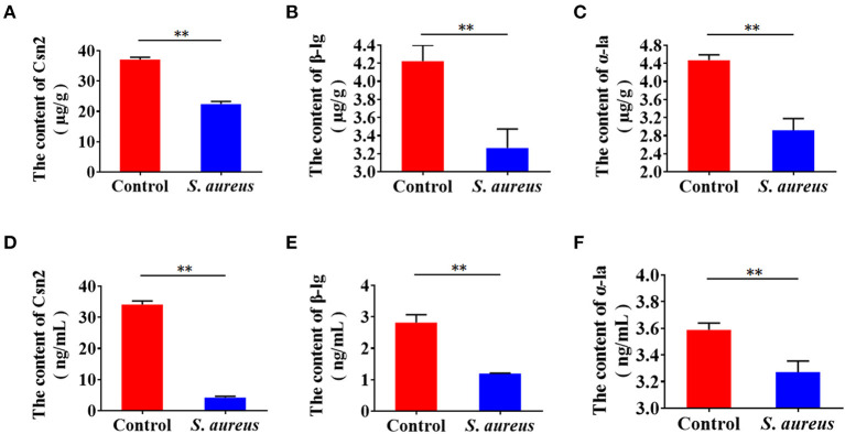
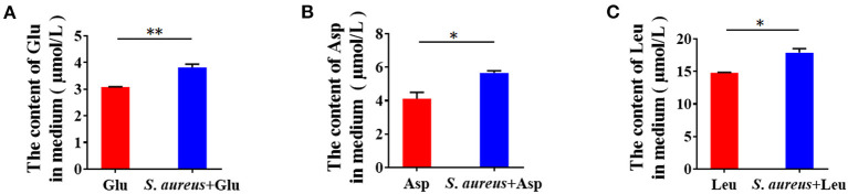
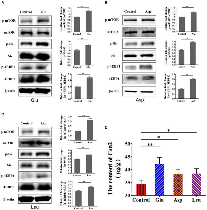
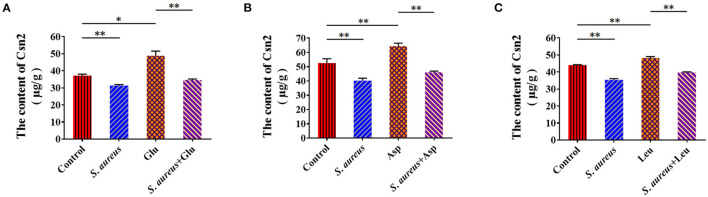
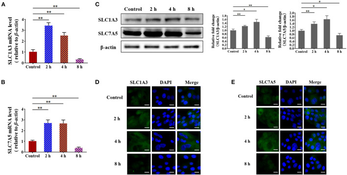
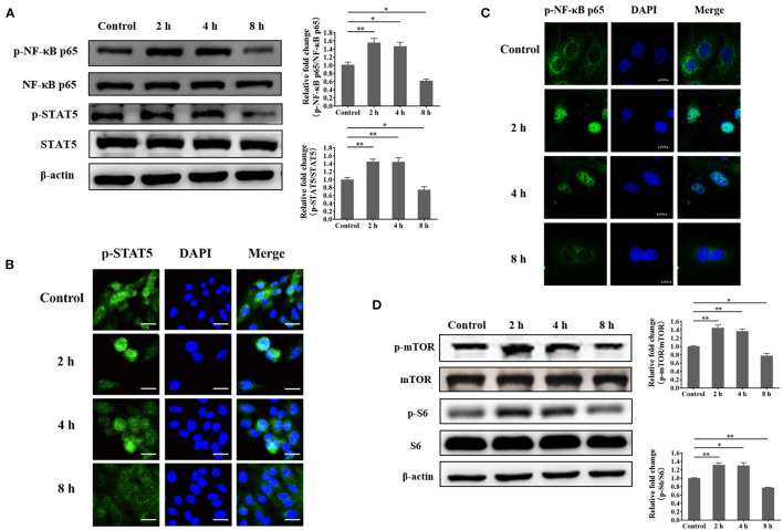
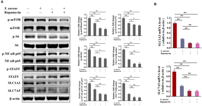
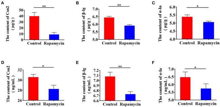
Staphylococcus aureus (S. aureus) is one of the main pathogens in cow mastitis, colonizing mammary tissues and being internalized into mammary epithelial cells, causing intracellular infection in the udder. Milk that is produced by cows that suffer from mastitis due to S. aureus is associated with decreased production and changes in protein composition. However, there is limited information on how mastitis-inducing bacteria affect raw milk, particularly with regard to protein content and protein composition. The main purpose of this work was to examine how S. aureus infection affects milk protein synthesis in bovine mammary epithelial cells (BMECs). BMECs were infected with S. aureus, and milk protein and amino acid levels were determined by ELISA after S. aureus invasion. The activity of mTORC1 signaling and the transcription factors NF-κB and STAT5 and the expression of the amino acid transporters SLC1A3 and SLC7A5 were measured by western blot or immunofluorescence and RT-qPCR. S. aureus was internalized by BMECs in vitro, and the internalized bacteria underwent intracellular proliferation. Eight hours after S. aureus invasion, milk proteins were downregulated, and the level of BMECs that absorbed Glu, Asp, and Leu from the culture medium and the exogenous amino acids induced β-casein synthesis declined. Further, the activity of mTORC1 signaling, NF-κB, and STAT5 was impaired, and SLC1A3 and SLC7A5 were downregulated. Eight hours of treatment with 100 nM rapamycin inhibited NF-κB and STAT5 activity, SLC1A3 and SLC7A5 expression, and milk protein synthesis in BMECs. Thus mTORC1 regulates the expression of SLC1A3 and SLC7A5 through NF-κB and STAT5. These findings constitute a model by which S. aureus infection suppresses milk protein synthesis by decreasing amino acids uptake in BMECs.

Keywords: Staphylococcus aureus; amino acid; amino acid transporters; bovine mammary epithelial cells; mTORC1; milk protein synthesis.


Conflict of interest statement

The authors declare that the research was conducted in the absence of any commercial or financial relationships that could be construed as a potential conflict of interest.

Figures

Figure 1
Figure 1
Staphylococcus aureus invasion suppresses milk protein synthesis and secretion in BMECs 8 h after infection. (A–C) Levels of intracellular Csn2 (β-casein), β-lg, and α-la. (D–F) Levels of Csn2 (β-casein), β-lg and α-la in cell culture medium. **p < 0.01. n = 3 independent experiments.
Figure 2
Figure 2
Staphylococcus aureus invasion prevents amino acid uptake in BMECs 8 h after infection. (A–C) Glu (A), Asp (B), and Leu (C) content in medium. *p < 0.05; **p < 0.01. n = 3 independent experiments.
Figure 3
Figure 3
Exogenous amino acid promotes mTORC1 activation and CSN2 (β-casein) synthesis. (A–C) Western blot of mTORC1 activation after BMEC stimulation with Glu (A), Asp (B), and Leu (C). Phosphorylation of mTOR, S6, and 4EBP1. (D) Intracellular Csn2 (β-casein). The resolved bands were quantified using Gel-Pro Analyzer 4.0 (Media Cybernetics, Inc., Rockville, MD, USA). *p < 0.05; **p < 0.01. n = 3 independent experiments.
Figure 4
Figure 4
Staphylococcus aureus invasion suppresses amino acid induced-casein synthesis. Intracellular Csn2 (β-casein) content (A) Glu, (B) Asp, and (C) Leu. *p < 0.05; **p < 0.01. n = 3 independent experiments.
Figure 5
Figure 5
Staphylococcus aureus infection impairs the expression of amino acid transporter genes at 8 h. (A,B) mRNA levels of SLC1A3 (A) and SLC7A5 (B) in S. aureus-infected BMECs. (C) Protein levels of SLC1A3 and SLC7A5 in S. aureus-infected BMECs. (D,E) Immunofluorescence assay of SLC1A3 (D) and SLC7A5 (E) in infected BMECs and control. Representative confocal microscopy images of SLC1A3 and SLC7A5 (Green) in cells that were co-stained with DAPI (blue). Scale bars represent 20 μm. The resolved bands were quantified using Gel-Pro Analyzer 4.0 (Media Cybernetics, Inc., Rockville, MD, USA). *p < 0.05; **p < 0.01. n = 3 independent experiments.
Figure 6
Figure 6
Staphylococcus aureus infection suppresses the phosphorylation of NF-κB p65, STAT5, mTOR, and S6 at 8 h. (A) Phosphorylation of NF-κB p65 and STAT5 in S. aureus-invaded BMECs. (B) Nuclear localization of phosphorylated STAT5 in S. aureus-invaded BMECs. Representative confocal microscopy images of the level of p-STAT5 (Green) in cells that were co-stained with DAPI (blue). Scale bars represent 20 μm. (C) Nuclear localization of phosphorylated NF-κB p65 in S. aureus-invaded BMECs. Representative confocal microscopy images of the level of p-NF-κB p65 (Green) in cells that were co-stained with DAPI (blue). Scale bars represent 10 μm. (D) mTORC1 signaling in S. aureus-invaded BMECs. The resolved bands were quantified using Gel-Pro Analyzer 4.0 (Media Cybernetics, Inc., Rockville, MD, USA). *p < 0.05; **p < 0.01. n = 3 independent experiments.
Figure 7
Figure 7
Inhibition of mTORC1 by S. aureus infection and rapamycin attenuates the phosphorylation of NF-κB p65 and STAT5 and the expression of SLC1A3 and SLC7A5. The phosphorylation of mTOR, S6, NF-κB p65, and STAT5 was detected by western blot, and SLC1A3 and SLC7A5 were examined by RT-qPCR and western blot. (A) Phosphorylation of mTOR, S6, NF-κB p65, and STAT5 and levels of SLC1A3 and SLC7A5 in different BMECs treatment groups. (B) mRNA levels of SLC1A3 and SLC7A5 in different BMECs treatment groups. mTORC1 pathway was inhibited during S. aureus infection. The resolved bands were quantified using Gel-Pro Analyzer 4.0 (Media Cybernetics, Inc., Rockville, MD, USA). *p < 0.05; **p < 0.01. n = 3 independent experiments.
Figure 8
Figure 8
Rapamycin suppresses milk protein synthesis. (A–C) Intracellular Csn2 (β-casein), β-lg, and α-la after rapamycin treatment for 8 h by ELISA. (D–F) Csn2 (β-casein), β-lg, and α-la in cell culture medium. *p < 0.05; **p < 0.01. n = 3 independent experiments.

Similar articles

Cited by

References

    1. Ramírez-Rivera EJ, Rodríguez-Miranda J, Huerta-Mora IR, Cárdenas-Cágal A, Juárez-Barrientos JM. Tropical milk production systems and milk quality: a review. Trop Anim Health Prod. (2019) 51:1295–305. 10.1007/s11250-019-01922-1 - DOI - PubMed
    1. Singhal S, Baker RD, Baker SS. A comparison of the nutritional value of cow's milk and nondairy beverages. J Pediatr Gastroenterol Nutr. (2017) 64:799–805. 10.1097/MPG.0000000000001380 - DOI - PubMed
    1. Farrell HM, Jr, Jimenez-Flores R, Bleck GT, Brown EM, Butler JE, Creamer LK, et al. . Nomenclature of the proteins of cows' milk–sixth revision. J Dairy Sci. (2004) 87:1641–74. 10.3168/jds.S0022-0302(04)73319-6 - DOI - PubMed
    1. Maurmayr A, Pegolo S, Malchiodi F, Bittante G, Cecchinato A. Milk protein composition in purebred Holsteins and in first/second-generation crossbred cows from Swedish Red, Montbeliarde and Brown Swiss bulls. Animal. (2018) 12:2214–20. 10.1017/S1751731117003640 - DOI - PubMed
    1. Bonfatti V, Grigoletto L, Cecchinato A, Gallo L, Carnier P. Validation of a new reversed-phase high-performance liquid chromatography method for separation and quantification of bovine milk protein genetic variants. J Chromatogr A. (2008) 1195:101–6. 10.1016/j.chroma.2008.04.075 - DOI - PubMed