. 2021 Nov 26:2021:5572915.
doi: 10.1155/2021/5572915. eCollection 2021.

Effects of Ozone on Hippocampus BDNF and Fos Expressions in Rats with Chronic Compression of Dorsal Root Ganglia

Affiliations

Effects of Ozone on Hippocampus BDNF and Fos Expressions in Rats with Chronic Compression of Dorsal Root Ganglia

Lingling Zhu et al. Biomed Res Int. .

Retraction in

Abstract

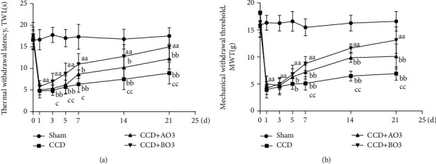
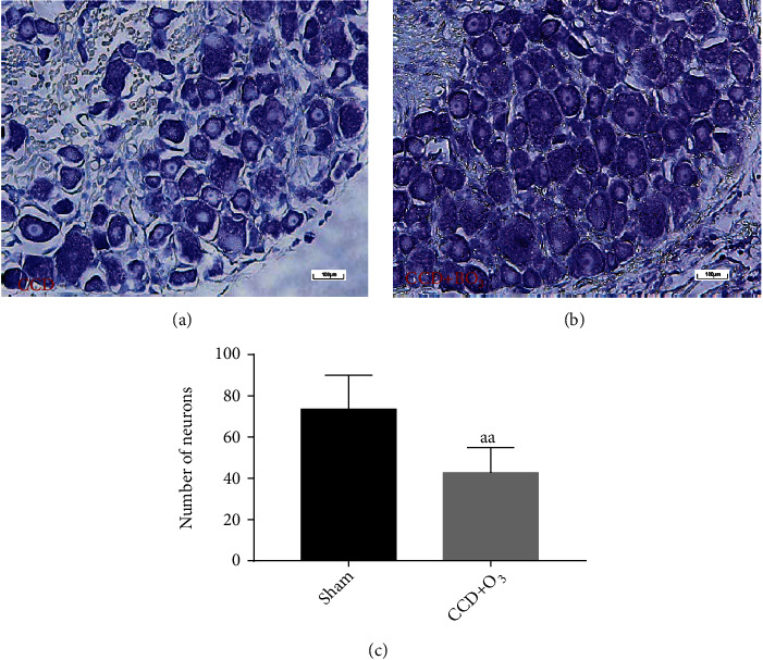
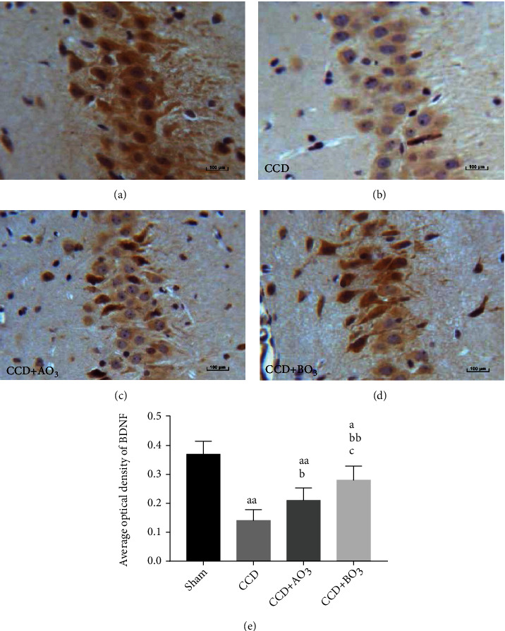
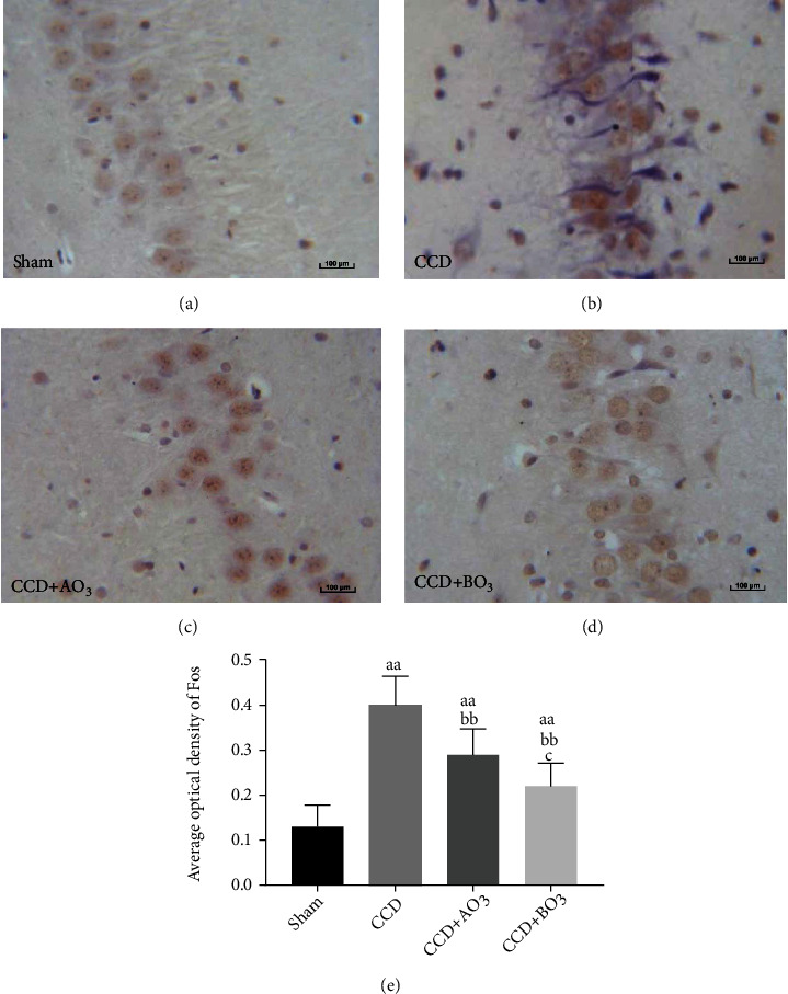
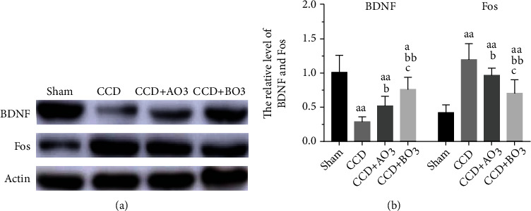
The effects of ozone on hippocampal expression levels of brain-derived neurotrophic factor (BDNF) and c-fos protein (Fos) were evaluated in rats with chronic compression of dorsal root ganglia (CCD). Forty-eight adult female Sprague-Dawley rats were randomly divided into the following 4 groups (n = 12): sham operation (sham group), CCD group, CCD with 20 μg/ml of ozone (CCD + AO3 group), and CCD with 40 μg/ml of ozone (CCD + BO3 group). Except the sham group, unilateral L5 dorsal root ganglion (DRG) compression was performed on all other groups. On days 1, 2, and 4 after the operation, the CCD + AO3 and CCD + BO3 groups were injected with 100 μl of ozone with concentrations of 20 and 40 μg/ml, respectively. Thermal withdrawal latencies (TWLs) and mechanical withdrawal thresholds (MWTs) were measured at various time points before and after the operation. BDNF and Fos expressions were examined in the extracted hippocampi using immunohistochemistry. The TWLs and MWTs of CCD model rats that received ozone were lower with decreased BDNF and increased Fos expression levels, on day 21 after the operation, compared to those of the sham group (P < 0.05). The TWLs and MWTs of the CCD + AO3 and CCD + BO3 groups were higher with increased BDNF and decreased Fos expression levels, on day 21 after the operation, compared to those of the CCD group (P < 0.05). The TWLs were longer and the MWTs were higher in the CCD + BO3 group at each time point with increased BDNF and decreased Fos expression levels, on day 21 after the operation, compared to those of the CCD + AO3 group (P < 0.05). Our results revealed that ozone can relieve the neuropathic pain caused by the pathological neuralgia resulting from DRG compression in rats. The mechanism of action for ozone is likely associated with changes in BDNF and Fos expression levels in the hippocampus.


Conflict of interest statement

All authors declare no conflicts of interest.

Figures

Figure 1
Figure 1
Effect of epidural ozone injection in alleviating pain in CCD model SD rats. With CCD, the thermal withdrawal latency (TWL) and mechanical withdrawal threshold (MWT) significantly reduced from day 1 to day 21 postsurgery (P < 0.001 for both) ((a) and (b)). The thermosensitive pain was alleviated between day 7 and day 21 after surgery in the CCD + AO3 group (P < 0.05 for all except day 10, where P = 0.002). The thermosensitive pain was significantly alleviated in the CCD + BO3 group between day 3 and day 21 after surgery (P < 0.001 for both). The thermosensitive pain-relieving effect between day 3 and day 21 postsurgery was more pronounced in the CCD + BO3 group than in the CCD + AO3 group (P < 0.001 for both) (a). The mechanical allodynia was alleviated between day 7 and day 21 in the CCD + AO3 group (P < 0.01 for all except day 7, where P = 0.046). The mechanical allodynia was also relieved significantly in the CCD + BO3 group between day 5 and day 21 (P < 0.001 for all except day 5, where P = 0.018). The thermosensitive pain-relieving between day 7 to day 21 was more pronounced in the CCD + BO3 group than in the CCD + AO3 group (P < 0.001) (b). Data are expressed as mean ± SD (aP < 0.05 and aaP < 0.01, vs. sham group; bP < 0.05 and bbP < 0.01, vs. CCD group; cP < 0.05 and ccP < 0.01, vs. CCD + AO3 group).
Figure 2
Figure 2
In the CCD group, DRG cells were shrunken with indistinct cytoplasm, loose arrangement, and faint staining (Figure 2(a)). After ozone intervention, the number of DRG cells increased, while the Nissl staining became darker compared to those of the CCD group (Figure 2(b)). The number of DRG neurons in the CCD + BO3 group was significantly higher than that in the CCD group (P < 0.01) (Figure 2(c)).
Figure 3
Figure 3
BDNF was mainly expressed in the cytoplasm and on the membrane of hippocampal cells in each group, as represented by brown staining. The average optical density of BDNF in the CCD group was lower than that in the sham group (P < 0.001), whereas those in the ozone intervention groups were higher than that in the CCD group (CCD + AO3, P < 0.05; CCD + BO3, P < 0.01). The average optical density of BDNF in the CCD + BO3 group was higher than that in the CCD + AO3 group (P < 0.05); however, it was still lower than that of the sham group (P < 0.05). Data are expressed as mean ± SD (aP < 0.05 and aaP < 0.01, vs. sham group; bP < 0.05 and bbP < 0.01, vs. CCD group; cP < 0.05 and ccP < 0.01, vs. CCD + AO3 group).
Figure 4
Figure 4
Fos staining, shown in brown, was mainly concentrated in the nucleus. Although it was lower than that in the CCD group (P < 0.01), the average optical density of Fos in hippocampal cells in the CCD + AO3 group was higher than that in the CCD + BO3 group (P < 0.05), which in turn was higher than that in the sham group (P < 0.01). Data are expressed as mean ± SD (aaP < 0.01 compared with the sham group; bP < 0.05 and bbP < 0.01, compared with the CCD group; cP < 0.05 and ccP < 0.01, compared with the CCD + AO3 group).
Figure 5
Figure 5
Western blot band images and quantitative determination of relative densities revealed that CCD of DRG significantly decreased the expression of BDNF in hippocampi compared to that in the sham group (n = 6/group). Epidural injection of ozone (20 or 40 μg/ml) increased the protein expression of BDNF compared with that in the sham group (n = 6/group) (P < 0.05). Quantitatively, BDNF relative densities were calculated as the ratio of BDNF protein band intensity to that of actin. Ozone reduced the hippocampal expression of Fos. Western blot images and quantitatively calculated relative densities revealed that CCD of DRG significantly increased the hippocampal expression of Fos compared with that in the sham group (n = 6/group). Epidural injection of ozone (20 or 40 μg/ml) decreased the protein expression of Fos compared to that in the CCD group (n = 6/group) (P < 0.05). Quantitatively, relative densities of Fos were calculated as the ratios of Fos protein band intensities to those of actin. Data are presented as mean ± SD (aaP < 0.01 vs. sham group; bP < 0.05 and bbP < 0.01, vs. CCD group; cP < 0.05 and ccP < 0.01, vs. CCD + AO3 group).

Similar articles

Cited by

References

    1. Song X. J., Hu S. J., Greenquist K. W., Zhang J. M., LaMotte R. H. Mechanical and thermal hyperalgesia and ectopic-neuronal discharge after chronic compression of dorsal root ganglia. Journal of Neurophysiology . 1999;82(6):3347–3358. doi: 10.1152/jn.1999.82.6.3347. - DOI - PubMed
    1. Kim H., Chen L., Lim G., et al. Brain indoleamine 2,3-dioxygenase contributes to the comorbidity of pain and depression. Journal of Clinical Investigation . 2012;122(8):2940–2954. doi: 10.1172/JCI61884. - DOI - PMC - PubMed
    1. Khanna S., Zheng F. Morphine reversed formalin-induced CA1 pyramidal cell suppression via an effect on septohippocampal neural processing. Neuroscience . 1999;89(1):61–71. doi: 10.1016/S0306-4522(98)00324-8. - DOI - PubMed
    1. Zhang M., Li W. B., Geng J. X., et al. The upregulation of glial glutamate transporter-1 participates in the induction of brain ischemic tolerance in rats. Journal of Cerebral Blood Flow & Metabolism . 2007;27(7):1352–1368. doi: 10.1038/sj.jcbfm.9600441. - DOI - PubMed
    1. Schmidt-Kastner R., Freund T. F. Genomic approach to selective vulnerability of the hippocampus in brain ischemia-hypoxia. Neuroscience . 2015;309(3):259–279. doi: 10.1016/j.neuroscience.2015.08.034. - DOI - PubMed

Publication types