. 2021 Oct 23:31:10-21.
doi: 10.1016/j.jpra.2021.10.004. eCollection 2022 Mar.

The long-term progression of macrodactyly

Affiliations

The long-term progression of macrodactyly

Merel L E Stor et al. JPRAS Open. .

Abstract

Background: Macrodactyly is a rare congenital disorder of overgrowth affecting the digits of the upper or lower extremity. Mostly, patients are surgically treated during childhood to reduce the digit or to stop growth. There are no standardized guidelines for the treatment and follow-up of macrodactyly. Consequently, follow-up may not be regularly scheduled into adulthood.

Methods: A retrospective, descriptive analysis of patients with the long-term progression of macrodactyly who presented at our tertiary referral hospital between July 2018 and March 2020 was performed. All patients from our local macrodactyly database were screened for progression of macrodactyly since adulthood; this resulted in four patients. The aim of these case series is to highlight the clinical features and disease course at long-term follow-up.

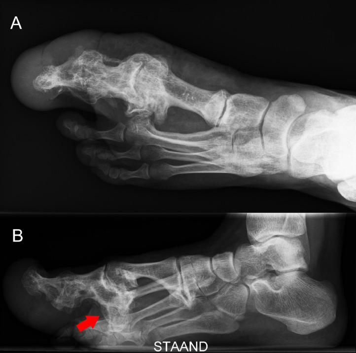
Results: All patients were surgically treated during childhood and showed progression of tissue overgrowth during adult life. All patients developed severe secondary degenerative bone changes in macrodactyly affected digits, such as ankyloses of joints, new bone formation, and bony spurs. Subsequently, tissue overgrowth and degenerative bone changes led to functional problems.

Conclusion: Patients with macrodactyly may experience growth during adult life, which may progress to deforming changes. Consequently, patients should be informed about the possible growth, and the progressive growth should be monitored.

Keywords: Macrodactyly; PIK3CA; macrodystrophia lipomatosa; overgrowth.


Figures

Figure 1
Figure 1
Lateral (A) and medial (B) view of the left foot with macrodactyly of case 1. Note the massive plantar swelling, hyperkeratosis, and psoriatic lesions.
Figure 2
Figure 2
Conventional A-P radiograph of the left foot of case 1 showing a synostosis between metatarsal I and II. Status after amputation of the first and second toe in 1962 and additional soft tissue debulking surgery in 1964, 1965, 1973, and 1978.
Figure 3
Figure 3
CT-scan of the left foot of case 1. Status after amputation of the first and second toe in 1962 and additional soft tissue debulking surgery in 1964, 1965, 1973, and 1978. 3A: Transversal view showing a synostosis between metatarsal I and II. 3B: Sagittal view with a visible bone spur from metatarsal II toward the plantar side (arrow).
Figure 4
Figure 4
Macrodactyly of the first digit of the left foot of case 2. Progression of overgrowth between age 26 years (4A) and age 31 years (4B).
Figure 5
Figure 5
Conventional radiographs of the left foot of case 2. Status after amputation of the second toe through the MTP joint and soft tissue debulking of the first toe in 1993. A: A-P radiograph showing significant bone overgrowth and deformation of the phalanxes and around the MTP joint of the first toe. B: Lateral radiograph in standing positions, notice the substantial bony formation on the plantar side of the foot (arrow) and thereby tilt of the foot.
Figure 6
Figure 6
Left foot of case 3 with macrodactyly of the second toe.
Figure 7
Figure 7
Conventional radiographs of the left foot of case 3. Status after amputation of the third toe through the MTP joint in 1980. 7A: A-P radiograph showing bone exostosis and osteoarthritic deformed bones of the second and third toe (arrow). 7B: Lateral radiograph showing elevation of the second toe because of the plantar swelling.
Figure 8
Figure 8
Conventional radiograph of the right hand of case 4. Status after soft tissue debulking surgery of the right thumb in 1989, additional soft tissue debulking, shortening of the proximal phalanx, and arthrodesis of the IP joint and MCP joint of the thumb in 2009, and correction osteotomy of the thumb in 2009. The current radiographs are showing progression of juxta-articular new bone formation between age 38 (8A) and age 47 (8B). In particular, the thumb and index finger (upper arrow) and the scaphoid, trapezium, and trapezoid (bottom arrow) show severe progression.

References

    1. Cerrato F, Eberlin KR, Waters P, Upton J, Taghinia A, Labow BI. Presentation and treatment of macrodactyly in children. The Journal of hand surgery. 2013;38(11):2112–2123. - PubMed
    1. Sudesh P, Raj N, Kumar R, lipomatosa Prakash S.Macrodystrophia. Foot (Edinb) 2012;22(3):172–174. - PubMed
    1. Prasetyono TO, Hanafi E, Astriana W. A Review of Macrodystrophia Lipomatosa: Revisitation. Arch Plast Surg. 2015;42(4):391–406. - PMC - PubMed
    1. Khan RA, Wahab S, Ahmad I, Chana RS. Macrodystrophia lipomatosa: four case reports. Ital J Pediatr. 2010;36:69. - PMC - PubMed
    1. Lindhurst MJ, Parker VE, Payne F, et al. Mosaic overgrowth with fibroadipose hyperplasia is caused by somatic activating mutations in PIK3CA. Nat Genet. 2012;44(8):928–933. - PMC - PubMed

LinkOut - more resources